Arq. Bras. Oftalmol. 2018;81 (3 )
:171-176
| DOI: 10.5935/0004-2749.20180037
Abstract
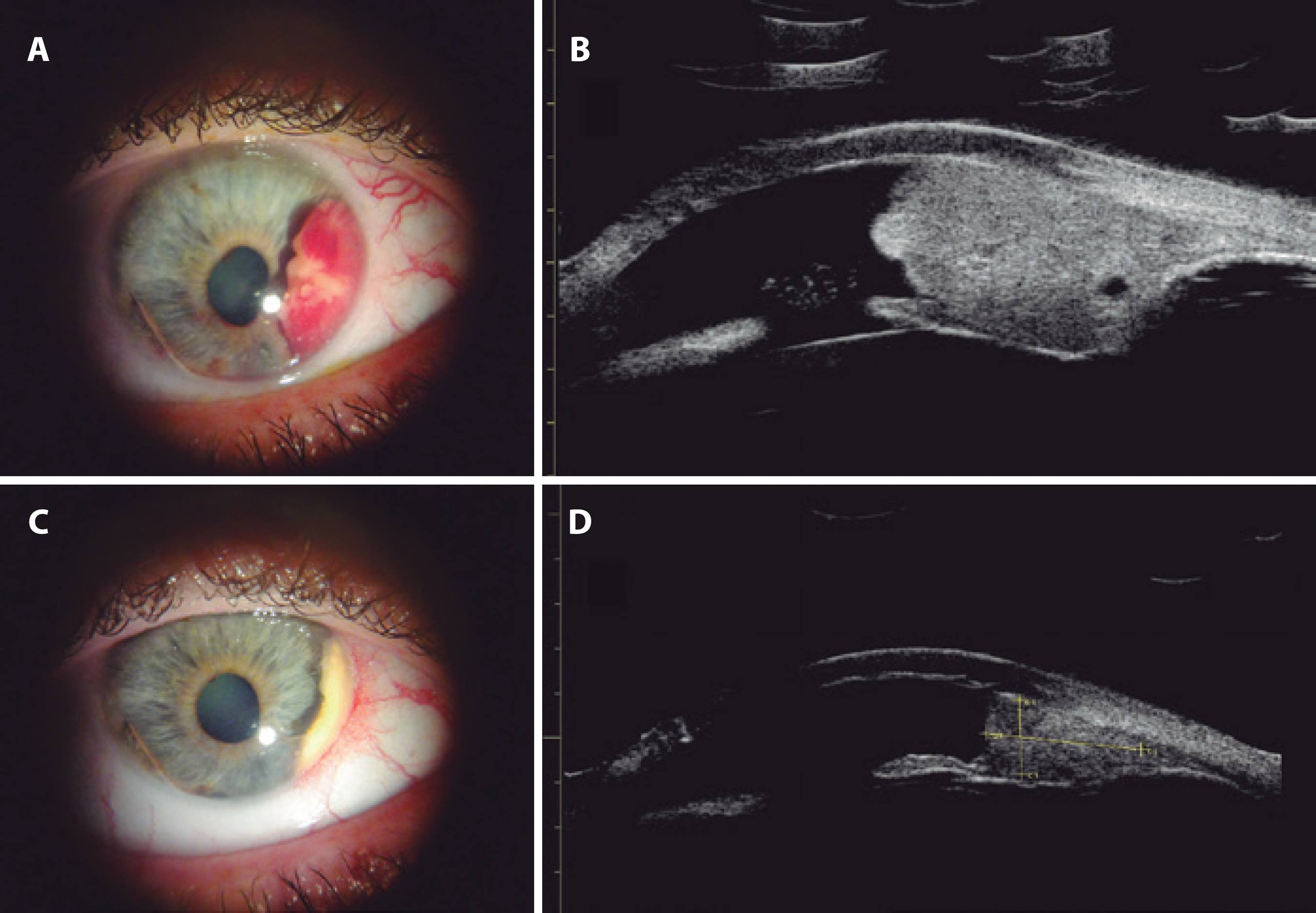
Objetivo: Avaliar o efeito do propranolol oral para hemangioma circunscrito da coroide.
Métodos: O estudo é do tipo prospectivo, quantitativo e descritivo. Propranolol oral na dose de 1.5 mg/kg/dia foi administrada em cinco pacientes com hemangioma circunscrito da coroide. Todos os pacientes foram avaliados com acuidade visual, oftalmoscopia binocular indireta, tomografia de coerência óptica, angiografia com tomografia de coerência óptica, angiografia com fluoresceína e indocianina verde e ultrassonografia ocular.
Resultados: Nenhuma mudança clínica ou no tamanho do hemangioma circunscrito da coroide foi vista através de métodos diagnósticos em qualquer momento do tratamento. Uma atenuação das complicações foi observada nos primeiros quatro meses de tratamento, com manutenção da condição e piora nos meses seguintes.
Conclusão: O estudo mostrou que o propranolol oral na dose de 1.5 mg/kg/dia não se mostrou efetivo como monoterapia no tratamento do hemangioma circunscrito da coroide.
Keywords: Hemangioma; Neoplasia da coroide; Propranolol; Verde de indocianina; Tomografia de coerência óptica
Arq. Bras. Oftalmol. 2025;88 (4 )
:1-5
| DOI: 10.5935/0004-2749.2024-0167
Abstract
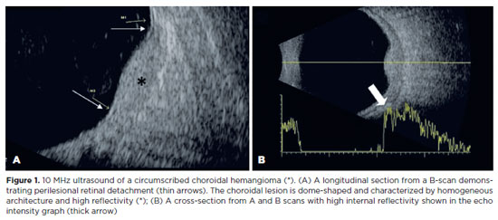
PURPOSE: To evaluate if color Doppler can detect internal blood flow in circumscribed choroidal hemangioma.
METHODS: This cross-sectional study examined seven eyes of seven participants with circumscribed choroidal hemangiomas, with or without prior treatment. B-scan ultrasound and color Doppler were used to assess the dimensions, topographical distribution, and internal blood flow of the affected eyes.
RESULTS: The sample included seven patients (five female) with a median age of 61 (62.29 ± 13.83) years. There were seven eyes with circumscribed choroidal hemangiomas in the patient sample. Color Doppler detected internal vascular flow in all cases (100%). The lesions had an average diameter/thickness ratio of >2 mm and an average thickness of <5 mm and were predominantly located superiorly and supero-temporally.
CONCLUSION: Internal blood flow was detected in circumscribed choroidal hemangiomas using color Doppler. Detection was unaffected by the patient's treatment status.
Keywords: Ultrasonography, doppler, collor; Choroidal neoplasms; Hemangioma
Arq. Bras. Oftalmol. 2024;87 (4 )
:1-7
| DOI: 10.5935/0004-2749.2023-0026
Abstract
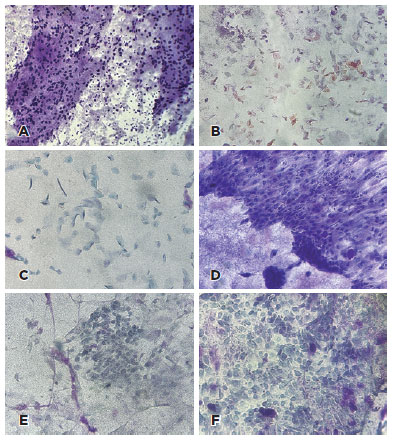
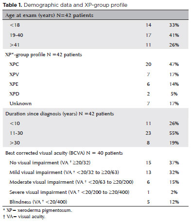
PURPOSE: To describe cellular alterations detected by impression cytology of the ocular surface in patients with xeroderma pigmentosum. The secondary objective was to assess the reliability of impression cytology in diagnosing ocular surface squamous neoplasia.
METHODS: Patients with xeroderma pigmentosum underwent a single-day complete ophthalmological examination and impression cytology for ocular surface evaluation using 13 mm diameter mixed cellulose esters membrane filters and combined staining with Periodic Acid Schiff, Hematoxylin and Eosin, and Papanicolaou stains followed by microscopic analysis. The cytological findings were correlated with the clinical diagnosis. The impression cytology findings at baseline and one-year follow-up were correlated with the clinical course (no tumor, treated tumor, residual tumor recurrent tumor, new tumor).
RESULTS: Of the 42 patients examined, impression cytology was performed in 62 eyes of 34 participants (65% females). The mean age of patients was 29.6 ± 17 years (range 7-62). Fifteen eyes had a clinical diagnosis of ocular surface squamous neoplasia. Impression cytology showed goblet cells (47, 75%), inflammatory cells (12, 19%), keratinization (5, 8%), and squamous metaplasia (30, 48%). Impression cytology was positive for atypical cells in 18 patients (12 with and 6 without ocular surface squamous neoplasia). The sensitivity, specificity, positive predictive value, and negative predictive value of impression cytology (at baseline) for diagnosis of ocular surface squamous neoplasia were 80%, 87%, 67%, and 93%, respectively, using clinical diagnosis of ocular surface squamous neoplasia as the reference standard.
CONCLUSION: Impression cytology has a moderate positive predictive value for the diagnosis of ocular surface squamous neoplasia in patients with xeroderma pigmentosum. However, the lack of detection of atypical cells on impression cytology has a high negative predictive value for ocular surface squamous neoplasia. Integration of impression cytology in the long-term management of high-risk patients, such as patients with xeroderma pigmentosum, can avoid unnecessary diagnostic biopsies.
Keywords: Xeroderma pigmentosum; Eye neoplasms; Conjunctiva/cytology; Cornea/cytology; Cytological techniques
Arq. Bras. Oftalmol. 2024;87 (2 )
:1-8
| DOI: 10.5935/0004-2749.2022-0319
Abstract
To assess Meibomian gland dysfunction using meibography in patients with xeroderma pigmentosum and correlate with ocular surface changes. This cross-sectional study evaluated patients with xeroderma pigmentosum. All patients underwent a comprehensive and standardized interview. The best-corrected visual acuity of each eye was determined. Detailed ophthalmic examination was conducted, including biomicroscopy examination of the ocular surface, Schirmer test type I, and meibography, and fundus examination was also performed when possible. Meibomian gland dysfunction was assessed by non-contact meibography using Oculus Keratograph® 5M (OCULUS Inc., Arlington, WA, USA). Saliva samples were collected using the Oragene DNA Self-collection kit (DNA Genotek Inc., Ottawa, Canada), and DNA was extracted as recommended by the manufacturer. Factors associated with abnormal meiboscores were assessed using generalized estimating equation models. A total of 42 participants were enrolled, and 27 patients underwent meibography. The meiboscore was abnormal in the upper eyelid in 8 (29.6%) patients and in the lower eyelid in 17 (62.9%). The likelihood of having abnormal meiboscores in the lower eyelid was 16.3 times greater than that in the upper eyelid.In the final multivariate model, age (p=0.001), mutation profile (p=0.006), and presence of ocular surface malignant tumor (OSMT) (p=0.014) remained significant for abnormal meiboscores. For a 1-year increase in age, the likelihood of abnormal meiboscores increased by 12%. Eyes with OSMT were 58.8 times more likely to have abnormal meiboscores than eyes without ocular surface malignant tumor.In the final model, age, xeroderma pigmentosum profile, previous cancer, and clinical alterations on the eyelid correlated with a meiboscore of ≥2.Meibomian gland dysfunction was common in patients with xeroderma pigmentosum, mainly in the lower eyelid. The severity of Meibomian gland dysfunction increases with age and is associated with severe eyelid changes.
Keywords: Meibomian glands/pathology; Meibomian glands/ diagnostic imaging; Photography; Xeroderma pigmentosum; Eyelid diseases/diagnostic imaging; Dry eye syndromes; DNA repair; Humans; Case report
 ABO is licensed under a Creative Commons Attribution-NonComercial 4.0 Internacional.
09-fig01.jpg)
02-fig01.jpg)