Arq. Bras. Oftalmol. 200871
| DOI: 10.1590/S0004-27492008000500002
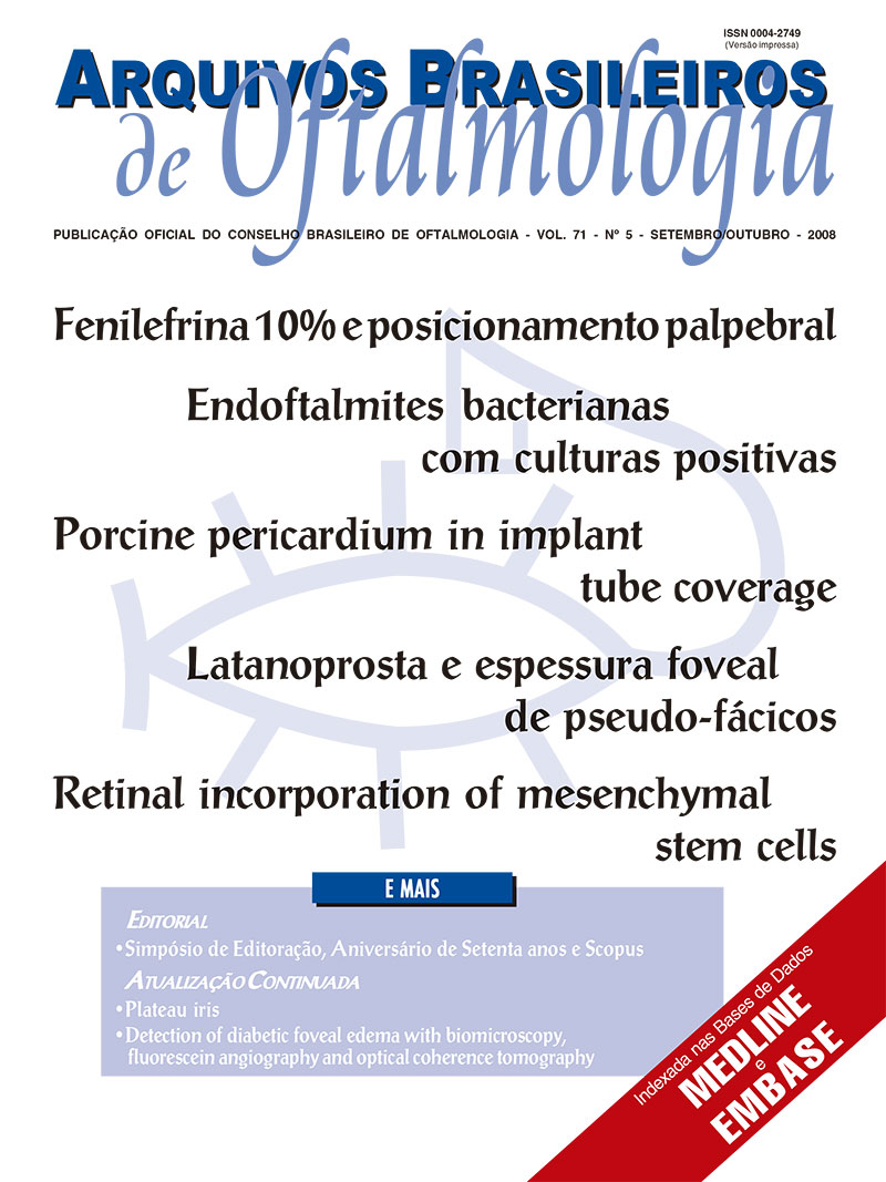
PURPOSE: To assess the distribution of microorganisms isolated from patients with bacterial endophthalmitis and their antimicrobial susceptibility.
METHODS: Retrospective analysis of medical and microbiological records of patients with suspected diagnosis of endophthalmitis and bacterial culture-proven at the Department of Ophthalmology, UNIFESP, between January 1 2000 and December 31 2005.
RESULTS: 153 (33.9%) of 451 patients showed positive bacterial culture. A total of 155 microorganisms were isolated, 79.35% were gram-positive and 20.65% gram-negative. Staphylococcus (CoNS) (41.94%) were the most frequently isolated. The antimicrobial susceptibility for gram-negative microorganisms was as follows: amikacin 87.10%, tobramycin 80.65%, ciprofloxacin 96.67%, levofloxacin, gatifloxacin and moxifloxacin 100%, ceftazidime 85.0%, and gentamicin 80.65%. Vancomycin sensitivity among gram-positive microorganisms was 100%. S. aureus and CoNS showed 83.33% of susceptibility to oxacillin, 89.61% to ciprofloxacin and 100% to gatifloxacin and moxifloxacin. The main acquisition mechanism was postoperative (60.65%).
CONCLUSION: We detected a low sensitivity of vitreous/aqueous culture for the etiologic diagnosis of endophthalmitis. The empiric antimicrobial therapy or prophylaxis should be active against gram-positive bacteria, particularly staphylococci. Surveillance studies of bacterial resistance are important for a better utilization of antimicrobials in this clinical setting.
Keywords: Eye infections, bacterial; Endophthalmitis; Endophthalmitis; Aqueous humor; Drug resistance, microbial; Microbial sensitivity tests
Arq. Bras. Oftalmol. 200871
| DOI: 10.1590/S0004-27492008000500003
PURPOSE: To evaluate the inflammatory response associated with the use of processed porcine pericardium and glycerin-preserved homologous sclera as silicone glaucoma drainage device coverage in a rabbit experimental model.
METHODS: Eight New Zealand white rabbits' eyes received an equal-sized glycerin-preserved homologous scleral patch or a double-layered processed porcine pericardium that was sutured to the bare sclera covering a silicone tube. Conjunctival hyperemia was graded using the double-blind method during the immediate postoperative period and during the first, third, and seventh postoperative weeks. After the seventh week, the enucleated eyes were histopathologically examined. They were also evaluated for signs of patch graft melting, tube erosion and chemosis.
RESULTS: There was no occurrence of graft melting or tube exposure, although porcine pericardium was associated with greater inflammation through clinical observation. Light microscopy revealed marked inflammation surrounding the porcine pericardium with foreign body granuloma formation. On the other hand, the sclera group presented milder inflammation with foreign body granulomas only around the sutures. CONCLUSIONS: Porcine pericardium is associated with significant inflammation when used as tube coverage in a rabbit model, at both histopathologic and clinical levels, when compared to glycerin-preserved homologous sclera during the seven-week follow-up period.
Keywords: Transplantation, heterologous; Pericardium; Glaucoma drainage implants; Glaucoma; Swine
Arq. Bras. Oftalmol. 200871
| DOI: 10.1590/S0004-27492008000500004
PURPOSE: To study prospectively using optical coherence tomography whether topical latanoprost induces retinal disorders in patients that underwent cataract surgery.
METHODS: Randomized, masked-observer, one-month clinical trial. Pseudophakic patients were treated with latanoprost (n=10) or lubricant drop q.d. (control group) (n=10). Half of the patients of each group presented ruptured posterior capsule (Nd:YAG laser). We evaluated the blood-retinal barrier status assessed by optical coherence tomography measurement of retinal thickness in the fovea. Before the beginning of the study and after 15 and 30 days of treatment, optical coherence tomography images were taken, and the visual acuity examination was performed.
RESULTS: There was no statistically significant increase in mean foveal thickness when patients instilled placebo (P>0.0610). A statistically significant increase in retinal thickness in the fovea was observed when patients instilled latanoprost (P<0.0004). No changes were observed in visual acuity in both groups. Mean retinal thickness in the fovea was significantly higher in the latanoprost group (P<0.0007). The mean foveal thickness in latanoprost treated eyes with ruptured posterior capsule was statistically greater when compared with that of intact posterior capsule (P<0.0461). When comparing only the patients with that of intact posterior capsule, there was a statistically significant difference in foveal thickness between patients treated with latanoprost (236.4 ± 29.4 mm) and placebo (197.8 ± 19.3 mm) only at 30 days of treatment. CONCLUSIONS: Latanoprost may lead to disruption of the blood-retinal barrier in pseudophakic patients, and is more probable to occur in patients with ruptured posterior capsule.
Keywords: Cataract extraction; Prostaglandins F, synthetic; Macular edema; Macular edema; Macular edema; Blood-retinal barrier; Tomography, optical coherence
Arq. Bras. Oftalmol. 200871
| DOI: 10.1590/S0004-27492008000500005
PURPOSE: To evaluate epidemiological aspects of eye injuries in a reference emergency center.
METHODS: The study had two surveys: in the first (A) victims of ocular trauma where 1,483 patients admitted along 2003 had their epidemiological data analyzed and the second (B), where 133 patients had clinical aspects and risk and protective factors analyzed by a standard questionnaire between May and June 2004.
RESULTS: In survey A, most patients were males (1,314 or 89%). Ocular surface foreign body was the most frequent occurrence with 863 (58%) cases. In survey B it was shown that ocular protection was used only by 17% (22) patients. The accidents occurred more frequently at the workplace 70% (93), followed by the home (22%). In survey B, 34% of the patients had previous accidents. CONCLUSIONS: The profile of patients victims of ocular trauma shows that there is a higher frequency of males and accidents involving cornea foreign body. The use of protective glasses is incipient and recurrence of this lesion is considerable. A preventive and educational strategy among the population with special focus on workplaces and homes is necessary to reduce ocular trauma occurrence.
Keywords: Emergencies; Eye foreign bodies; Eye injuries; Emergency medical services; Emergency service, hospital; Accidents, occupational; Eye diseases
Arq. Bras. Oftalmol. 200871
| DOI: 10.1590/S0004-27492008000500006
PURPOSE: To determine the upper and lower eyelid position with a single drop of 10% phenylephrine in normal individuals and observe positional lid changes in upper and lower lids of the opposite eye.
METHODS: Prospective observational study including 24 healthy subjects of both genders (age range: 20-39 yrs) submitted to instillation of a single drop of 10% phenylephrine in the upper fornix of one randomly chosen eye. A video camera recorded the images of both eyes before and after eye drop application at 3, 10, 15, 30, 45 and 60 minutes. The images were digitally processed and edited to analyze lid measurements (upper lid height: distance between the highest upper margin lid point and a horizontal line traced from the medial to the lateral canthus; lower lid height: distance between the lowest margin lid point and the above-mentioned line).
RESULTS: Before and after drug instillation, upper lid height was significantly different (p<0.001) at all moments with a mean elevation of 0.79 mm. The lower lid height changed too with a mean retraction of 0.37 mm. In the fellow eye upper lid drop was observed at almost all moments with the lowest value at 3 minutes (mean reduction of 0.68 mm) and elevation (0.37 mm) of lower lid. CONCLUSIONS: This experiment showed that a single drop of 10% phenylephrine acted not only on the upper eyelid but also on lower lid of the tested eye. Control mechanism of lid opening and closing changed upper and lower lid positioning of the opposite eye in healthy subjects.
Keywords: Eyelids; Pupil; Phenylephrine; Phenylephrine; Ophthalmic solutions
Arq. Bras. Oftalmol. 200871
| DOI: 10.1590/S0004-27492008000500007
PURPOSE: To evaluate the pattern of retinal integration and differentiation of mesenchymal stem cells (MSCs) injected into the vitreous cavity of rat eyes with retinal injury.
METHODS: Adult rat retinas were submitted to laser damage followed by transplantation of DAPI-labeled BM-MSCs grafts. To assess the integration and differentiation of BM-MSCs in laser-injured retina, host retinas were evaluated 2.4 and 8 weeks after injury/transplantation.
RESULTS: Our results demonstrated that the grafted cells survived in the retina for at least 8 weeks and almost all BM-MSCs migrated and incorporated into the neural retina, specifically in the outer nuclear layer (ONL), inner nuclear layer (INL) and ganglion cell layer (GCL) while a subset of grafted cells were found in the subretinal space posttransplantation. At 8 weeks immunohistochemical analysis with several retinal specific markers revealed that the majority of the grafted cells expressed rhodopsin, a rod photoreceptor marker, followed by parvalbumin, a marker for bipolar and amacrine cells. A few subsets of cells were able to express a glial marker, glial fibrillary acidic protein. However, grafted cells failed to express pan-cytokeratin, a retinal pigment epithelium marker. CONCLUSIONS: These results suggest the potential of BM-MSCs to differentiate into retinal neurons. Taken together, these findings might be clinically relevant for future mesenchymal stem cell therapy studies concerning retinal degeneration repair.
Keywords: Mesenchymal stem cells; Cell survival; Retinal degeneration; Retina; Injections; Vitreous body; Rats
Arq. Bras. Oftalmol. 200871
| DOI: 10.1590/S0004-27492008000500008
PURPOSE: To evaluate refractive outcomes of wavefront-guided LASIK (Zyoptix, Bausch & Lomb) versus standard LASIK (PlanoScan, Bausch & Lomb) in retreatment of primary LASIK for myopia and myopic astigmatism.
METHODS: A prospective, randomized trial with paired eye control of 74 eyes from 37 patients who underwent LASIK retreatment was performed. Each patient underwent retreatment using Zyoptix LASIK in 1 eye and Planoscan LASIK in the contralateral eye. A complete ophthalmologic examination was performed, including evaluation of high contrast visual acuity and manifest refraction, with a follow-up of 6 months.
RESULTS: Both Zyoptix and Planoscan groups had similar values of high contrast visual acuity, gain and loss of best spectacle-corrected visual acuity, safety and efficacy indexes, percentage of eyes with spherical equivalent within ± 0,50 D and ± 1,00 D in the postoperative period of 6 months. In the Zyoptix group, the values of spherical component and spherical equivalent of manifest refraction were more hyperopic than in the Planoscan group in the postoperative periods of 1 week, 1, 3 and 6 months. The predictability of spherical equivalent correction was better for the Zyoptix group than for the Planoscan group at 6 months. CONCLUSIONS: Wavefront-guided LASIK with Zyoptix produces superior refractive outcomes than standard LASIK with Planoscan in retreatment of refractive errors after primary LASIK. High contrast visual acuity measures are not sensitive in showing the best technique.
Keywords: Refractive errors; Cornea; Keratomileusis, laser in situ; Retreatment; Lasers
Arq. Bras. Oftalmol. 200871
| DOI: 10.1590/S0004-27492008000500009
PURPOSE: In order to assess the occurrence of adverse ocular effects of antipsychotic drugs, we sought to evaluate intraocular pressure of schizophrenic patients treated with psychiatric medications.
METHODS: Twenty-eight outpatients with DSM-IV diagnosis of schizophrenia who met both the inclusion and exclusion criteria were submitted to an ophthalmic evaluation for ocular abnormalities which included intraocular pressure measurement with Goldmann applanation tonometry.
RESULTS: Raised intraocular pressure was found in three patients (11%). Abnormality in cup-disc ratio was seen in only one patient with cup-disc ratio asymmetry of 0.4. All these four patients were taking only ziprasidone. CONCLUSIONS: Patients using ziprasidone were found to have abnormalities in both intraocular pressure and cup-disc ratio.
Keywords: Intraocular pressure; Schizophrenia; Polypharmacy; Antipsychotic agents; Glaucoma
Arq. Bras. Oftalmol. 200871
| DOI: 10.1590/S0004-27492008000500010
PURPOSES: To describe a surgical technique utilized in a group of Oriental patients to create a crease in the superior eyelid, associated or not with a blepharoplasty, and to review the main factors to attain a satisfactory final result.
METHODS: A retrospective study was made in 12 data of patients submitted to a surgery for creation of an eyelid crease during the period of April/2004 to April/2007. The surgical technique consisted of the removal of excess skin and retroseptal fat when present, and the placement of cutaneous sutures on the epitarsus to form an eyelid crease. A second surgery was done in two patients to improve the symmetry of the eyelid crease.
RESULTS: All patients demonstrated satisfaction with the final result, symmetrical eyelids and natural look being observed.
CONCLUSION: The surgical technique utilized in this group of patients for the formation of an eyelid crease showed good esthetical results in all cases.
Keywords: Blepharoplasty; Eyelids; Asian continental ancestry group; Surgery plastic; Japan
Arq. Bras. Oftalmol. 200871
| DOI: 10.1590/S0004-27492008000500011
PURPOSE: To determine the efficacy of freeze-dried amniotic membrane (AM) for reconstruction of the ocular surface in rabbit eyes.
METHODS: The sterilized, freeze-dried amniotic membrane (lyophilized or FD-AM) is a preservative method that uses the drying by freezing process to maintain the AM well preserved for a long time even at room temperature. This paper is an experimental animal interventional study. One eye of each of 15 male New Zealand rabbits (1.5 - 3.0 kg) had the central cornea marked with a 6.0 mm trephine. The marked area was deepithelialized with a No.15 blade. The denuded corneal surface was covered as follows: Group 1: cryopreserved AM (n=6); Group 2: freeze-dried AM (n=6); and Group 3: not covered (control group, n=3). The AM in group 1 and 2 and the periphery of the denuded area in group 3 were secured with continuous 10-0 nylon sutures. The clinical evaluation was made by a blinded observer and graded on a four-point scale (1= minimal, 4= marked) for conjunctival and ciliary hyperemia, eyelid edema, corneal neovascularization, corneal opacity and reepithelialization on postoperative (PO) days 1, 7 and 30 . After PO day 30, the rabbits were euthanized and their corneas were sent for histopathological and ultrastructural analysis to evaluate tissue inflammation, reepithelialization, and basement membrane integrity.
RESULTS: Two eyes in group 2 had a corneal infection and were excluded from the analysis. No statistically significant differences among the three groups were found (p>0.05) regarding the clinical evaluation on 1st, 7th and 30th PO days. On transmission electron microscopy, the basement membrane in lyophilized and control groups was more continuous and homogeneous than in the glycerol group. CONCLUSIONS: The freeze-drying method seems to be a good option to preserve human amniotic membrane to be used in ocular surface reconstruction. This preservative method reduces the preservation costs and may enhance the use of AM, facilitating its storage and transport.
Keywords: Amnion; Freeze-drying; Cryopreservation; Tissue preservation; Rabbits
Arq. Bras. Oftalmol. 200871
| DOI: 10.1590/S0004-27492008000500012
PURPOSE: To determine plasma and erythrocyte zinc concentrations in elderly with and without senile cataract in a tertiary eye care center at Teresina-Piauí.
METHODS: A quantitative, transversal and controlled study was developed at the Hospital de Olhos Francisco Vilar, Piauí, Brasil. Fifty-six elderly subjects (37 females, 19 males) with no known conditions that modify zinc blood levels or increase risk of cataract were included. A score >II was used to define cataract, according to Lens Opacities Classification System II. Plasma and erythrocyte zinc concentrations were determined by flame atomic absorption spectrometry. Statistical tests included Student's t and chi-square tests, with a probability level of 0.05 as significant.
RESULTS: Senile cataract was present in 58.9% of the subjects. The most common type was nuclear (51.8%), followed by cortical (26.8%) and posterior subcapsular (8.9%). Zinc deficiencies in plasma (<70 μg/dL) and erythrocyte (<40 μg/gHb) were found in 49.1% and 30.4% of participants, respectively. There were no significant differences between elderly with or without cataract, regardless of type, in relation to plasma (p=0.165) or erythrocyte (p=0.426) zinc concentrations.
CONCLUSION: Zinc deficiency in plasma or erythrocyte were common among the elderly. However, the data suggest that no significant differences exist between elderly with or without senile cataract, regardless of type, in relation to the referred parameters of zinc evaluation.
Keywords: Zinc; Zinc deficiency; Cataract; Nutritional status; Aged
Arq. Bras. Oftalmol. 200871
| DOI: 10.1590/S0004-27492008000500013
PURPOSE: To evaluate reproducibility and precision of ocular measurements by digital photograph analysis, in addition to the transformation of the measures according to the individual iris diameter as an oculometric reference.
METHODS: Twenty-four eyes have been digitally photographed in a standardized way at two distances. Two researchers have analyzed these printed images using a caliper and these digital forms by ImageJ 1.37™. Several external ocular parameters were estimated (mm and as iris diameter) and methods of measurement compared regarding their precision, agreement and correlation.
RESULTS: Caliper and digital analysis of oculometric measures provided significant agreement and correlation, nevertheless the precision of digital measures was higher. The estimates of numeric transformation from oculometric measures according to individual iris diameter resulted in great correlation to caliper measures and high agreement when compared to different distances of taking the photographs. CONCLUSIONS: Facial digital photographs allowed oculometric precise and reproducible estimates, endorsing clinical research usefulness. Using iris diameter as individual oculometric reference disclosed high reproducibility when facial photographs were taken at different distances.
Keywords: Eye; Photography; Biometry; Iris; Measures; Image processing, computer-assisted; Eyelids
Arq. Bras. Oftalmol. 200871
| DOI: 10.1590/S0004-27492008000500014
PURPOSE: To investigate if there is any difference in volume and drop weight of prostaglandin analogs when adopting drip angles of 45º and 90º, regarding a horizontal line.
METHODS: An experimental study was conducted using the follow ophthalmic solutions: latanoprost, travoprost and bimatoprost. In this study the ophthalmic solutions were dripped according to an angle of 45º or 90º. Prostaglandin analogs were chosen due to their common use in ophthalmology and their cost. The first drop and other ten drops were weighed, alternating the drip angle (45º or 90º). Statistical analysis was done with SPSS® 12.0 (Microsoft), using quantitative comparisons with the ANOVA test. An odds value (P) below 0.001 was considered a statistical significant difference.
RESULTS: We verified differences in weight and size of the travoprost and bimatoprost drops instilled at 45º and 90º. The drip at 45º produces a smaller drop of travoprost (P<0.001), and the inverse occurs for bimatoprost. There were no statistical significant differences in weight of latanoprost drops according to the instillation angle. CONCLUSIONS: Once there was statistical difference in weight - which has direct relationship with size - of the drops of two kinds of prostaglandin analogs and in another one we did not observe this variation, we infer that cost and therapy duration estimates should be analyzed carefully, especially if this kind of drip measure is used.
Keywords: Glaucoma; Drug price; Ophthalmic Solutions; Ophthalmic solutions; Instillationm, drug; Prostaglandins F, synthetic; Health care costs; Health expenditures; Comparative study
Arq. Bras. Oftalmol. 200871
| DOI: 10.1590/S0004-27492008000500015
PURPOSE: To evaluate the importance of the presence of 3T3 fibroblasts for establishing limbal epithelial cultures from cell suspension obtained from corneo-scleral rims (CSR).
METHODS: Corneo-scleral rims from different donors (n=6) had their posterior stroma and endothelium stripped away. Each corneo-scleral rim was divided into three equal segments that were set up in tissue culture in three different conditions: one of the segments was placed with the epithelial side up on the bottom of a 6-well culture plate (Group A). The other two fragments were trypsinized and the obtained cell suspension was cultured with (Group B) or without (Group C) irradiaded 3T3 cells. The cells were cultured in supplemental hormonal epithelial medium (SHEM), the epithelial migration and clone formation in groups A, B and C were evaluated with phase contrast microscopy and rodamine B staining.
RESULTS: Epithelial cell growth was observed in 4/6 rims (Group A). All epithelial cell suspensions that were cultured with 3T3 cells (Group B) formed clones. No adhesion or true clone formation (holo- or meroclones) was observed in the cell suspensions that were cultivated without 3T3 (Group C) (p=0.009). CONCLUSIONS: Epithelial cell suspension obtained from corneo-scleral rims in this model needs to be cultivated with 3T3 cells in order to form clones and establish limbal epithelial cell colonies with the potential to be used for ocular surface reconstruction.
Keywords: Epithelial cells; Cell culture techniques; Cell differentiation; Stem cells; Limbus corneae
Arq. Bras. Oftalmol. 200871
| DOI: 10.1590/S0004-27492008000500016
PURPOSE: To compare the 14% and 67% duty cycles of ultrasound power, continuous module, manual pulse control in the Sovereign® phacoemulsification system with WhiteStarTM power modulation for cataract surgery. The phacoemulsification nuclear preslice technique was used to evaluate effective ultrasound time, ultrasound percentage, salt solution amount and endothelial cell loss.
METHODS: This was a prospective and comparative clinical trial in 32 patients (38 eyes) with senile nuclear cataract NO3 NC3 and NO4 NC4 (LOCS III) divided into two groups: A and B. To perform the cataract surgery phacoemulsification with nuclear preslice technique, manual pulse control was used; for group A with 14% duty cycle (n=21) and group B with 67% duty cycle (n=17). Both groups were subdivided according to crystalline opaque level in two subgroups. Preoperative measures included complete ophthalmologic examination, biometry, pachymetry and specular microscopy and 3 months after surgery specular microscopy.
RESULTS: The surgeries that were used with the 67% duty cycle had significantly more ultrasound effective time than the 14% group in more dense cataracts. The ultrasound percentage in the 67% duty cycle, in different cataract densities, was significantly higher than in the 14% duty cycle. There was no difference in the salt solution amount used in the surgeries. There was no statistically significant difference of endothelial cell loss between both ultrasound cycles. There was a statistically significant correlation between the endothelial cell loss and the variables: ultrasound effective time (EPT) and salt solution amount used in surgeries in both cycles. CONCLUSIONS: This clinical trial showed less effective time of ultrasound and ultrasound percentage in the 14% duty cycle for more dense cataracts. The salt solution amount used and the endothelial cell loss were the same in both cycles. The ultrasound effective time and the salt solution amount used in the surgery were correlated to endothelial cell loss.
Keywords: Phacoemulsification; Cataract extraction; Prospective studies; Endothelial cells; Visual acuity; Comparative study
Arq. Bras. Oftalmol. 200871
| DOI: 10.1590/S0004-27492008000500017
PURPOSE: To demonstrate an innovative technique of gold weight implantation through a posterior approach and evaluate its effectiveness and possible complications. The results will be compared to the other existing technique through anterior approach which has been unchanged for a long time, according to the literature.
METHODS: A prospective study (sequence of cases) was undertaken with patients who presented paralytic lagophthalmos for over 6 months. These patients presented many different etiologies and were assisted at the Department of Ophthalmology of Governador Celso Ramos Hospital-SC from February 2006 to February 2007. The implantation through posterior approach was performed by only 2 surgeons.
RESULTS: Thirteen patients with paralytic lagophthalmos, 9 men and 4 women, with an average age of 53.07 (range from 17 to 73) were submitted to a gold weight implantation through posterior approach. The follow-up period of those patients varied from 2 months to 1 year, with an average of 6.3 months. In 3 of the patients, the implanted weight caused asymmetry to the margin-reflex distance (MRD) on the primary look position with ptosis of 2 mm in 2 patients and 4 mm in 1 patient. CONCLUSIONS: Although such technique has shown a satisfactory result, the authors believe that it is essential to extend the follow-up a little longer, so that the sustainability of its efficacy can be proved.
Keywords: Facial palsy; complications; Eyelid diseases; etiology; Eyelid diseases; surgery; Gold
Arq. Bras. Oftalmol. 200871
| DOI: 10.1590/S0004-27492008000500018
PURPOSE: To analyze and compare the effects on intraocular pressure (IOP) of several topic hypotensive agents after posterior capsulotomy with Nd:YAG laser in non glaucoma patients.
METHODS: 145 pseudophakic eyes underwent to Nd:YAG laser posterior capsulotomy. Before capsulotomy. 21 were treated with apraclonidine, 20 with brimonidine, 23 with dorzolamide, 20 with latanoprost, 20 with pilocarpine, and 20 with timolol. Controls (21 eyes) received placebo. IOP measurements (Goldmann applanation tonometry) were taken under masked conditions 1 hour before procedure and after 1 and 2 hours. If postoperative PIO was above 20 mmHg its measurements were extended to 4 and 24 hours. Capsulotomy was performed with Abraham lens, under topic anesthetic, using Nd:YAG laser. Ocular hypertension would be considered if the IOP had suffered an increase of 4 mmHg above the initial. Mean total energy used was 2.1 ± 1 mJ.
RESULTS: The preoperative IOP did not differ statistically among groups. Mean IOPs of treated eyes 1h (11.9 ± 3.8) and 2h (11.5 ± 3.0) were statistically lower than IOP compared with control group (12,6 ± 2,8) (p=0.001). There were no statistically significant differences for the other measurements. Control and pilocarpine had a percentual IOP increase after 2 hours of 8.7 ± 19.1% (13.5 ± 3.2 mmHg) and 1.2 ± 26.3% (12.5 ± 3.6 mmHg) respectively. Mean percentual postoperative IOP reduction was detected in the apraclonidine group -24.7 ± 15.5% (9.8 ± 2.6 mmHg), in the brimonidine group -8.9 ± 15.5% (10.1 ± 1.7 mmHg), in the dorzolamide group -6.9 ± 20.3% (12.1 ± 2.8 mmHg), in the latanoprost group -0.4 ± 25.9% (12.1 ± 2.9 mmHg) and in timolol group -16.2 ± 14.1% (10.3 ± 1.7 mmHg). These differences were statistically significant (p=0.001). There was no significant difference between frequencies of hypertension (p=0.148).
CONCLUSION: Apraclonidine caused higher hypotensive effect after capsulotomy with YAG laser when compared with brimonidine, dorzolamide, latanoprost, pilocarpine, timolol and control group.
Keywords: Ocular hypertension; Lens capsule, crystalline; Lasers; Antihypertensive agents
Arq. Bras. Oftalmol. 200871
| DOI: 10.1590/S0004-27492008000500019
PURPOSE: 1) To verify the average number of the authors per article and its geographic origin; 2) To evaluate the language of publications; 3) To evaluate the bibliographical references with their number per article, type and most cited periodicals, besides the behavior of the growth of the references along the years.
METHODS: Systematic revision was done of 876 publications of Arquivos Brasileiros de Oftalmologia (ABO) in the period of January of 1986 to December of the year 2000. These scientific publications were separated in to three quinquennial groups.
RESULTS: The average number of authors per article was 3.73 ± 1.7. According to the geographic origin, 83.6% of the publications came from the Southeastern region. Portuguese was the language of the majority of articles (94.7%). The number of references per article was 12.7 ± 7.5, and a scientific periodical (81.8%) was the most reported document. American Journal of Ophthalmology, Archives of Ophthalmology and Ophthalmology were the most cited periodicals and their growth (R²=0.6075), as well as of the national ones (R²=0.7861) was increasing.
CONCLUSION: The average number of authors per article did not present significant variation and the geographical origin was mostly the Southeastern region. The Portuguese language was the most frequent. The mostly quoted document was the scientific periodical. The quotations of international publications as well as national presented a significant increase.
Keywords: Periodicals; Journal article; Indexing; Databases; Impact factor
Arq. Bras. Oftalmol. 200871
| DOI: 10.1590/S0004-27492008000500020
In this report, we describe an unusual patient with a choreiform movement disorder, misdiagnosed as Huntington disease, who later developed dense vitreitis leading to the identification of Treponema pallidum as the underlying pathogen of both abnormalities.
Keywords: Vitreous body; Neurosyphilis; Treponema pallidum; HIV infections; Eye diseases
Arq. Bras. Oftalmol. 200871
| DOI: 10.1590/S0004-27492008000500021
We report two cases of true capsular exfoliation occurring in elderly patients with no known history of prolonged heat exposure or intraocular inflammation. The biomicroscopic aspects, histopathologic findings and possible surgical implications are discussed.
Keywords: Lens capsule, crystalline; Lens capsule, crystalline; Lens disease; Lens disease; Lens disease; Case reports
Arq. Bras. Oftalmol. 200871
| DOI: 10.1590/S0004-27492008000500022
The purpose of this case report is to correlate clinical features of granular dystrophy using 820 nm optical coherence tomography (OCT) in order to determine the depth of intrastromal corneal deposits. We report a case of a 38-year-old man with granular dystrophy. After complete ophthalmologic examination the patient was submitted to OCT, using the Stratus OCT III, originally proposed for studying disorders of the posterior segment. Best-corrected visual acuity was 20/50 in both eyes. Biomicroscopy revealed bilateral "snowflake"opacities in different levels of the stromal layer. OCT disclosed multiple hiperreflective dots in anterior and deeper stromal layers. It was possible to assess the morphology of the cornea and to determine the depth of the corneal deposits with serial cross-sectional OCT scans using the Stratus OCT III.
Keywords: Cornea; Corneal dystrophies, hereditary; Corneal stroma; Tomography, optical coherence; Case reports
Arq. Bras. Oftalmol. 200871
| DOI: 10.1590/S0004-27492008000500023
To report a case of an optic nerve sheath meningioma located at the optic canal area mimicking the progression of a glaucomatous axonal damage. A 60-year-old female patient developed progressive visual field loss and enlargement of the optic disc cup in the left eye while in treatment for primary open-angle glaucoma. Because of the rapid progression of axonal loss, the asymmetry of the visual field defect and the fact that intraocular pressure had been under control, a compressive optic neuropathy was suspected. A computerized tomography scan of the orbit was normal but a high-resolution magnetic resonance imaging demonstrated an optic nerve sheath meningioma located at the optic canal area. Compressive lesions of the anterior visual pathways are uncommon causes of optic disc cupping but should be considered in patients suspected of or undergoing treatment for glaucoma that present with atypical progression of visual loss.
Keywords: Meningioma; Neoplasms; pathology; Optic nerve; Glaucoma; Tomography, x-ray computed; Orbit; Visual acuity; Human; Female; Adult; Case reports
Arq. Bras. Oftalmol. 200871
| DOI: 10.1590/S0004-27492008000500024
Hyperhomocysteinemia is a risk factor for thromboembolic events of the retina associated with vascular venous or arterial occlusion. We describe a patient with occlusion of the peripheral arteriolar network without active vasculitis, associated with neovascular proliferation, peripheral vitreous-retinal traction and relapsing vitreous hemorrhage. The high serum homocysteine level resulting from vitamin B12 and folic acid deficiency, without further changes in the coagulation cascade including the test for Leiden's Factor V, indicates hyperhomocysteinemia as a direct causal factor in this clinical condition. Despite a high PPD, Eales Disease, a major differential diagnosis, was not fully considered, since it is established by exclusion. The patient was treated with photocoagulation and vitamin supplements and the condition was successfully controlled. Patients with retinal vascular obstruction should have their total plasma homocysteine levels measured, since this modifiable risk factor can be easily treated with dietary approaches including vitamin supplementation.
Keywords: Retinal artery; Retinal artery occlusion; Homocysteine; Retinal vasculitis; Retinal vessels; Case reports
Arq. Bras. Oftalmol. 200871
| DOI: 10.1590/S0004-27492008000500025
The authors report a rare association of uveal effusion with presumed ocular tuberculosis in a male patient who presented clinical improvement with the systemic use of prednisone 40 mg/d and rifampicin 600 mg/d. In addition, relevant aspects of the uveal effusion such as the pathogenesis, evolution of the disease, differential diagnosis and treatment are described through a revision of the literature.
Keywords: Choroid diseases; Uveal diseases; Retinal detachment; Uveitis; Tuberculosis, ocular; Case reports
Arq. Bras. Oftalmol. 200871
| DOI: 10.1590/S0004-27492008000500026
Traumatic optic nerve avulsion is a rare event. A case of complete unilateral optic nerve avulsion after blunt ocular trauma without major damage to other ocular structures is reported. B-scan ultrasonography with 10 and 20 MHz probes proved to be helpful in diagnosing the optic nerve lesion, whereas orbital computed tomography was inconclusive. The prognosis is ominous and it is dependent on the initial visual acuity, which was no light perception in this case. There is no efficient treatment for complete optic nerve avulsion, and no specific treatment was performed in this case.
Keywords: Ultrasonography; Optic nerve injuries; Accidents, occupational; Diagnostic techniques, ophthalmological; Tomography, emission-computed; Orbit; Case reports
Arq. Bras. Oftalmol. 200871
| DOI: 10.1590/S0004-27492008000500027
A 22-year-old man developed a central scotoma in the left eye during an episode of severe headache 6 months previously. He had a diagnosis of migraine headaches with aura since age 15. Ophthalmic examination showed retinal nerve fiber layer thickness reduction on the papilomacular bundle in the left eye and unremarkable in the right. Optical coherence tomography documented severe macular thickness reduction and a mild and localized retinal nerve fiber layer loss on the temporal side of the optic disc in the left eye. Systemic investigation, neuroimaging and cardiovascular studies were non-revealing. The cause of his visual loss was possibly related to retinal migraine, although an ischemic episode of another origin could not be excluded. Our case is interesting because it draws attention to the fact that optical coherence tomography may be able to identify localized neural loss not only by measuring peripapillary retinal nerve fiber layer but also by means of macular thickness measurements.
Keywords: Tomography, optical coherence; Nerve fibers; Retina; Macula lutea; Vision, low; Visual acuity; Human; Male; Adult; Case reports
Arq. Bras. Oftalmol. 200871
| DOI: 10.1590/S0004-27492008000500028
Essential blepharospasm is a facial dystonia characterized by spontaneous, spasmodic and involuntary contractions of the eyelid muscles. In advanced cases, blepharospasm patients develop severe eyelid spasms that render them functionally blind, socially reclusive, and unable to work or care for themselves. Oculoplastic surgeons frequently have to deal with patients with blepharospasm. The decrease in quality of life caused by this pathology drives all the attention to the resolution of the spasms. However, other conditions may be associated with them and must be kept in mind during the ophthalmological examination. Four patients with essential blepharospasm were diagnosed as glaucomatous during their follow-up at the Oculoplastic Service. All of them showed glaucomatous optic neuropathy and corresponding visual field defect and no clinically apparent secondary cause for their glaucoma. Forced eyelid closure may lead to intraocular pressure peaks. These patients with blepharospasm present repetitive and spasmodic eyelid contractions and the intraocular pressure rise observed during eyelid squeezing could be an additional risk factor for glaucomatous damage. Our case series suggest that patients with blepharospasm should be seriously evaluated for glaucoma.
Keywords: Eye; Blepharospasm; Glaucoma; Dystonia; Eyelid; Intraocular pressure; Water; Drinking
Arq. Bras. Oftalmol. 200871
| DOI: 10.1590/S0004-27492008000500029
The term plateau iris was first coined in 1958 to describe the iris configuration of a patient. Two years later the concept of plateau iris was published. In 1977, the plateau iris configuration was classically defined as presurgical changes of an eye with a relative normal central anterior chamber depth, flat iris by conventional biomicroscopy, but displaying an extremely narrow or closed angle on gonioscopic examination. On the other hand, the plateau iris syndrome was defined as an acute glaucoma crisis in one eye with a relative normal central anterior chamber depth and patent iridotomy on direct examination, presenting angle closure confirmed by gonioscopic examination after mydriasis. In 1992, the anatomic aspects of plateau iris were studied using ultrasound biomicroscopy. Finally, plateau iris has been considered an anatomic variant of iris structure in which the iris periphery angulates sharply forward from its insertion point and then again angulates sharply and centrally backward, along with an anterior positioning of the ciliary processes seen on ultrasound biomicroscopy. The clinical treatment of plateau iris syndrome is carried out with topical use of pilocarpine. However, the definitive treatment should be fulfilled by performing an argon laser peripheral iridoplasty.
Keywords: Glaucoma, angle-closure; Anterior chamber; Gonioscopy; Iris diseases; Syndrome
Arq. Bras. Oftalmol. 200871
| DOI: 10.1590/S0004-27492008000500030
To describe different techniques for the detection of diabetic foveal edema. A retrospective review of the detection of diabetic foveal edema. Noncontact lens biomicroscopy is relatively insensitive in mild foveal thickening apparent on optical coherence tomography. Optical coherence tomography measurements can detect early retinal damage in diabetic retinopathy patients.
Keywords: Fovea centralis; Tomography, optical coherence; Retina; Diabetic, retinopathy; Visual acuity


 ABO is licensed under a Creative Commons Attribution-NonComercial 4.0 Internacional.

