Arq. Bras. Oftalmol. 200265
| DOI: 10.1590/S0004-27492002000500002
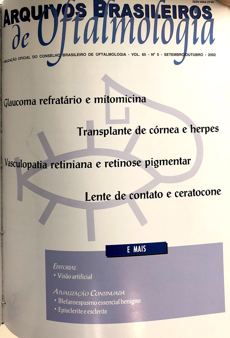
Purpose:To evaluate the behavior of the intraocular pressure (IOP) and possible complications with the use of mitomycin-C (MMC) in trabeculectomies of patients with refractory glaucoma.
Methods:108 eyes of 92 patients who underwent mitomycin-C trabeculectomy (0.2 mg/ml for 5 minutes) were studied. Mean follow up time was 11.92 ± 5.13 months (range, 6 to 24 months). Mean age was 39.59 ± 24.05 years (range, 3 to 87 years). Mean preoperative intraocular pressure was 32.36 ± 9.90 mmHg, median 30 mmHg (range, 18 to 68 mmHg).
Results:Mean postoperative intraocular pressure was 13.74 ± 9.68 mmHg, median 12 mmHg (range, 1 to 60 mmHg). Success rate was 90.7%, 76.8% of patients needed no additional treatment. There was no statistical difference as to intraocular pressure and complication rate between primary and secondary cases. Twenty-five percent of the eyes presented complications; the most frequent were flat chamber (11.11%) and hypotony (7.41%).
Conclusion:mitomycin-C trabeculectomy yields good results in refractory glaucoma, and has few complications.
Keywords: Trabeculectomy; Mitomycin; Mitomycin; Open-angle glaucoma; Intraocular pressure
Arq. Bras. Oftalmol. 200265
| DOI: 10.1590/S0004-27492002000500003
Purpose: To evaluate the results of corneal transplantation for herpes simplex keratitis.
Methods: The records of thirty-eight patients who underwent penetrating keratoplasty for herpes simplex keratitis, from 1993 to 1998 were retrospectively reviewed. Postoperative follow-up periods ranged from 6 to 68 months (mean 21).
Results: The survival rate of a clear graft was 81.6%. Allograft rejection was observed in 36.8% and herpetic recurrence occurred in 10.5%. Visual acuities of 20/80 or better were achieved in 60%. Prophylactic antiviral treatment was not associated with increase in corneal graft survival rate.
Conclusion: Better results have been achieved on keratoplasty for herpetic keratitis.
Keywords: Corneal transplantation; Keratitis herpetic; Graft rejection; Postoperative complications; Recurrence; Retrospective studies
Arq. Bras. Oftalmol. 200265
| DOI: 10.1590/S0004-27492002000500004
Purpose: To introduce a practical method based on the topographic values for rigid gas-permeable contact lens (RGPCL) fitting in keratoconus patients.
Methods: Thirty-three eyes of 17 consecutive patients with keratoconus were studied between July 1997 and April 1999. The initial ophthalmologic examination consisted of visual acuity examination, refraction, biomicroscopy, corneal topography with Eye Sys system. The tested contact lenses were selected based on results of corneal topography 1.5 mm superiorly from the visual axis at 90º the base curve.
Results: Rigid gas-permeable contact lens fitting was successful in 30 eyes (91%). There was improvement in visual acuity in all eyes, ranging from 20/20 to 20/60. A mean of 3 ±1 tests were performed to select the final contact lenses parameters. Refitting was necessary in 3 cases (10%). The majority of the rigid gas-permeable contact lenses (57%) had their base curves similar to K 1.5 mm superiorly from the visual axis compared to K value. This finding was confirmed with statistical analysis of the individual measurements and the mean differences between the two topographic measurements and the final base curve of the lens. Conclusions: Rigid gas-permeable contact lens fitting based on the corneal topography 1.5 mm superior from the visual axis at 90º showed to be easy and efficient for eyes with keratoconus.
Keywords: Keratoconus; Contact lenses; Corneal topography; Astigmatism; Ocular adaptation
Arq. Bras. Oftalmol. 200265
| DOI: 10.1590/S0004-27492002000500005
Purpose: To evaluate the onset time and quality of peribulbar anesthesia with 1% ropivacaine associated or not with hyaluronidase 100 tru/ml for cataract extraction.
Methods: Prospective, randomized, double-blind and controlled study including fifty-seven patients, scheduled to undergo peribulbar anesthesia for cataract extraction, allocated to two groups. Group C: 1% ropivacaine with addition of 100 tru/ml hyaluronidase, and Group S 1% ropivacaine, without hyaluronidase. The onset time for globe akinesia was studied at intervals of 2 minutes, using Nicoll's score. We evaluated pain by analogic score during the surgery and the necessity of complementing the anaesthesia. The peribulbar block was considered satisfactory when the Nicoll's score was less than 4.
Results: The mean time of onset of block in group C was 4.07 minutes (± 3.24), and in group S 5.03 (± 3.28). There was no statistically significant difference between the groups. Both were similar regarding pain score, no pain was observed in 57.14% of group C, and in 68.97% of group S. The supplementary anesthetic was necessary in 2 cases of group C and in 3 cases of group S. Two cases of bradycardia (heart rate < 50 bpm) were observed during the surgery, and in one case administration of atropine IV was necessary.
Conclusion: 1% ropivacaine provided a good quality of anesthesia for cataract extraction, with a faster onset of action in the group with hyaluronidase 100 iu/ml, although without significant difference.
Keywords: Cataract extraction; Ropivacaine; Hyaluronoglucosaminidase; Local anesthesia; Comparative study; Efficacy
Arq. Bras. Oftalmol. 200265
| DOI: 10.1590/S0004-27492002000500006
Purpose: To study the effects of pupillary constriction on frequency doubling perimetry in a group of normal subjects.
Methods: Eighteen healthy volunteers participated in the study. Only one eye per patient (right eye) underwent frequency doubling perimetry (Full Threshold C-20 strategy). For the second session, one drop of 2% pilocarpine was administered to the volunteers' right eye and the examination was repeated after 60 minutes.
Results: Sixty minutes after administration of 2% pilocarpine, there was a significant reduction of the pupillary diameter from 4.22 ± 0.17 mm to 1.55 ± 0.51 mm (p<0.05). There was a significant reduction of the mean retinal sensibility after pupillary constriction. The threshold sensitivity of the central 5º worsened by 5.67 ± 2.49 dB; the area between 2.5º and 10º worsened by 4.49 ± 2.73 dB; and the area between 10º and 20º worsened by 5.10 ± 3.55 dB (p<0.01). A reduction of 4.06 ± 2.67 dB was observed in the mean deviation, as well as an increase of 0.64 ± 0.94 dB in the pattern standard deviation (p<0.01). No differences were observed regarding the number of fixation losses, false-positive and false-negatives responses, and duration of the examination.
Conclusion: Changes in pupillary diameter may produce significant declines in threshold sensitivities of the 20º visual field tested by frequency doubling perimetry. These results suggest that is important to maintain a constant pupillary diameter in seriate examinations.
Keywords: Miosis; Miotics; Perimetry; Pupil; Muscle contraction; Pilocarpine
Arq. Bras. Oftalmol. 200265
| DOI: 10.1590/S0004-27492002000500007
Purpose: To evaluate the corneal abnormalities which occurred after pterygium (Pt) surgery.
Methods: A prospective study in 49 eyes with primary Pt was performed. Age, sex, lesion extension [grade (G) I, GII, GIII and GIV] and morphology (atrophic or fleshy) at slip lamp examination were evaluated. All patients were submitted to computer videokeratoscopy and keratometric examination. The data were submitted to statistical analysis.
Results: 63% of the sample were males and 80% were older than 41 years. The most commontly observed lesion was atrophic Pt (77%), GII and GIII (39% and 28%, respectively). Astigmatism (Astg) on the 60th postoperative day was similar to that observed in the preoperative period and a small keratometric change was detected from the 1st to the 2nd month after surgery, mainly in the smaller lesions. The observed variations were related to the pterygium grade rather than to the age of the patients or the morphologic type of the lesion.
Conclusion: The keratometric and topographic corneal evaluations in patients with pterygium showed that those with smaller lesions (GI and GII) had less astigmatism and fewer changes in corneal curvature in the postoperative period. Two months after surgery, the corneal curvature has the same pattern as that observed before in most patients.
Keywords: Pterygium; Corneal topography; Prospective studies; Human
Arq. Bras. Oftalmol. 200265
| DOI: 10.1590/S0004-27492002000500008
Purpose: Methods of management for choroidal melanomas include basically observation, radiotherapy, surgery, and laser therapy. A reliable measure of the size of the tumor is very important to follow tumor growth. The purpose of this experimental study is to verify the reliability of a low-cost mathematical model to measure areas of tumors of the fundus.
Methods: Twenty-five eyes of pigmented rabbits were used in this study. Experimental Greene hamster amelanotic choroidal melanomas were implanted into the suprachoroidal space. When the tumors reached 3 to 4 papillary diameters, we performed retinography and fluorescein angiograms with a fixed focus retinal camera without Imaginet. Through a mathematical formula we found the real values of tumor areas.
Results: It was possible to verify the real value of tumor areas and it was possible to verify by a comparative analysis that there was no statistically significant difference between them (p=0.717). Conclusions: We concluded that with a non-Imaginet retinal camera and a mathematical formula, it was possible to measure accurately the real areas of the hamster amelanotic melanomas implanted into the suprachoroidal space of rabbits.
Keywords: Melanoma, amelanotic; Melanoma; amelanotic; Melanoma; amelanotic; Choroid neoplasms; Laser therapy; low-level; Mathematical models; Animal; Rabbits
Arq. Bras. Oftalmol. 200265
| DOI: 10.1590/S0004-27492002000500009
Purpose: To evaluate the applicability of the equine fetal membrane (amniotic membrane, chorion and alantoid) as graft in lamelar keratoplasty in dogs.
Methods: 9 mixed breed dogs were used, according to the ARVO statement for use of animals in ophthalmic and vision research. Superficial keratectomy was performed with a 5 mm trephine and a 6 mm diameter fragment of the fetal membrane that was sutured in place with 8-0 nylon simple interrupted stitches. Clinical examination were performed during 2, 7, 15 and 60 days and, afterwards, the animals were euthanized; the eyes were enucleated for histological study.
Results: Clinically, one could observe slight corneal edema near the implant since the early phases, until the middle stage of the investigation. Neovascularization appeared progressively; its higher intensity was observed in the intermediate phase, disappearing gradually. At 60 days, one could notice a macula at the surgery site. The histological findings showed epithelialization and perfect integration of the graft to the receptor tissue at early stages; the cellular and vascular reactions were more intense in this phase. On day 15, the vascular elements were reduced in relation to the matrix and cellular elements. At any stage, inflammatory infiltrate in the graft, as well at the site of the suture were not observed. Conclusions: The equine fetal membrane (amniotic membrane, chorion and alantoid) can be useful as graft in lamellar keratoplasty in dogs.
Keywords: Corneal transplantation; Fetal membranes; Animal; Dogs; Horses
Arq. Bras. Oftalmol. 200265
| DOI: 10.1590/S0004-27492002000500010
Purpose: To identify the main ocular diseases in a neonate population attended by the "Sistema Único de Saúde" in reference maternities of Pernambuco State, advising the follow-up and necessary treatment.
Methods: A transversal study was done among newborn infants in three public maternities, during the period of April to October 2000. An ophthalmological examination was performed during weekly visits, advising the follow-up and treatment of the cases with ocular alterations or risk factors.
Results: 3280 newborns were examined: 1403 (42.8%) in the Encruzilhada maternity (CISAM), 1232 (37.5%) in the maternity of the Hospital Barão de Lucena (MHBL) and 645 (19.7%) in the maternity of the Hospital Agamenon Magalhães (MHAM). 387 were pre-term (11.8%). 701 neonates (21.4%) with ocular alterations or risk factors were referred to the specialized service. Of these, 46.4% were pre-term and 37.9% received oxygen therapy, with a higher percentage in MHBL (45.4%). Conjunctivitis was found in 3.0% of the eyes, leucocoria in 0.4% and subconjunctival hemorrhage in 2.0%. At fundoscopy, retinal hemorrhages were found in 7.8% of cases, with 4.3% affecting the macula. Conclusions: The most frequent diseases were retinal hemorrhages and conjunctivitis. The main risk factors seen were prematurity and neonatal infectious diseases. The authors suggest that ocular examinations should be done routinely in neonates.
Keywords: Eye diseases; Eye; Infant; newborn; Risk factors
Arq. Bras. Oftalmol. 200265
| DOI: 10.1590/S0004-27492002000500011
Purpose: To evaluate which kind of medical care cataract patients have.
Methods: The authors of the survey conducted 299 interviews with patients who took part in the "Cataract Campaign" on April 15, 2000.
Results: Among the interviewees, 58.9% were males, and 41.1% were females, with an average age of 69.9 years. Our population consisted largely of low educational level people (89.5% between illiteracy and elementary school level), and 70.9% are economically inactive (retired, 50.2%; unemployed, 8.3%; housewives 12.5%). The kind of medical care most frequently used was public health service (87.0%); followed by health insurance companies (8.0%) and private care (4.9%). The main reasons reported by patients who had health insurance but had not used it for the surgery were: the companies would not pay for the surgery (25.0%), would not provide the intraocular lens (33.0%).
Conclusion: We conclude that the target public in the "Cataract Campaign" comprises low educational level individuals, most of them economically inactive. Even those who have health insurance seek for help in the campaign due to the lack of economical conditions. In the conditions of this study, the "Cataract Campaign" does not attract people who can pay for the surgery.
Keywords: Cataract; Visual acuity; Medical assistance; Socioeconomic factors; Community health services; Human; Aged
Arq. Bras. Oftalmol. 200265
| DOI: 10.1590/S0004-27492002000500012
The authors presents two cases of Coats' type exsudative vasculopathy associated with retinitis pigmentosa. These are the first cases in siblings described in Brazil and our purpose is to add two more cases, to the 47 described in the world literature.
Keywords: Retinal diseases; Retinitis pigmentosa; Consanguinity; Retinal vessels; Light coagulation; Case report
Arq. Bras. Oftalmol. 200265
| DOI: 10.1590/S0004-27492002000500013
A 25-year-old man presented with diplopia in all fields of gaze, associated with painful ductions of the right eye, after suffering a blunt trauma in the region of the right orbit. Computadorized tomography (CT) showed a right medial isolated blowout orbital fracture. Surgical correction eliminated his complaint of diplopia and improved his binocular field of vision.
Keywords: Orbital fractures; Eye injuries; Diplopia; Strabismus; Eye movements; Binocular vision; Visual fields; Case report
Arq. Bras. Oftalmol. 200265
| DOI: 10.1590/S0004-27492002000500014
The authors present an unusual case of bilateral retinoblastoma, showing early appearance (bilateral leukocoria at 1 month of age), "phthysis bulbi" in OD and massive calcification of the vitreous cavity.
Keywords: Eye neoplasms; Retinal neoplasms; Retinoblastoma; Case report
Arq. Bras. Oftalmol. 200265
| DOI: 10.1590/S0004-27492002000500015
Purpose: Clinicopathological report of three cases of multifocal paracoccidioidomycosis involving the lid and in two of them also the conjunctiva.
Methods: Review of clinical charts and of histopathologic sections obtained by biopsy.
Results: The histopathologic study of the three cases showed inflammatory reaction of the granulomatous type. A special staining technique (Grocott) revealed the presence of the Paracoccidioides brasiliensis.
Conclusion: It is important to include paracoccidioidomycosis in the differential diagnosis of tumoral lesions occurring in the eyelids.
Keywords: Paracoccidioidomycosis; Eyelid diseases; Differential diagnosis; Eyelids; Conjunctiva; Case report
Arq. Bras. Oftalmol. 200265
| DOI: 10.1590/S0004-27492002000500016
The authors present a case of a large congenital coloboma of the upper eyelid which was reconstructed through a transposition flap from the lower eyelid (Cutler-Beard technique), with good esthetic and functional result. The patient presented as a complication, development of deprivation amblyopia, (as a result of the time of visual axis occlusion between the first and second operation), which was solved utilizing appropriate therapy. A bibliographic review was made and the principles that rule the treatment of upper eyelid defects were commented, showing the advantages and disadvantages of each technique. Discussion was raised about the best method of reconstruction in newborns, facing the risks of occurrence of amblyopia.
Keywords: Coloboma; Ambliopia; Eyelids; Plastic surgery; Surgical flaps; Case report
Arq. Bras. Oftalmol. 200265
| DOI: 10.1590/S0004-27492002000500017
Essential blepharospasm is a focal dystonia characterized by progressive involuntary spasms of the orbicularis oculi and upper facial (corrugator and procerus) muscles. Chronic, forceful contractions of the periocular muscles is debilitating for the patient and leads to functional and cosmetic eyelid deformities. Treatment has included a variety of modalities and oral medications that are of limited efficacy. Botulinum-A injections have yielded the best temporary relief from this disorder, while the periorbital myectomy operation has been shown to give the best long-term results.
Keywords: Botulinum toxin type A; Blepharospasm; Eyelids; Dystonia; Oculomotor muscles; Differential diagnosis
Arq. Bras. Oftalmol. 200265
| DOI: 10.1590/S0004-27492002000500018
The most frequent scleral diseases are inflammatory, divided into episcleritis and scleritis. Episcleritis is a benign disease, with inflammatory response of the superficial episcleral tissue. On the other hand, scleritis is a progressive severe disease, with inflammatory response of superficial, deep epis cleral and scleral tissues. The association with systemic rheumathological diseases requires systematic clinical approach of patients with scleral inflammation.
Keywords: Scleritis; Scleritis; Inflammation; Sclera; Bacterial eye infections; Bacterial eye infections; Autoimmune diseases; Rheumatoid arthritis; Adrenal cortex hormones; Prednisolone; Differential diagnosis


 ABO is licensed under a Creative Commons Attribution-NonComercial 4.0 Internacional.

