Arq. Bras. Oftalmol. 2025;88 (4 )
:1-5
| DOI: 10.5935/0004-2749.2024-0167
Abstract
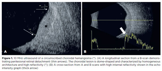
PURPOSE: To evaluate if color Doppler can detect internal blood flow in circumscribed choroidal hemangioma.
METHODS: This cross-sectional study examined seven eyes of seven participants with circumscribed choroidal hemangiomas, with or without prior treatment. B-scan ultrasound and color Doppler were used to assess the dimensions, topographical distribution, and internal blood flow of the affected eyes.
RESULTS: The sample included seven patients (five female) with a median age of 61 (62.29 ± 13.83) years. There were seven eyes with circumscribed choroidal hemangiomas in the patient sample. Color Doppler detected internal vascular flow in all cases (100%). The lesions had an average diameter/thickness ratio of >2 mm and an average thickness of <5 mm and were predominantly located superiorly and supero-temporally.
CONCLUSION: Internal blood flow was detected in circumscribed choroidal hemangiomas using color Doppler. Detection was unaffected by the patient's treatment status.
Keywords: Ultrasonography, doppler, collor; Choroidal neoplasms; Hemangioma
Arq. Bras. Oftalmol. 2022;85 (5 )
:498-505
| DOI: 10.5935/0004-2749.20220066
Abstract
Objetivo: Avaliar alterações da coroide através de imagens de tomografia de coerência óptica (OCT) com profundidade realçada na doença por coronavírus de 2019 (COVID-19).
Métodos: Foram incluídos no estudo 32 pacientes com COVID-19 moderada e 34 indivíduos saudáveis. A espessura da coroide foi medida em 3 pontos: subfoveal, a 1500 mm da fóvea na direção nasal e a 1500 mm da fóvea na direção temporal. A área total da coroide, a área luminal, a área estromal e o índice vascular da coroide foram medidos com o programa ImageJ. Todas as medições foram feitas durante a doença ativa e 4 meses após a remissão.
Resultados: No grupo de pacientes, as espessuras subfoveal, nasal e temporal da coroide mostraram-se reduzidas em comparação com os controles. A diferença não foi estatisticamente significativa (respectivamente, p=0,534, p=0,437 e p=0,077). As médias das áreas total da coroide, estromal e luminal, bem como o índice vascular da coroide, mostraram-se diminuídos com significância estatística no grupo de pacientes (respectivamente, p<0,001, p=0,001, p=0,001 e p=0,003). Aos 4 meses após a remissão, os parâmetros estruturais e o índice vascular da coroide revelaram um aumento estatisticamente significativo em pacientes com COVID-19, em comparação com as medidas iniciais (todos os valores de p<0,001 para os parâmetros estruturais e p=0,047 para o índice vascular da coroide).
Conclusão: Os parâmetros vasculares da coroide e do estroma mostraram uma diminuição transitória, mas significativa em pacientes com COVID-19 durante a doença.
Keywords: Coróide; COVID-19; Infecções por coronavirus; Tomografia de coerência óptica
Arq. Bras. Oftalmol. 2024;87 (4 )
:1-7
| DOI: 10.5935/0004-2749.2023-0047
Abstract
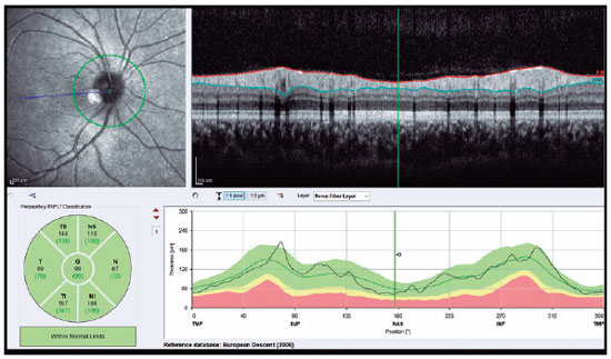
PURPOSE: We aimed to evaluate retinal nerve fiber and choroidal layer alterations in adolescents with anorexia nervosa using spectral-domain optical coherence tomography.
METHODS: Thirty patients with anorexia nervosa and 30 healthy adolescents aged 12-18 years were included in this study. Their age, sex, body mass index, anorexia nervosa type, disease duration, and spectral-domain optical coherence tomography data were recorded.
RESULTS: Central macular thickness and retinal nerve fiber layer thickness in the temporal and inferior regions were significantly lesser in patients with anorexia than in healthy controls (p<0.05). Moreover, significant choroidal thinning around the foveal and subfoveal regions in patients with anorexia was observed (p<0.05). In addition, a statistically significant relation between the increase in disease duration and the thinning of the inferior retinal nerve fiber layer was detected (p<0.05).
CONCLUSION: The retinal nerve fiber layer and choroidal layer thicknesses were lesser in patients with anorexia than in healthy controls. Screening for retinal indices might prevent the development of irreversible retinal pathologies in adolescents with anorexia nervosa. In addition, thinning of the retinal nerve fiber and choroidal layers could reflect structural or functional changes in the brain of adolescents with anorexia nervosa.
Keywords: Tomography; Optical coherence; Nerve fibers; Choroid; Adolescents


 ABO is licensed under a Creative Commons Attribution-NonComercial 4.0 Internacional.


12-fig01tb.jpg)
08-fig01.jpg)
13-fig01.jpg)
13-fig01.jpg)
11-fig01.jpg)
13-fig01.jpg)
15-fig01.jpg)
02-fig01.jpg)
02-fig01.jpg)
06-fig01.jpg)