Arq. Bras. Oftalmol. 2020;83 (5 )
:361-365
| DOI: 10.5935/0004-2749.20200044
Abstract
OBJETIVOS: Comparar as taxas de sucesso anatômico da vitrectomia e tamponamento de gás SF6 na cirurgia de buraco macular com e sem a postura pronada pós-operatória.
MÉTODOS: Foi realizado um estudo observacional, longitudinal e retrospectivo de séries de casos. O estudo incluiu 52 olhos de 52 pacientes submetidos à vitrectomia posterior via pars-plana com peeling de membrana limitante interna auxiliada por azul trypan e tamponamento com gás SF6 a 25% para os estágios 2, 3 e 4 dos buracos maculares. Após a cirurgia, todos os pacientes foram orientados a manter um regime postural pós-operatório: 31 pacientes foram orientados a não realizar posição pronada de cabeça, enquanto 21 foram orientados a manter uma pronada pós-operatória por 7 dias. O objetivo principal foi a análise da taxa de fechamento do buraco macular. A análise estatística foi realizada usando Epi-Info 7.1.
RESULTADOS: Um total de 47 (90,3%) pacientes obtiveram fechamento do buraco macular. O grupo de postura não pronada e o grupo de postura pronada obtiveram taxas de fechamento de 90,3%, e 90,4%, respectivamente; essas taxas não foram significativamente diferentes. A análise estatística revelou que não houve diferenças significativas relacionadas ao gênero, idade, duração do buraco macular, estágio do buraco macular, acuidade visual corrigida pré e pós-operatória entre os dois grupos.
CONCLUSÃO: Nossos resultados sugerem que a cirurgia para buraco macular com o uso de gás de curta duração (SF6) é segura e eficaz e que a manutenção de uma orientação pós-operatória de não-pronada também é segura. No entanto, essas recomendações devem ser avaliadas em um estudo prospectivo e randomizado para delinear de forma abrangente os riscos e benefícios associados.
Keywords: Perfurações retinianas; Vitrectomia; Cirurgia vitreorretiniana; Hexafluoreto de enxofre/administração & dosagem; Fluorocarbonetos/administração & dosagem; Decúbito dorsal; Cuidados pós-operatórios
Arq. Bras. Oftalmol. 2021;84 (4 )
:316-323
| DOI: 10.5935/0004-2749.20210045
Abstract
OBJETIVO: O objetivo deste estudo foi analisar a segurança do implante de lente intraocular primária em um grande número de olhos em crianças <24 meses.
MÉTODOS: Foram revisados os prontuários de pacientes com idade entre 5-24 meses, submetidos a implante primário de lente intraocular no saco capsular. Uma lente intraocular acrílica de três peças dobrável foi implantada pelo mesmo cirurgião usando uma única técnica cirúrgica. Pacientes que tiveram <1 ano de acompanhamento após a cirurgia foram excluídos. Os principais resultados incluíram medidas de acuidade visual, mudança miópica, complicações pós operatórias e cirurgias adicionais.
RESULTADOS: Foram analisados 68 pacientes (93 olhos). A média de idade dos pacientes no momento da cirurgia foi de 15,06 ± 6,19 (5 a 24) meses, e o equivalente esférico 1 mês após a cirurgia foi de 3,62 ± 2,32 D. Após 5,67 ± 3,10 anos, o equivalente esférico foi de -0,09 ± 3,22 D, e a acuidade visual corrigida à distância foi de 0,33 ± 0,33 e 0,64 ± 0,43 logMAR em casos bilaterais e casos unilaterais, respectivamente (p=0,000). A maior mudança míopica foi observado em bebês submetidos à cirurgia aos 5 e 6 meses de idade. As complicações mais frequentes incluíram opacificação do eixo visual e corectopia. Glaucoma e descolamento de retina não foram relatados.
CONCLUSÃO: O implante primário de lente intraocular no saco capsular em crianças de 5-24 meses é seguro e está associado à baixas taxas de eventos adversos e cirurgias adicional.
Keywords: Catarata pediátrica; Lente intraocular; Implante primário LIO; Mudança miópica; Catarata congênita
Arq. Bras. Oftalmol. 2025;88 (6 )
:1-8
| DOI: 10.5935/0004-2749.2025-0077
Abstract
PURPOSE: Standard intravitreal medication dosages are based on an assumed vitreous cavity volume of 4.0-4.5 mL. However, individual variations in vitreous cavity volume may influence both the efficacy and safety of these medications. This study proposes dosage adjustments for intravitreal medications and gases according to axial length and the corresponding vitreous cavity volume.
METHODS: This descriptive study employed reference guidelines that use axial length to estimate the Axial Length-based Volume of the Vitrectomized Space and the Vitreous Volume EXact table for determining dose adjustments across varying eye sizes. Small eyes (axial length 19-22 mm) have an average vitreous cavity volume of 3.5 mL at an axial length of 20.5 mm; standard-sized eyes (22-25 mm) have 4.8 mL at 23.5 mm; large eyes (25-28 mm) have 6.4 mL at 26.5 mm; and extra-large eyes (28-32 mm) have 8.4 mL at 29.5 mm. The medications considered included anti-infectives, anti-VEGFs, complement inhibitors, recombinant proteases, chemotherapy agents, corticosteroids, and medical gases.
RESULTS: Analysis of intravitreal drug concentrations relative to vitreous cavity volume demonstrated notable variability when a standard dose was administered. Small eyes received about 135% of the concentration intended for a standard-sized eye; large eyes received around 75%; and extra-large eyes received under 60%. The recommended dose adjustments are as follows: for small eyes, administer 70-80% of the standard dose; for large eyes, 130-140%; and for extra-large eyes, 170-180%.
CONCLUSIONS: Tailoring intravitreal drug and gas dosages according to axial length and vitreous cavity volume may enhance intraocular drug distribution, potentially improving both safety and therapeutic outcomes.
Keywords: Intravitreal injections; Axial length; Vitreous body; Drug dosage calculations; Pharmacokinetics; Anti-infective agents
Arq. Bras. Oftalmol. 2020;83 (5 )
:396-401
| DOI: 10.5935/0004-2749.20200078
Abstract
Objetivo: Comparar a eficácia de três injeções intravítreas mensais iniciais de aflibercept, seguidas de dosagem de pro re nata (3+PRN) versus cinco injeções mensais iniciais intravítreas de aflibercept, seguidas de doses de pro re nata (5+PRN) em pacientes com edema macular diabético.
Métodos: Foram analisados neste estudo retrospectivo e comparativo 60 pacientes que não receberam tratamento prévio com edema macular e foram submetidos a injeções intravítreas de aflibercept (2 mg/0,05 mL) com pelo menos um ano de acompanhamento. Os pacientes foram divididos em dois grupos de acordo com o número de injeções intravítreas de aflibercept administradas na fase inicial. O grupo 3+PRN compreendeu 27 pacientes, enquanto o grupo 5+PRN compreendeu 33 pacientes. Os resultados visuais e anatômicos foram comparados entre os dois grupos no período inicial e aos 3, 6, 9 e 12 meses.
Resultados: Tanto os grupos 3+PRN quanto 5+PRN mostraram melhoras estatisticamente significativas na acuidade visual melhor corrigida e na espessura macular central ao longo do período de estudo (p<0,001 e p <0,001, respectivamente). Não houve diferenças significativas entre os dois grupos em termos de alterações na acuidade visual melhor corrigida e na espessura macular central (p=0,453 e p=0,784, respectivamente). O número médio de injeções intravítreas de aflibercept foi significativamente maior no grupo 5+PRN (6,1 ± 0,8) do que no grupo 3+PRN (3,9 ± 0,8) (p <0,001).
Conclusão: Os regimes 3+PRN e 5+PRN mostraram resultados visuais e anatômicos semelhantes em 12 meses após o tratamento com injeções intravítreas de aflibercept em pacientes com edema macular.
Keywords: Retinopatia diabética; Edema macular; Injeções intravítreas; Receptores de fatores de crescimento do endotélio vascular/administração & dosagem
Arq. Bras. Oftalmol. 2023;86 (5 )
:1-7
| DOI: 10.5935/0004-2749.20230066
Abstract
Objetivo: Descrever os resultados anatômicos e visuais associados à injeção intravítrea de perfluoropropano seguida de tratamento a laser para descolamento de retina macular secundário à fosseta do disco óptico.
Métodos: Estudo retrospectivo em um único centro. Foram revisados os prontuários médicos dos pacientes com descolamento macular associado a fosseta do disco óptico congênito em um centro de referência terciário de retina entre 2011 e 2018. Todos receberam como estratégia de tratamento inicial injeção intravítrea de perfluoropropano 100% seguido por fotocoagulação a laser ao longo da margem temporal do disco óptico.
Resultados: Foram identificados seis pacientes com descolamento macular associado a fosseta do disco óptico durante o período do estudo. O seguimento pós-operatório variou de 13 a 52 meses, com média de 28 meses. SD-OCT demonstrou resolução completa do fluido em cindo dos seis casos, sem recorrência. Quatro casos apresentaram reabsorção completa após perfluoropropano intravítreo associado a laser, e um paciente necessitou de procedimento adicional (vitrectomia via pars plana com peeling da membrana limitante interna e inversão do retalho do pedículo sobre a margem temporal do disco óptico) para obter reabsorção completa de fluidos. Um paciente apresentou fluido intrarretiniano persistente e negou tratamentos adicionais. O tempo entre o procedimento inicial e a resolução completa do fluido variou entre 6,5 a 41 meses, com média de 19,5 meses. A acuidade visual corrigida melhorou após a cirurgia, considerando a última consulta de acompanhamento em todos os casos.
Conclusão: A injeção intravítrea de perfluoropropano 100% seguida de fotocoagulação ao longo da margem temporal da margem do disco óptico foi associada à melhora anatômica e visual na maioria dos casos e representa uma abordagem terapêutica alternativa para o descolamento macular associado a fosseta do disco óptico.
Keywords: Disco óptico/anormalidades; Doenças do nervo óptico/complicações; Descolamento retiniano; Terapia a laser; Injeções intravítreas; Fluorcarbonetos/administração & dosagem; Gases/administração & dosagem.
Arq. Bras. Oftalmol. 2025;88 (6 )
:1-5
| DOI: 10.5935/0004-2749.2025-0085
Abstract
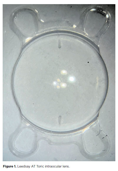
PURPOSE: The purpose of this study was to assess visual outcomes and patient satisfaction following cataract surgery involving the implantation of quad-loop intraocular lenses, including trifocal, bifocal, and toric variants.
METHODS: Information was obtained from both physical and electronic medical records of patients who underwent phacoemulsification cataract surgery with implantation of different intraocular lenses between January 1, 2022, and December 31, 2023. The study included individuals aged over 18 who received bilateral implantation of bifocal, trifocal, or monofocal toric intraocular lenses. Visual acuity was assessed at various postoperative time points using the logMAR scale. Quantitative variables were analyzed using mean and standard deviation.
RESULTS: A total of 92 eyes received premium intraocular lenses: 4 bifocal, 32 trifocal, 52 toric monofocal, and 4 trifocal toric lenses. The average preoperative corrected visual acuity was logMAR 0.478 ± 0.259. On the first postoperative day, the average uncorrected visual acuity was logMAR 0.301 ± 0.207. By day 30, 67.4% of eyes achieved uncorrected distance visual acuity of logMAR 0.2 or better. Patient satisfaction was high, with few reports of glare or halos.
CONCLUSION: Quad-loop intraocular lenses-including trifocal, bifocal, and toric models-demonstrated effective improvement in visual acuity and high levels of patient satisfaction. These lenses represent a suitable option for enhancing visual outcomes after cataract surgery. Additional studies with larger cohorts are recommended to confirm these results.
Keywords: Cataract extraction; Aberrometry/methods; Lenses, intraocular; Lens implantation, intraocular; Prosthesis design
Arq. Bras. Oftalmol. 2026;89 (2 )
:1-8
| DOI: 10.5935/0004-2749.2025-0175
Abstract
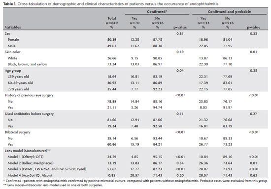
PURPOSE: Endophthalmitis is one of the most important adverse events after cataract surgery, as it can lead to total vision loss. This study aimed to describe the occurrence of endophthalmitis after phacoemulsification with intraocular lens implantation in patients treated in a community setting in Porto Velho, Rondônia, Brazil.
METHODS: This retrospective cohort study was conducted using a database of 649 medical records of patients who underwent surgery and were followed for three months. Poisson regression analysis was used to estimate relative risks and 95% confidence intervals (95% CIs).
RESULTS: The incidence of confirmed endophthalmitis was 11.94% (95% CI, 9.50-14.76), while the incidence of confirmed and probable cases was 20.50% (95% CI, 17.52-23.73). For confirmed cases, bilateral surgery and the use of lens model 3 were identified as risk factors for endophthalmitis, whereas age over 70 yr and preoperative antibiotic use were protective factors. For confirmed and probable cases, brown and yellow skin color, bilateral surgery, and the use of lens model 3 were also identified as risk factors. Gram-negative bacteria were the predominant etiological agents, and corneal edema was the main clinical manifestation. The mean duration of treatment was eight days, and 27.12% of patients used antibiotics.
CONCLUSION: The incidence observed was substantially higher than that reported in the literature, with a predominance of Gram-negative agents and an association with bilateral surgeries and the Eyeol intraocular lens model. These findings reinforce the need for continuous epidemiological surveillance and the implementation of specific biosafety and infection control protocols during cataract surgery campaigns.
Keywords: Endophthalmitis; Disease outbreaks; Phacoemulsification; Lens implantation, intraocular; Lenses, intraocular; Cataract; Risk factors; Anti-bacterial agents
Arq. Bras. Oftalmol. 2025;88 (2 )
:1-6
| DOI: 10.5935/0004-2749.2023-0229
Abstract
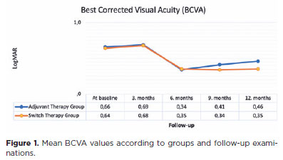
PURPOSE: To compare the outcomes of intravitreal dexamethasone implant used as either an adjuvant or a switching therapy for diabetic macular edema in patients with poor anatomic response after three consecutive monthly injections of ranibizumab.
METHODS: This retrospective study included patients with diabetic macular edema who received three consecutive doses of ranibizumab as initial therapy and demonstrated poor response. A single dose of intravitreal dexamethasone implant was administered to these patients. The patients were divided into two groups according to the treatment modalities: the adjuvant therapy group, consisting of patients who continued treatment with ranibizumab injection after receiving intravitreal dexamethasone implant, and the switch therapy group, consisting of patients who were switched from ranibizumab treatment to intravitreal dexamethasone implant as needed. The main outcome measurements were best corrected visual acuity and central retinal thickness at baseline and at 3, 6, 9, and 12 months of follow-up.
RESULTS: In this study that included 64 eyes of 64 patients, the best corrected visual acuity and central retinal thickness values did not significantly differ between the groups at baseline and at 6 months of follow-up (p>0.05). However, at 12 months, the best corrected visual acuity values in the adjuvant and switch therapy groups were 0.46 and 0.35 LogMAR, respectively (p=0.012), and the central retinal thickness values were 344.8 and 270.9, respectively (p=0.007).
CONCLUSIONS: In a real-world setting, it seems more reasonable to use intravitreal dexamethasone implant as a switch therapy rather than an adjuvant therapy for diabetic macula edema refractory to ranibizumab despite three consecutive monthly injections of ranibizumab. Patients switched to intravitreal dexamethasone implant were found to have better anatomic and visual outcomes at 12 months than those who continued ranibizumab therapy despite their less-than-optimal responses.
Keywords: Diabetic retinopathy; Macular edema/drug therapy; Dexamethasone/administration & dosage; Drug implants; Intravitreal injections; Ranibizumab/administration & dosage; Tomography, optical coherence; Endothelial growth factors
Arq. Bras. Oftalmol. 2024;87 (3 )
:1-7
| DOI: 10.5935/0004-2749.2021-0514
Abstract
Objetivo: Comparar os achados oculares em longo prazo de crianças que se submeteram à cirurgia de catarata congênita antes dos dois anos de idade e receberam uma injeção intracameral de triancinolona no intraoperatório ou usaram prednisolona oral no pós-operatório para modular a inflamação ocular.
Métodos: Neste estudo prospectivo de coorte, todos os pacientes que participaram de um ensaio clínico anterior, que analisou os resultados cirúrgicos de 1 ano da cirurgia de catarata congênita usando triancinolona intracameral (Grupo de Estudo) ou prednisolona oral (Grupo Controle), eram elegíveis para participar. Os prontuários médicos dos pacientes foram revisados e as crianças foram submetidas a um exame oftalmológico completo no acompanhamento final. As principais medidas de desfecho foram: achados biomicroscópicos, pressão intraocular, espessura central da córnea, a necessidade de intervenções cirúrgicas adicionais e achados compatíveis com glaucoma.
Resultados: Vinte e seis olhos (26 pacientes) foram incluídos (Grupo de Estudo = 11 olhos; Grupo de Controle = 15 olhos). O seguimento médio foi de 8,2 ± 1,2 anos e 8,1 ± 1,7 anos nos Grupos de Estudo e Controle, respectivamente (p=0,82). Todos os olhos apresentavam lente intraocular centrada. Não houve diferença estatisticamente significativa entre os grupos com relação à presença de sinéquia posterior (p=0,56), pressão intraocular (p=0,49) ou espessura central da córnea (p=0,21). Nenhum dos olhos preencheu os critérios diagnósticos para glaucoma, apresentou opacificação secundária do eixo visual ou foi reoperado.
Conclusão: Os achados oculares em longo prazo de crianças que se submeteram à cirurgia de catarata congênita e receberam uma injeção intracameral de triancinolona no intraoperatório foram semelhantes aos que usaram prednisolona oral no pós-operatório para modular a inflamação ocular, sugerindo que a triancinolona intracameral pode substituir a prednisolona oral na cirurgia de catarata congênita, facilitando o tratamento pós-operatório e a adesão ao mesmo.
Keywords: Catarata congênita; Triancinolona; Prednisolona; Esteroides; Complicações pós-operatórias; Criança
Arq. Bras. Oftalmol. 2025;88 (5 )
:1-8
| DOI: 10.5935/0004-2749.2024-0328
Abstract
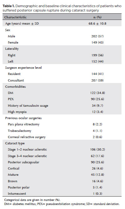
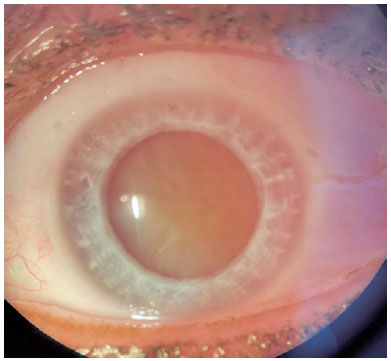
PURPOSE: Posterior capsule rupture is defined as an intraoperative posterior capsule tear resulting in vitreous loss. This study aimed to analyze the clinical characteristics, preoperative risk factors, intraoperative management strategies, and postoperative complications associated with posterior capsule rupture during phacoemulsification surgery.
METHODS: This was a retrospective observational cohort study of the medical records for 25,224 phacoemulsification surgeries performed at our tertiary eye care center between 2017 and 2022. We collected and collated the demographic characteristics and clinical findings of the patients in our cohort. Intraoperative management strategies and postoperative outcomes over a 1-year followup period were also recorded.
RESULTS: Posterior capsule rupture occurred in 351 eyes (351 patients), giving an overall posterior capsule rupture rate of 1.3%. The mean patient age was 68.6 ± 10.8 years. Pseudoexfoliation syndrome, mature cataracts, brown cataracts, and surgery performed by a resident were identified as risk factors for posterior capsule rupture (p<0.05 for each; the risk ratios were 2.70, 2.15, 2.44, 1.34, respectively). The most common intraoperative complications were dislocated lens fragments in the vitreous (8%) and iris damage (7.1%). The mean best-corrected visual acuity improved from 1.31 ± 0.84 (logMAR) postoperatively to 0.51 ± 0.56 at the end of the 1-year follow-up period (p<0.001). Corneal edema (55.6%) and elevated intraocular pressure (33.3%) were the most common early postoperative complications. Persistently elevated intraocular pressure (11.1%) and cystoid macular edema (5.1%) were the most common late postoperative complications.
CONCLUSION: Posterior capsule rupture is a common complication of phacoemulsification surgery that requires prolonged postoperative follow-up and a multidisciplinary approach. Despite the increased incidence of complications when rupture occurs, appropriate intraoperative and postoperative management can lead to satisfactory visual outcomes.
Keywords: Cataract extraction; Phacoemulsification; Posterior capsule rupture; Corneal edema; Risk factors; Postoperative complications; Intraoperative complications
Arq. Bras. Oftalmol. 2025;88 (5 )
:1-7
| DOI: 10.5935/0004-2749.2024-0368
Abstract
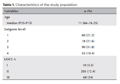
PURPOSE: To compare endothelial corneal cell changes following cataract surgery performed by phacoemulsification with intraocular lens implantation, conducted by surgeons with varying levels of experience.
METHODS: Two hundred and eighty-three eyes diagnosed with cataract were included. Lens opacity was classified into three categories (I, II, and III). Surgeons were categorized into four experience levels (1, 2, 3, and 4), based on years of practice and lifetime surgeries performed. Corneal endothelial characteristics were assessed using non-contact specular microscopy, with measurements taken before surgery and 30-60 days post-surgery.
RESULTS: Pre- and postoperative endothelial analysis showed no significant differences between surgeon levels regarding visual acuity achieved, corneal thickness, and endothelial hexagonality. However, the central endothelial cell density index showed a significantly greater reduction among level 1 surgeons (p=0.026). Grade II cataracts exhibited significant variations in the central endothelial cell density (p=0.011) and average cell size, with level 1 surgeons showing the largest increases (p=0.024).
CONCLUSIONS: The analysis revealed significant differences in visual acuity and endothelial indices between surgeon experience levels, with less experienced surgeons showing greater variations and poorer performance. Clinical protocols should consider these data to establish safer training protocols.
Keywords: Cataract extraction; Phacoemulsification; Endothelium; corneal; Lens implantation, intraocular; Visual acuity; Internship and residency; Surgeons
 ABO is licensed under a Creative Commons Attribution-NonComercial 4.0 Internacional.
01-tab01.jpg)
02-tab01tb.jpg)

07-tab01.jpg)
08-tab01Atb.jpg)
12-fig01tb.jpg)
01-fig01.jpg)