Showing of 1 until 15 from 121 result(s)
Search for: Biometry; Intraocular lens; Cataract; Artificial intelligence
02-fig01.jpg)
Abstract
OBJETIVO: Determinar o efeito da blefaroplastia superior na topografia corneana e no cálculo do poder das lentes intraoculares usando Galilei e IOLMaster.
MÉTODOS: Trinta pacientes submetidos a blefaroplastia superior de maio de 2014 a março de 2017 no Hospital Oftalmológico de Sorocaba, São Paulo, Brasil foram incluídos neste estudo de série de casos observacional. Todos os pacientes foram submetidos a sessões de imagem com Galilei e IOLMaster antes da cirurgia (exame de base) e no 1º e 6º mês pós-operatório. Os resultados primários utilizando os dois aparelhos incluíram ceratometria, astigmatismo corenano e astigmatismo corneano induzido pela blefaroplastia. O comprimento axial e o cálculo do poder da lente intraocular foram realizados unicamente com o IOLMaster (fórmula de Holladay). Teste-t pareado e análise vetorial foram usados na análise estatística.
RESULTADOS: Sessenta olhos de 30 pacientes foram incluídos prospectivamente. A análise vectorial mostrou que após 6 meses da cirurgia, a blefaroplastia superior induziu na média 0,39 D de astigmatismo corneano medido com o Galilei e 0,31 D com IOLMaster. As medidas com o IOLMaster mostraram que a ceratometria média (44,56 vs 44,64 D, p=0,01), ceratometria máxima (45,17 vs 45,31, p=0,01) e o astigmatismo corneano (1,22 vs 1,34, p=0,03) foram maiores após 6 meses da blefaroplastia. As medidas com IOLMaster mostraram que o poder da lente intraocular foi significativamente menor 6 meses após a blefaroplastia (22,07 vs 21,93, p=0,004). Todos os outros parâmetros não mostraram mudanças entre o pré-operatório e o 6º mês da cirurgia (p>0,05 para todas as comparações).
CONCLUSÕES: A blefaroplastia superior influenciou o cálculo da lente intraocular utilizando o IOLMaster. Contudo, a influência não foi clinicamente significativa. Não foram encontradas mudanças topográficas com o Galilei.
Keywords: Blefaroplastia; Lentes intraoculares; Ceratometria; Topografia da córnea; Biometria
02-tab01tb.jpg)
Abstract
OBJETIVO: O objetivo deste estudo foi analisar a segurança do implante de lente intraocular primária em um grande número de olhos em crianças <24 meses.
MÉTODOS: Foram revisados os prontuários de pacientes com idade entre 5-24 meses, submetidos a implante primário de lente intraocular no saco capsular. Uma lente intraocular acrílica de três peças dobrável foi implantada pelo mesmo cirurgião usando uma única técnica cirúrgica. Pacientes que tiveram <1 ano de acompanhamento após a cirurgia foram excluídos. Os principais resultados incluíram medidas de acuidade visual, mudança miópica, complicações pós operatórias e cirurgias adicionais.
RESULTADOS: Foram analisados 68 pacientes (93 olhos). A média de idade dos pacientes no momento da cirurgia foi de 15,06 ± 6,19 (5 a 24) meses, e o equivalente esférico 1 mês após a cirurgia foi de 3,62 ± 2,32 D. Após 5,67 ± 3,10 anos, o equivalente esférico foi de -0,09 ± 3,22 D, e a acuidade visual corrigida à distância foi de 0,33 ± 0,33 e 0,64 ± 0,43 logMAR em casos bilaterais e casos unilaterais, respectivamente (p=0,000). A maior mudança míopica foi observado em bebês submetidos à cirurgia aos 5 e 6 meses de idade. As complicações mais frequentes incluíram opacificação do eixo visual e corectopia. Glaucoma e descolamento de retina não foram relatados.
CONCLUSÃO: O implante primário de lente intraocular no saco capsular em crianças de 5-24 meses é seguro e está associado à baixas taxas de eventos adversos e cirurgias adicional.
Keywords: Catarata pediátrica; Lente intraocular; Implante primário LIO; Mudança miópica; Catarata congênita
15-tab01tb.jpg)
Abstract
Objetivo: Avaliar o implante de lente intraocular primária para tratamento da afacia pediátrica no Sistema Único de Saúde (SUS) e comparar os resultados em diferentes faixas etárias.
Métodos: Foram incluídas crianças com catarata congênita e do desenvolvimento unilateral ou bilateral de 0-12 anos de idade e submetidas a implante de lente intraocular primária.
Resultados: Cento e oito olhos de 68 crianças divididas em quatro grupos de idade (<7m; 7m-2a; 2-5a e > 5a) foram avaliados. Dezenove olhos (17,59%) apresentaram opacificação do eixo visual como complicação pós-operatória. Essa complicação foi mais frequente na faixa etária <7 meses (37,93%). A diferença foi significativa entre os grupos de idade <7 meses e > 5 anos (p=0,002). A opacificação do eixo visual foi dividida em duas categorias: membrana pupilar e proliferação de células do cristalino. Oito olhos apresentaram membrana pupilar e 14 proliferação de células do cristalino. Dos oito olhos com membrana pupilar, sete ocorreram na faixa etária <7 meses. A diferença entre o grupo de idade <7 meses e os grupos de 2-5 anos e > 5 anos foi significativa (p=0,01). A proliferação de células do cristalino foi mais frequente nos grupos de idade <7 meses e 2-5 anos, mas significativa apenas quando comparados o grupo de idade <7 meses com o grupo> 5 anos de idade (p=0,040). Glaucoma e suspeitos de glaucoma não foram observados durante o acompanhamento.
Conclusões: A principal complicação encontrada no estudo foi a opacificação do eixo visual. Sua incidência foi maior em crianças operadas antes dos 7 meses de idade.
Keywords: Extração de catarata; Lentes intraoculares; Complicações intraoperatórias; Glaucoma; Segmento anterior do olho; Criança.
08-fig01.jpg)
Abstract
Objetivo: Desenvolver um aplicativo (TopEye) na plataforma iOS para dispositivos móveis que possibilite a captação e interpretação do mapa de cores gerados por qualquer topógrafo corneano através da inteligência artificial (IA).
Métodos: A execução, acompanhamento e avaliação do projeto foi utilizada a metodologia Scrum, processo de desenvolvimento interativo e incremental para gerenciamento de projetos e desenvolvimento ágil de software. O banco de padrões de diagnóstico gerado consiste em 1172 exemplos, divididos em: 275 padrões esféricos, 302 regulares simétricos, 295 regulares assimétricos e 300 irregulares (ceratocone). Para o desenvolvimento da inteligência artificial do aplicativo, foi estabelecido o treinamento da rede com 240 imagens de cada tipo de padrão, totalizando 960 (81,91%) padrões. O restante das imagens, 212 (18,09%), foram utilizadas para testar o aplicativo e usadas para gerar os resultados. O processo é semiautomático, assim a captação da imagem topográfica é realizada com smartphone, o examinador realiza o contorno do relevo corneano manualmente para em seguida a rede neural realizar o diagnóstico.
Resultados: O aplicativo diagnosticou 201 (94,81%) imagens corretamente. De um total de 212 imagens, o algoritmo errou a classificação de apenas 11 (5,19%). A principal ocorrência de erro foi na distinção das classes simétrica e assimétrica. No rastreio do ceratocone o aplicativo alcançou 95,00% de sensibilidade e 98,68% especificidade.
Conclusão: O trabalho resultou na obtenção de um aplicativo eficiente na captura da imagem topográfica pela câmera do smartphone e na interpretação da mesma através da inteligência artificial aplicada.
Keywords: Dispositivos móveis; Inteligência artificial; Topografia corneana; Astigmatismo
07-tab01tb.jpg)
Abstract
Objetivo: Criar modelos, em catarata pediátrica, para estimar valores futuros de ceratometria e comprimento axial, com base na ceratometria e no comprimento axial medidos na cirurgia, para previsão do poder da lente intraocular para emetropia em idades futuras.
Métodos: Olhos com catarata bilateral, ceratometria e comprimento axial medidos na cirurgia e pelo menos um exame pós-operatório com medidas de ceratometria e comprimento axial foram considerados para este estudo. Os modelos para estimar futuras ceratometrias e comprimentos axiais foram criados considerando (1) ceratometria e comprimento axial medidos na cirurgia, (2) a inclinação média da regressão logarítmica da ceratometria e comprimento axial criada para cada olho e (3) a idade na cirurgia. A lente intraocular para emetropia em idades futuras pode ser estimada usando esses valores em fórmulas de terceira geração. Os erros de estimativa da ceratometria, comprimento axial e poder da lente intraocular, usando os modelos, também foram calculados.
Resultados: 57 olhos de 29 pacientes preencheram os critérios de inclusão. A idade média na cirurgia e acompanhamento foram de 36,96 ± 32,04 meses e 2,39 ± 1,46 anos, respectivamente. A inclinação média da regressão logarítmica criada para cada olho foi de -3.286 para ceratometria e + 3.189 para o comprimento axial. Os erros médios de estimativa absoluta para ceratometria e comprimento axial foram respectivamente: 0,61 ± 0,54 D e 0,49 ± 0,55 mm, e para o poder da lente intraocular usando as fórmulas SRK-T, Hoffer-Q e Holladay I foram: 2,04 ± 1,73 D, 2,49 ± 2,10 D e 2,26 ± 1,87 D, respectivamente.
Conclusões: Os modelos apresentados podem ser utilizados para estimar o poder da lente intraocular que levaria a emetropia em idades futuras e orientar a escolha do poder da lente intraocular a ser implantada na catarata pediátrica.
Keywords: Catarata; Biometria/métodos; Emetropia; Comprimento axial do olho; Lentes intraoculares; Criança
Abstract
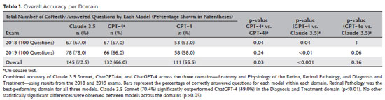
PURPOSE: Natural language models and chatbots, particularly OpenAI’s Generative Pre-Trained Transformer architecture, have transformed human interaction with digital interfaces. The latest versions, including ChatGPT-4o, offer enhanced functionalities compared to their predecessors. This study evaluates the accuracy of ChatGPT-4, ChatGPT-4o, and Claude 3.5 Sonnet in answering questions from the Brazilian Retina and Vitreous Society certification exam.
METHODS: We compiled 200 multiple-choice questions from the Brazilian Retina and Vitreous Society 2018 and 2019 exams. Questions were categorized into three domains: Anatomy and Physiology of the Retina, Retinal Pathology, and Diagnosis and Treatment. Using a standardized prompt developed according to prompt design guidelines, we tested ChatGPT-4, ChatGPT-4o, and Claude 3.5 Sonnet, recording their first responses as final. Three retina specialists performed a qualitative analysis of the answers. Accuracy was determined by comparing responses to the official correct answers. Statistical analysis was conducted using chi-square tests and Cohen’s Kappa.
RESULTS: Claude 3.5 Sonnet achieved the highest overall accuracy (72.5%), followed by ChatGPT-4o (66.0%) and ChatGPT-4 (55.5%). Claude 3.5 Sonnet and ChatGPT-4o significantly outperformed ChatGPT-4 (p<0.01 and p=0.03, respectively), while no significant difference was observed between Claude 3.5 Sonnet and ChatGPT-4o (p=0.16). Model responses agreed 74.5% of the time, with a Cohen’s κ of 0.47. Retinal Pathology was the best-performing domain for all models, whereas Anatomy and Physiology of the Retina and Diagnosis and Treatment were the weakest domains for Claude 3.5 Sonnet and ChatGPT-4, respectively.
CONCLUSIONS: This study is the first to assess Claude 3.5 Sonnet, ChatGPT-4, and ChatGPT-4o in retina specialist certification exams. Claude 3.5 Sonnet and ChatGPT-4o significantly outperformed ChatGPT-4, highlighting their potential as effective tools for studying retina specialist board exams. These findings suggest that the enhanced functionalities of Claude 3.5 Sonnet and ChatGPT-4o offer substantial improvements in medical education contexts.
Keywords: Artificial intelligence; ChatGPT; Retina; Medical education; Ophthalmology, Large language model; Natural language processing
Abstract
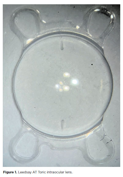
PURPOSE: The purpose of this study was to assess visual outcomes and patient satisfaction following cataract surgery involving the implantation of quad-loop intraocular lenses, including trifocal, bifocal, and toric variants.
METHODS: Information was obtained from both physical and electronic medical records of patients who underwent phacoemulsification cataract surgery with implantation of different intraocular lenses between January 1, 2022, and December 31, 2023. The study included individuals aged over 18 who received bilateral implantation of bifocal, trifocal, or monofocal toric intraocular lenses. Visual acuity was assessed at various postoperative time points using the logMAR scale. Quantitative variables were analyzed using mean and standard deviation.
RESULTS: A total of 92 eyes received premium intraocular lenses: 4 bifocal, 32 trifocal, 52 toric monofocal, and 4 trifocal toric lenses. The average preoperative corrected visual acuity was logMAR 0.478 ± 0.259. On the first postoperative day, the average uncorrected visual acuity was logMAR 0.301 ± 0.207. By day 30, 67.4% of eyes achieved uncorrected distance visual acuity of logMAR 0.2 or better. Patient satisfaction was high, with few reports of glare or halos.
CONCLUSION: Quad-loop intraocular lenses-including trifocal, bifocal, and toric models-demonstrated effective improvement in visual acuity and high levels of patient satisfaction. These lenses represent a suitable option for enhancing visual outcomes after cataract surgery. Additional studies with larger cohorts are recommended to confirm these results.
Keywords: Cataract extraction; Aberrometry/methods; Lenses, intraocular; Lens implantation, intraocular; Prosthesis design
Abstract
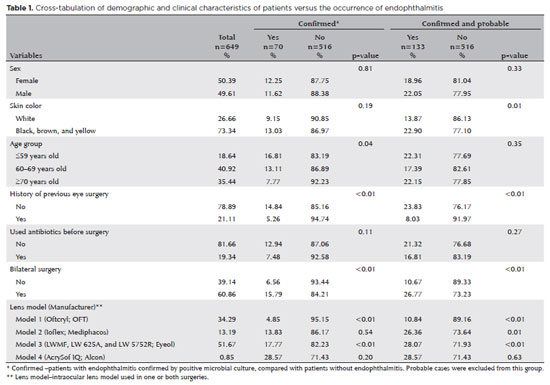
PURPOSE: Endophthalmitis is one of the most important adverse events after cataract surgery, as it can lead to total vision loss. This study aimed to describe the occurrence of endophthalmitis after phacoemulsification with intraocular lens implantation in patients treated in a community setting in Porto Velho, Rondônia, Brazil.
METHODS: This retrospective cohort study was conducted using a database of 649 medical records of patients who underwent surgery and were followed for three months. Poisson regression analysis was used to estimate relative risks and 95% confidence intervals (95% CIs).
RESULTS: The incidence of confirmed endophthalmitis was 11.94% (95% CI, 9.50-14.76), while the incidence of confirmed and probable cases was 20.50% (95% CI, 17.52-23.73). For confirmed cases, bilateral surgery and the use of lens model 3 were identified as risk factors for endophthalmitis, whereas age over 70 yr and preoperative antibiotic use were protective factors. For confirmed and probable cases, brown and yellow skin color, bilateral surgery, and the use of lens model 3 were also identified as risk factors. Gram-negative bacteria were the predominant etiological agents, and corneal edema was the main clinical manifestation. The mean duration of treatment was eight days, and 27.12% of patients used antibiotics.
CONCLUSION: The incidence observed was substantially higher than that reported in the literature, with a predominance of Gram-negative agents and an association with bilateral surgeries and the Eyeol intraocular lens model. These findings reinforce the need for continuous epidemiological surveillance and the implementation of specific biosafety and infection control protocols during cataract surgery campaigns.
Keywords: Endophthalmitis; Disease outbreaks; Phacoemulsification; Lens implantation, intraocular; Lenses, intraocular; Cataract; Risk factors; Anti-bacterial agents
Abstract
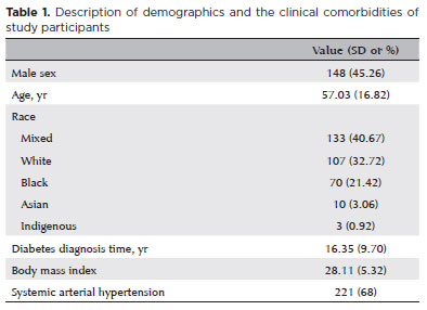
PURPOSE: Diabetic retinopathy screening in low- and middle-income countries is limited by restricted access to specialized care. Portable retinal cameras offer a practical alternative; however, image quality – affected by mydriasis – directly influences the performance of artificial intelligence models. This study evaluated the effect of mydriasis on image gradability and AI-based diabetic retinopathy detection in real-world, resource-limited settings.
METHODS: The proportions of gradable images were compared between mydriatic and non-mydriatic groups. Generalized estimating equations were used to identify factors associated with image gradability, including age, sex, race, diabetes duration, and systemic hypertension. A ResNet-200d model was trained on the mobile Brazilian Ophthalmological dataset and externally validated on both mydriatic and non-mydriatic images. Model performance was evaluated using accuracy, F1 score, area under the curve, and confusion matrix metrics. Sensitivity differences were assessed using the McNemar test, and area under the curves were compared using DeLong's test. The Youden index was used to determine optimal classification thresholds. Agreement between macula- and disc-centered images was analyzed using Cohen's κ.
RESULTS: The mydriatic group demonstrated a higher proportion of gradable images compared with the non-mydriatic group (82.1% vs. 55.6%; p<0.001). In non-mydriatic images, lower gradability was associated with systemic hypertension, older age, male sex, and longer diabetes duration. The AI model achieved better performance in mydriatic images (accuracy, 85.15%; area under the curve, 0.94) than in non-mydriatic images (accuracy, 79.68%; area under the curve, 0.93). The McNemar test showed a significant difference in sensitivity (p=0.0001), whereas DeLong's test revealed no significant difference in area under the curve (p=0.4666). The Youden index indicated that optimal classification thresholds differed based on mydriasis status. Agreement between image fields was moderate to substantial and improved with mydriasis.
CONCLUSION: Mydriasis significantly improves image gradability and enhances AI performance in diabetic retinopathy screening. Nonetheless, in low- and middle-income countries where pharmacologic dilation may be impractical, optimizing model calibration and thresholding for non-mydriatic images is essential to ensure effective AI implementation in real-world clinical environments.
Keywords: Artificial intelligence; Bias; Diabetic retinopathy; Portable camera; Retina
Abstract
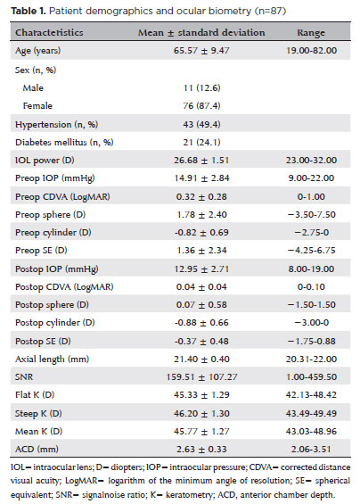
PURPOSE: To compare the refractive prediction error of Hill-radial basis function 3.0 with those of 3 conventional formulas and 11 combination methods in eyes with short axial lengths.
METHODS: The refractive prediction error was calculated using 4 formulas (Hoffer Q, SRK-T, Haigis, and Hill-RBF) and 11 combination methods (average of two or more methods). The absolute error was determined, and the proportion of eyes within 0.25-diopter (D) increments of absolute error was analyzed. Furthermore, the intraclass correlation coefficients of each method were computed to evaluate the agreement between target refractive error and postoperative spherical equivalent.
RESULTS: This study included 87 eyes. Based on the refractive prediction error findings, Hoffer Q formula exhibited the highest myopic errors, followed by SRK-T, Hill-RBF, and Haigis. Among all the methods, the Haigis and Hill-RBF combination yielded a mean refractive prediction error closest to zero. The SRK-T and Hill-RBF combination showed the lowest mean absolute error, whereas the Hoffer Q, SRK-T, and Haigis combination had the lowest median absolute error. Hill-radial basis function exhibited the highest intraclass correlation coefficient, whereas SRK-T showed the lowest. Haigis and Hill-RBF, as well as the combination of both, demonstrated the lowest proportion of refractive surprises (absolute error >1.00 D). Among the individual formulas, Hill-RBF had the highest success rate (absolute error ≤0.50 D). Moreover, among all the methods, the SRK-T and Hill-RBF combination exhibited the highest success rate.
CONCLUSIONS: Hill-radial basis function showed accuracy comparable to or surpassing that of conventional formulas in eyes with short axial lengths. The use and integration of various formulas in cataract surgery for eyes with short axial lengths may help reduce the incidence of refractive surprises.
Keywords: Cataract; Lenses, intraocular; Axial length, eye; Refractive errors; Artificial intelligence
Abstract
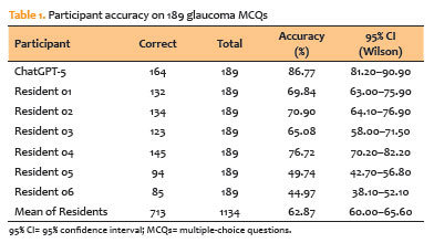
PURPOSE: To assess the performance of a contemporary large language model (ChatGPT-5) against ophthalmology residents on a standardized set of glaucoma multiple-choice questions.
METHODS: We conducted a cross-sectional comparative study with 189 text-only glaucoma multiple-choice questions from the Cybersight question bank. ChatGPT-5 was tested under standardized conditions, with each item placed in a new chat and limited to letter-only outputs. Six ophthalmology residents from a Brazilian training program (two Postgraduate Year 1, two Postgraduate Year 2, and two Postgraduate Year 3) answered the same questions under supervision. Accuracy was calculated using the official key. McNemar’s exact test was used to compare items between ChatGPT-5 and residents, and matched odds ratios and 95% confidence intervals (95% CIs) were calculated using the Haldane–Anscombe correction.
RESULTS: ChatGPT-5 received 164 of 189 correct responses (86.8%; 95% CI, 81.2–90.9). Residents’ overall accuracy was 62.9% (713/1,134; 95% CI, 60.0–65.6). The top-performing resident earned 76.7%. ChatGPT-5 outperformed all residents in head-to-head comparisons, with odds ratios ranging from 1.84 (95% CI, 1.10–3.08) to 13.15 (95% CI, 5.93–29.20), all p≤0.023. ChatGPT-5 correctly answered 17/189 items (9.0%), but fewer than half of residents were correct (“large language model-only wins”), whereas residents were more successful on items that ChatGPT-5 overlooked.
CONCLUSIONS: ChatGPT-5 outperformed ophthalmology residents on text-based glaucoma multiple-choice questions, indicating its potential as a subspecialty education and assessment tool. Generalizability is limited by the single question bank, text-only items, a small resident cohort, and the evaluation of one large language model version at a single time point. Before incorporating these findings into clinical decision-making, larger, multimodal, and longitudinal studies are required.
Keywords: Glaucoma; Artificial intelligence; Large language models; Education, medical; Medical staff, hospital
Abstract
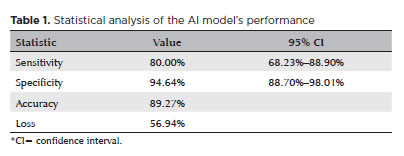
PURPOSE: Standard automated perimetry has been the standard method for measuring visual field changes for several years. It can measure an individual’s ability to detect a light stimulus from a uniformly illuminated background. In the management of glaucoma, the primary objective of perimetry is the identification and quantification of visual field abnormalities. It also serves as a longitudinal evaluation for the detection of disease progression. The development of artificial intelligence-based models capable of interpreting tests could combine technological development with improved access to healthcare.
METHODS: In this observational, cross-sectional, descriptive study, we used an artificial intelligence-based model [Inception V3] to interpret gray-scale crops from standard automated perimetry that were performed in an ophthalmology clinic in the Brazilian Amazon rainforest between January 2018 and December 2022.
RESULTS: The study included 1,519 standard automated perimetry test results that were performed using Humphrey HFA-II-i-750 (Zeiss Meditech). The Subsequently, 70%, 10%, and 20% of the dataset were used for training, validation, and testing, respectively. The model achieved 80% (68.23%–88.9%) sensitivity and 94.64% (88.8%–98%) specificity for detecting altered perimetry results. Furthermore, the area under the receiver operating characteristic curve was 0.93.
CONCLUSIONS: The integration of artificial intelligence in the diagnosis, screening, and monitoring of pathologies represents a paradigm shift in ophthalmology, enabling significant improvements in safety, efficiency, availability, and accessibility of treatment.
Keywords: Glaucoma; Disease progression; Perimetry; Visual Fields; Visual field tests; Artificial intelligence; Neural networks, computers; Machine learning
Abstract
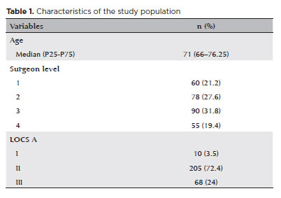
PURPOSE: To compare endothelial corneal cell changes following cataract surgery performed by phacoemulsification with intraocular lens implantation, conducted by surgeons with varying levels of experience.
METHODS: Two hundred and eighty-three eyes diagnosed with cataract were included. Lens opacity was classified into three categories (I, II, and III). Surgeons were categorized into four experience levels (1, 2, 3, and 4), based on years of practice and lifetime surgeries performed. Corneal endothelial characteristics were assessed using non-contact specular microscopy, with measurements taken before surgery and 30-60 days post-surgery.
RESULTS: Pre- and postoperative endothelial analysis showed no significant differences between surgeon levels regarding visual acuity achieved, corneal thickness, and endothelial hexagonality. However, the central endothelial cell density index showed a significantly greater reduction among level 1 surgeons (p=0.026). Grade II cataracts exhibited significant variations in the central endothelial cell density (p=0.011) and average cell size, with level 1 surgeons showing the largest increases (p=0.024).
CONCLUSIONS: The analysis revealed significant differences in visual acuity and endothelial indices between surgeon experience levels, with less experienced surgeons showing greater variations and poorer performance. Clinical protocols should consider these data to establish safer training protocols.
Keywords: Cataract extraction; Phacoemulsification; Endothelium; corneal; Lens implantation, intraocular; Visual acuity; Internship and residency; Surgeons
Abstract
We present a case report detailing the successful phacoemulsification surgery with artificial iris implantation for two individuals with oculocutaneous albinism. These women suffered from cataracts, resulting in reduced visual acuity and heightened photophobia due to iris pigmentary epithelium deficiency. The patients underwent phacoemulsification along with prosthetic artificial iris implantation into the posterior chamber. This intervention resulted in improved visual acuity, reduced photophobia and glare, and an overall enhanced quality of life. Our report highlights two cases of successful phacoemulsification and artificial iris implantation in patients with oculocutaneous albinism and cataracts, leading to improved visual acuity, reduced photophobia, and enhanced quality of life. Notably, there are no prior records in South American literature of cataract surgery combined with artificial iris implantation for oculocutaneous albinism patients up to the time of this publication.
Keywords: Cataract extraction; Albinism, oculocutaneous; Lens implantation, intraocular
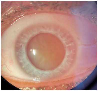
01-fig01.jpg)
Abstract
Um homem de 59 anos apresentou embaçamento visual unilateral no olho esquerdo. Sua acuidade visual nesse olho era no nível de movimentos da mão. O paciente havia se submetido a uma cirurgia de facoemulsificação em que foi feita a implantação intraestromal de uma lente intraocular de câmara posterior. Foi feita a extração dessa lente intraestromal intraocular e uma nova lente intraocular foi implantada. A melhor acuidade visual corrigida final foi de 20/40 pela tabela de Snellen. Com este relato de caso, os autores desejam apontar que uma incisão de degrau único em córnea clara, quando combinada com a injeção de uma lente ocular através da incisão, pode levar a um direcionamento incorreto da lente intraocular para dentro do estroma corneano. Portanto, recomenda-se uma construção cuidadosa da incisão ao se remover uma lente intraocular direcionada incorretamente.
Keywords: Implante de lente intraocular; Lentes intraocular; Facoemulsificação; Cicatrização; Catarata; Acuidade visual
 ABO is licensed under a Creative Commons Attribution-NonComercial 4.0 Internacional.
About
Issues
Editorial Board
Submission
Arquivos Brasileiros de Oftalmologia
Official publication of Brazilian Council of Ophthalmology - Conselho Brasileiro de Oftalmologia (CBO)
Rua Casa do Ator, 1.117 - 2nd floor - Zip Code: 04546-004
São Paulo - SP, Brazil
TEL: +55 11 3266-4000
E-mail: [email protected]