Abstract
Objetivo: Avaliar os resultados clínicos da ceratectomia fotorrefrativa associada à ceratotomia lamelar pediculada (LASIK), para miopia moderada e alta, com ou sem secagem do leito estromal durante a foto-ablação. Pacientes e Métodos: Estudo retrospectivo de 39 olhos de 30 pacientes submetidos a LASIK sem secagem do leito estromal durante a foto-ablação no período de abril de 1996 a abril de 1997 (Grupo I) e 42 olhos de 28 pacientes com secagem do leito estromal durante a foto-ablação a cada 80 pulsos, no período de abril de 1997 a setembro de 1997 (Grupo II). Após a ceratotomia lamelar pediculada corneana com um microcerátomo automatizado (Chiron Corneal Shaper ®), foi realizada a foto-ablação com excimer laser de fluoreto de argônio de 193 nm da Summit modelo Apex Plus®. Em 2 olhos do Grupo I, a foto-ablação não foi realizada, devido a complicações durante a ceratotomia lamelar. Resultados: O tempo médio de seguimento pós-operatório foi de 8,7 meses no Grupo I e 7,7 meses no Grupo II. A média de tratamento foi de -10,81D (±2,38) no grupo I e -8,73D (±2,82) no grupo II. As médias das variações dos equivalentes esféricos obtidos em relação ao tratamento desejado nos meses 1, 3, 6 e 12 foram respectivamente, -0,96D (±1,19), -1,19D (±1,37), -1,06D (±1,41) e -1,10D (±0,66) no Grupo I e -0,23D (±1,02), -0,41D (±1,34), -0,75D (±1,16) e -1,03D (±1,31) no Grupo II. Três olhos do total (3,7%), todos do Grupo I, perderam 2 ou mais linhas de visão. Na visita mais recente, 17 olhos (45,9%) do Grupo I e 31 olhos (73,8%) do Grupo II apresentaram Acuidade Visual sem correção de 20/40 ou melhor. Ocorreram 4 complicações intra-operatórias no grupo I, sendo que 2 casos tiveram a foto-ablação não-efetuada e uma complicação intra-operatória no Grupo II. Conclusão: A secagem do leito estromal possibilitou recuperação visual mais rápida, devido a menor hipocorreção primária. O seguimento a longo prazo não evidenciou diferenças estatisticamente significantes entre os dois tratamentos.
Keywords: Miopia, cirurgia p.; Laser, cirurgia de; LASIK
 ABO is licensed under a Creative Commons Attribution-NonComercial 4.0 Internacional.
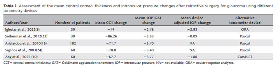
07-tab01.jpg)
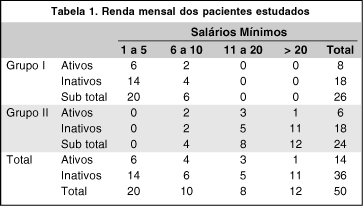
06-tab01.jpg)
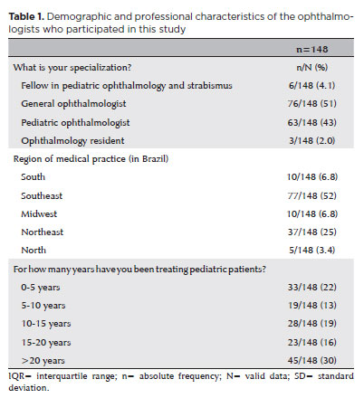
08-tab01tb.jpg)
08-fig01.jpg)