Arq. Bras. Oftalmol. 1999;62 (6 )
:705-711
| DOI: 10.1590/S0004-27491999000600010
Abstract
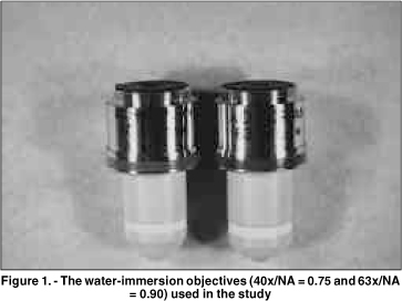
Objetivo: Este estudo prospectivo compara as imagens de microscopia confocal do epitélio corneano do coelho e do homem, obtidas através de 2 objetivas com aberturas numéricas (AN) diferentes. Métodos: Dez olhos de coelhos foram enucleados e fixados através de um suporte pneumático para garantir o melhor desempenho de cada objetiva. Cinco pacientes normais foram selecionados após consentimento. Os olhos de coelhos e dos pacientes foram previamente examinados na lâmpada de fenda. O exame de microscopia confocal (Tomey, Erlangen-Tennenlohe, Alemanha) foi realizado com as objetivas Achroplan 40x/AN = 0,75 e 63x/AN = 0,9 (Zeiss, Oberkochen, Alemanha). Imagens selecionadas do epitélio corneano foram avaliadas qualitativamente com relação ao tamanho, forma e refletividade das células. Resultados: As células no epitélio superficial dos coelhos e dos pacientes, previamente à descamação, tiveram uma refletividade maior que as células adjacentes. Este aspecto foi claramente observado somente com a objetiva 63x/AN = 0,9. As camadas basal e intermediária do epitélio em coelhos foram visualizadas somente através desta objetiva. Estas camadas nos pacientes tornaram-se mais nítidas com a objetiva de abertura numérica maior (63x/AN = 0,9). Conclusão: Uma objetiva de abertura numérica elevada produz melhor resolução dos cortes ópticos, facilitando a análise das camadas do epitélio no coelho e no homem.
Keywords: Corneal epithelium; Confocal microscopy; Epitélio corneano; Microscopia confocal
Arq. Bras. Oftalmol. 2022;85 (1 )
:1-6
| DOI: 10.5935/0004-2749.20220010
Abstract
OBJETIVO: Relatar as alterações no plexo nervoso corneano subbasal em pacientes com ceratite infecciosa de origem bacteriana utilizando a microscopia confocal in vivo.
MÉTODOS: Treze olhos de 13 pacientes com ceratite bacteriana unilateral e 12 indivíduos saudáveis como grupo controle foram incluídos prospectivamente no estudo. A microscopia confocal in vivo foi realizada em todos os pacientes em 2 momentos: na fase aguda da ceratite infecciosa e após 28 ± 0,6 meses da resolução da infecção.
RESULTADOS: A densidade dos nervos no plexo subbasal foi de 5,15 ± 1,03 mm/mm2 na fase aguda da
ceratite infecciosa (comparada com o grupo controle: 19,02 ± 1,78 mm/mm2, p<0,05). Apesar de significativa regeneração dos nervos corneanos ao longo de um intervalo de 28 meses após a resolução da infecção, a densidade dos nervos se manteve significativamente reduzida (9,73 ± 0,93 mm/mm2) quando comparada com o grupo controle (19,02 ± 1,78 mm/mm2, p<0,05). Além disso, as imagens obtidas com a microscopia confocal mostraram áreas de hiperreflectividade referente ao tecido corneano cicatricial com ramos de nervos, afinados e tortuosos, se regenerando nessas áreas.
CONCLUSÕES: Foi observado regeneração parcial dos nervos do plexo corneano subbasal durante os primeiro 28 meses após a resolução da fase aguda da ceratite infecciosa. Além disso, os nervos corneanos regenerados se mantiveram morfologicamente alterados quando comparados ao grupo controle. Esses resultados podem ser relevantes para o acompanhamento clínico e planejamento cirúrgico desses pacientes.
Keywords: Córnea/inervação; Nervo oftálmico; Infecções oculares virais; Ceratite herpética; Microscopia confocal
Arq. Bras. Oftalmol. 2021;84 (3 )
:230-234
| DOI: 10.5935/0004-2749.20210037
Abstract
OBJETIVO: Investigar o efeito do uso de uma substância viscoelástica na ruptura da membrana de Descemet em casos de ceratoplastia lamelar anterior profunda em “bolha dupla”.
MÉTODOS: Foram avaliados retrospectivamente prontuários e vídeos de cirurgias de 40 pacientes operados entre janeiro de 2014 e julho de 2015. Os pacientes foram divididos em dois grupos: 20 pacientes nos quais a parede posterior do estroma foi puncionada sem a colocação de nenhuma substância viscoelástica (grupo 1) e 20 pacientes nos quais uma substância viscoelástica foi aplicada sobre o estroma posterior ao ser puncionada a parede posterior do estroma (grupo 2). A taxa de perfuração da membrana de Descemet foi comparada entre os grupos.
RESULTADOS: Observou-se perfuração da membrana de Descemet em 12 casos (60,0%) no grupo 1 e em apenas 3 casos (15,0%) no grupo 2. Essa diferença foi estatisticamente significativa (p=0,003). Apenas um caso (5%) no grupo 2 teve macroperfuração durante o procedimento, sendo a cirurgia então convertida em uma ceratoplastia penetrante. Onze casos (55,0%) no grupo 1 tiveram macroperfuração da membrana de Descemet e essas cirurgias foram convertidas em ceratoplastias penetrantes. Essa diferença entre os grupos foi estatisticamente significativa (p=0,001).
CONCLUSÕES: A aplicação de substância viscoelástica sobre o lado posterior do estroma logo antes da punção é um método eficaz para diminuir o risco de perfuração da membrana de Descemet na ceratoplastia lamelar anterior profunda.
Keywords: Lâmina limitante posterior/cirurgia; Substâncias viscoelásticas; Transplante de córnea; Substância propria; Ceratoplastia penetrante
Arq. Bras. Oftalmol. 2026;89 (2 )
:1-5
| DOI: 10.5935/0004-2749.2025-0208
Abstract
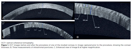
PURPOSE: To assess the reliability and penetration depth of an automated micropuncture system using a tattoo machine.
METHODS: Twenty human corneas were obtained and subjected to intrastromal micropuncture using a tattoo machine. Each cornea was divided into two halves: one received pigment, while the other received saline solution as a control. The Cheyenne tattoo machine was operated at 60 Hz, with standardized needle exposure (six passes per application). The machine used cartridges containing five microneedles. The study was registered with Agência Nacional de Vigilância Sanitária ANVISA (numbers 80281110015, 80281110016, and 80281110019). The pigment used was Electric Ink black ink, with a density of 1,271,460 μg/mL. Puncture depth was measured before and after the procedure using both anterior segment optical coherence tomography and histopathological analysis. Puncture depth measurements were analyzed using ImageJ software. Each cornea was measured thrice, and the results were subsequently compared.
RESULTS: No corneal perforations were observed with the use of the tattoo machine, and puncture depth measurements ranged from 107 to 486 µm.
CONCLUSIONS: The use of a tattoo machine represents a viable and accessible approach for keratopigmentation, with potential for both cosmetic and therapeutic applications. Its adaptation for controlled intrastromal drug delivery may enable the targeted treatment of deep infectious keratitis, corneal neovascularization, and stromal inflammatory disorders, representing a promising approach for corneal stromal diseases. Further research is needed to optimize techniques and evaluate long-term safety and efficacy, particularly for the delivery of antimicrobial, anti-inflammatory, and anti-vascular endothelial growth factor agents.
Keywords: Eye banks; Cadaver; Cornea; Corneal stroma; Drug delivery systems; Tissue donors; Tattooing/instrumentation; Punctures
Arq. Bras. Oftalmol. 2025;88 (6 )
:1-8
| DOI: 10.5935/0004-2749.2025-0118
Abstract
PURPOSE: Using advanced imaging techniques, this study aimed to evaluate corneal stability, epithelial remodeling, and tear film changes over a one-year period in first-time soft-contact lens wearers.
METHODS: A retrospective study was conducted on 100 eyes of 50 first-time daily soft-contact lens users aged 21–65 years with no prior rigid gas-permeable lens wear. The Sirius Scheimpflug imaging system was used to assess corneal topography, epithelial thickness, and non-invasive tear break-up time at baseline, 3, 6, and 12 months. Corneal warpage was evaluated using symmetry indices and Baiocchi Calossi Versaci indices. We performed statistical analysis using repeated-measures analyses of variance with Greenhouse-Geisser correction.
RESULTS: The mean baseline central corneal thickness was 537.83 (±7.92) µm, with no significant thinning after one year. The average simulated keratometry values remained stable, indicating no progressive corneal steepening or flattening. There were no significant changes in warpage indices over time, suggesting corneal shape preservation. Higher-order aberrations (coma, trefoil, and spherical aberrations) and non-invasive tear break-up time remained unchanged throughout the study period.
CONCLUSIONS: Modern silicone hydrogel soft-contact lenses do not induce significant corneal warpage, epithelial remodeling, or optical aberrations over a one-year period. We found that corneal morphology and tear film stability were preserved, supporting the safety of soft-contact lens use. These findings provide clinically relevant insights into the long-term impact of contact lens wear. They may facilitate improved lens fitting strategies and preoperative refractive surgery assessments.
Keywords: Contact lenses, hydrophilic; Cornea/surgery; Corneal diseases; Corneal topography; Adaptation, ocular/physiology; Endothelium, corneal/pathology; Refractive errors; Tears/metabolism.
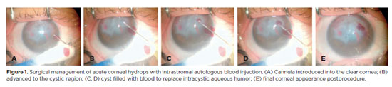
Arq. Bras. Oftalmol. 2025;88 (6 )
:1-7
| DOI: 10.5935/0004-2749.2025-0120
Abstract
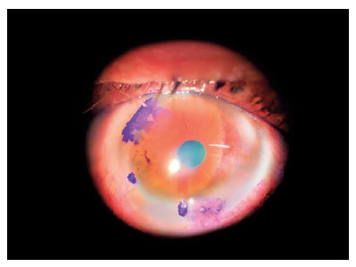
PURPOSE: To describe the technique and outcomes of intrastromal autologous blood injection in patients with severe corneal hydrops.
METHODS: Nineteen patients with corneal hydrops underwent intrastromal autologous blood injection. Postoperative assessments included best-corrected visual acuity and time to resolution of corneal edema
RESULTS: Corneal edema resolved within 1 week in 5 patients, within 1 month in 11, and within 3 months in 3. The mean duration of edema persistence was 37.94 ± 33.05 days (range, 6–124). Corneal thickness decreased from 2.06 ± 0.71-mm preoperatively to 1.34 ± 0.65-mm at day 7, 0.85 ± 0.56-mm at day 30, and 0.57 ± 0.13-mm at day 90 (p<0.001). Descemet’s membrane (DM) detachment decreased from 1.01 ± 0.75-mm to 0.44 ± 0.57-mm, 0.24 ± 0.36-mm, and 0.08 ± 0.11-mm on postoperative days 7, 30, and 90, respectively (p<0.001). DM break size decreased from 1.12 ± 1.19-mm to 0.62 ± 0.84-mm at 3 months (p<0.005). Three patients developed hematocornea; no other major complications were observed. At 3 months, mean best-corrected visual acuity improved from 2.37 ± 0.66 to 0.41 ± 0.17 logMAR with hard contact lenses (p<0.001).
CONCLUSIONS: Intrastromal autologous blood injection is an effective treatment for severe corneal hydrops, promoting faster edema resolution and visual improvement with minimal complications.
Keywords: Corneal edema; Corneal diseases; Edema; Visual acuity; keratoconus.
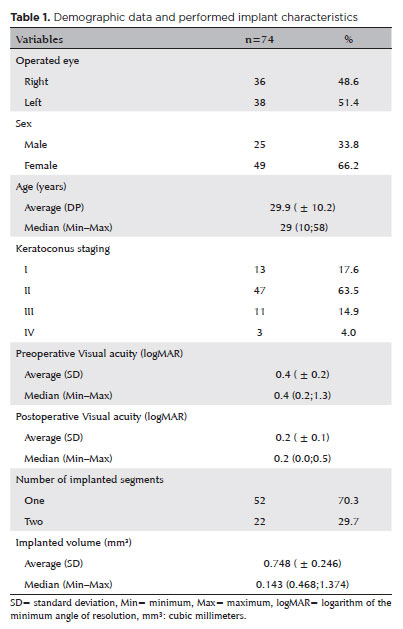
Arq. Bras. Oftalmol. 2025;88 (5 )
:1-7
| DOI: 10.5935/0004-2749.2024-0217
Abstract
PURPOSE: This study aimed to evaluate the influence of intrastromal corneal ring segment implants on the intraocular pressure measurements using Goldmann applanation tonometry, rebound tonometry, and noncontact tonometry in keratoconic corneas and analyze the intertonometer agreement.
METHODS: We enrolled 74 eyes in this observational and prospective study. Each participant had a complete eye examination, corneal analysis with Scheimpflug Tomography (Pentacam®), and intraocular pressure evaluation with Goldmann applanation tonometry, rebound tonometry, and noncontact tonometry, before and after intrastromal corneal ring segment implantation (on postoperative days 1, 7, 45, and 90). Intertonometer agreement was assessed using Bland-Altman analysis.
RESULTS: The mean age was 29.9 ± 10.2 years, and 47 (63.5%) eyes had keratoconus grade II. Intraocular pressures were higher for noncontact tonometry preoperatively and on 90 postoperative day (mean ± SD: 12.4 ± and 12.1 ± 2.2 mmHg, respectively), followed by Goldmann applanation tonometry (11.1 ± 3.0 and 11.2 ± 2.7 mmHg, respectively), and were lower for rebound tonometry (9.7 ± and 9.4 ± 3.2 mmHg, respectively). The variation from the Goldmann tonometry on 7 postoperative day to the baseline (p=0.022) and that of noncontact tonometry on 90 postoperative day to the baseline (p=0.021) were statistically significant. The rebound tonometry underestimated intraocular pressure when compared with the Goldmann applanation tonometry by a mean of 1.47 ± 5.19 mmHg. Noncontact tonometry, when compared with Goldmann applanation tonometry, overesti-mated intraocular pressure by a mean of 1.23 ± 4.15 mmHg.
CONCLUSION: Despite statistically significant differences between some postoperative periods, the intraocular pressure measurement differences may not be clinically relevant.
Keywords: Keratoconus; Intraocular pressure; Cornea; Corneal stroma; Postoperative period; Tonometry ocular; Prostheses and implants
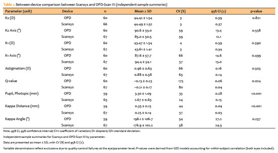
Arq. Bras. Oftalmol. 2026;89 (3 )
:1-9
| DOI: 10.5935/0004-2749.2025-0259
Abstract
PURPOSE: To evaluate the reliability and comparability of a Scheimpflug-based tomographer relative to a Placido-based topographer and specular microscopy in healthy eyes.
METHODS: This cross-sectional study included 40 patients (80 eyes). Each eye underwent randomized imaging with a Scheimpflug-based tomographer, a Placido-based topographer, and Tomey EM-4000 specular microscopy. Three acquisitions per device were obtained. For interdevice comparisons, the best-quality scan per eye/device was selected, whereas all three scans were used for intradevice repeatability analyses. Unreliable scans were repeated (up to five attempts) and excluded if acceptable quality was not achieved, resulting in variable denominators. Between-device comparisons were performed using generalized estimating equations
with participant-level clustering and robust standard errors and were supplemented by Bland–Altman analysis.
RESULTS: The effective sample size varied by parameter (independent summaries: 59–67 eyes; paired comparisons: 48–51 eyes). In paired-eye analyses, the Scheimpflug-based tomographer measured slightly higher keratometry values than the Placido-based topographer (K1: 43.95 vs. 43.78 D, p=0.003; K2: 44.91 vs 44.73 D, p=0.002), more negative Q-values (p=0.001), smaller photopic pupil diameter (p<0.001), and shorter kappa distance (p<0.001). Mean absolute differences were 0.32 D for K1 and 0.30 D for K2, with high dispersion for angular metrics (kappa angle coefficient of variation: 195%).
CONCLUSIONS: The Scheimpflug-based tomographer provides reproducible corneal measurements in healthy eyes. However, systematic differences relative to the Placido-based topographer—particularly for keratometry, asphericity, and pupil and kappa metrics—suggest limited interchangeability. Consistent device use is recommended when these parameters inform clinical decision-making.
Keywords: Scheimpflug tomography; Placido topography; Specular microscopy; keratometry; Corneal imaging; Refractive surgical procedures; Lenses, intraocular
Arq. Bras. Oftalmol. 2024;87 (4 )
:1-6
| DOI: 10.5935/0004-2749.2022-0128
Abstract
Objetivo: Relatar um experimento projetado para determinar alterações anatômicas em córneas porcinas após a colocação de um novo implante depolímero na córnea.
Métodos: Foi utilizado olho de porco ex vivo. Um novo agente modelador biocompatível, de colágeno tipo 1, com 6mm de diâmetro foi moldado com excimer laser em sua face posterior, para criar três formatos planocôncavos. Os implantes foram inseridos dentro de um bolsão, dissecado manualmente, a 200 micrômetros (µm). Foram definidos três grupos de tratamento: grupo A (n=3), teve a profundidade máxima de ablação de70 µm; o grupo B (n=3), profundidade máxima de ablação de 64 µm; e o grupo C (n=3), profundidade máxima de ablação de 104 µm, com buraco central. O grupo controle, D (n=3), foi incluído, com a criação do bolsão estromal, porém sem inserir o material. A avaliação desses olhos foi realizada por tomografia de coerência óptica (OCT) e por tomografia corneana.
Resultados: A tomografia corneana mostrou uma tendência para diminuição da ceratometria média em todos os 4 grupos. A tomografia de coerência óptica mostrou córneas com implantes localizados no estroma anterior e aplanamento visível, enquanto as córneas não mudaram qualitativamente o formato no grupo controle.
Conclusões: O novo implante de biomaterial planocôncavo descrito aqui foi capaz de remodelar a córnea em modelo de animal ex vivo, resultando no aplanamento corneano. Novos estudos são necessários usando modelos animais in vivo para confirmar tais achados.
Keywords: Córnea; Cirurgia da córnea a laser; Substância própria; Proteses e implantes; Lasers de excimer; Materiais biocompatíveis; Animais; Suínos
 ABO is licensed under a Creative Commons Attribution-NonComercial 4.0 Internacional.
03-fig01.jpg)
06-fig01.jpg)
09-fig01.jpg)
02-fig01.jpg)
05-fig01tb.jpg)
02-fig01.jpg)
03-fig01.jpg)
01-fig01.jpg)