Arq. Bras. Oftalmol. 2020;83 (4 )
:269-276
| DOI: 10.5935/0004-2749.20200039
Abstract
Objetivo: Avaliar a influência do comprimento axial ocular na espessura da camada de fibras nervosas da retina peripapilar e na espessura da camada de células ganglionares-plexiforme interna em olhos saudáveis após correção para efeito de magnificação ocular.
Métodos: Neste estudo transversal, avaliamos 120 olhos de 60 participantes voluntários (míopes, emétropes e hipermétropes). A espessura da camada de fibras nervosas da retina peripapilar e da camada de células ganglionares-plexiforme interna foram medidas usando a tomografia de coerência óptica espectral (OCT)-Cirrus HD-OCT e correlacionada com o comprimento axial ocular. O ajuste para a magnificação ocular foi realizado aplicando a fórmula de Littmann.
Resultados: Antes do ajuste para magnificação ocular, a análise de modelos mistos ajustada por idade demonstrou uma correlação negativa significante entre o comprimento axial e a espessura média da camada de fibras nervosas da retina peripapilar (r=-0,43; p<0,001), espessura da camada de fibras nervosas da retina peripapilar inferior (r=-0,46; p <0,001), espessura da camada de fibras nervosas da retina peripapilar superior (r=-0,31; p<0,05), espessura da camada de fibras nervosas da retina peripapilar nasal (r=-0,35; p<0,001) e espessura média das células ganglionares-plexiforme interna (r=-0,35; p<0,05). No entanto, após a correção do efeito de magnificação, os resultados foram consideravelmente diferentes, revelando apenas uma correlação positiva entre o comprimento axial e a espessura temporal da camada de fibras nervosas da retina(r=0,42; p<0,001). Além disso, demonstramos uma correlação positiva entre o comprimento axial e a espessura média das células ganglionares-plexiforme interna (r=0,48; p<0,001). Todas as outras correlações não foram consideradas estatisticamente significativas.
Conclusão: Antes do ajuste para o efeito de magnificação ocular, o comprimento axial estava negativamente correlacionado com a espessura da camada de fibras nervosas da retina peripapilar e das células ganglionares-plexiforme interna medido pelo Cirrus-OCT. Atribuimos esse efeito à magnificação ocular associada a comprimentos axiais maiores, o que foi corrigido com a fórmula de Littman. Mais estudos são necessários para investigar o impacto da correção da magnificação ocular na acurácia diagnóstica do Cirrus-OCT.
Keywords: Tomografia de coerência óptica; Células ganglionares da retina; Comprimento axial do olho
Arq. Bras. Oftalmol. 2024;87 (4 )
:1-5
| DOI: 10.5935/0004-2749.2022-0335
Abstract
PURPOSE: To clarify the postoperative incidence of macular edema in patients undergoing surgery to repair rhegmatogenous retinal detachment and identify the associated risk factors.
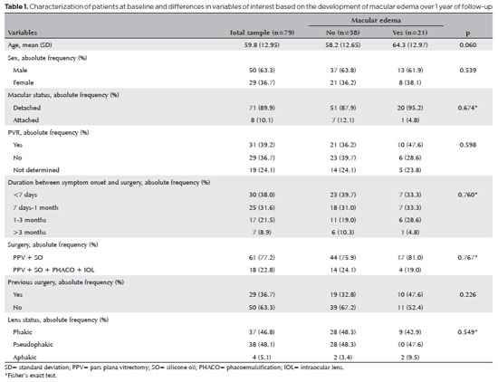
METHODS: In this prospective, observational study, 79 patients who underwent surgery to correct rhegmatogenous retinal detachment using pars plana vitrectomy with silicone oil injection were analyzed. Patients were followed up postoperatively at 7, 30, 90, 180, and 365 days. At each visit, optical coherence tomography was performed to assess the presence or absence of macular edema. were analyzed as possible risk factors for macular edema: age, sex, macular status (attached or detached), presence of vitreoretinal proliferation, history of previous intraocular surgery, reported time of symptoms suggestive of rhegmatogenous retinal detachment up to the date of surgery, and the surgical modality performed.
RESULTS: The 1-year macular edema prevalence rate was 26.6%. In the adjusted analysis, older patients had a higher risk of macular edema, and each 1-year increase in age increased the risk of macular edema by 6% (95% confidence interval = 1.00-1.12). The macular status, vitreoretinal proliferation, the surgical technique used, prior intraocular surgery, and the intraocular lens status were not identified as risk factors. However, the incidence of macular edema increased up to 180 days after surgery, peaking at 10.6%, and then decreased until 365 days after surgery.
CONCLUSION: Macular edema was a common complication after surgery to treat rhegmatogenous retinal detachment, with its incidence peaking between 30 and 180 days after surgery. Age was an important risk factor for macular edema in this cohort.
Keywords: Macular edema; Retinal detachment; Vitrectomy; Tomography, optical coherence; Incidence; Risk factors
Arq. Bras. Oftalmol. 2024;87 (3 )
:1-6
| DOI: 10.5935/0004-2749.2022-0369
Abstract
PURPOSE: To evaluate the choroidal vascular alterations and effect of surgical treatment in the setting of idiopathic epiretinal membranes.
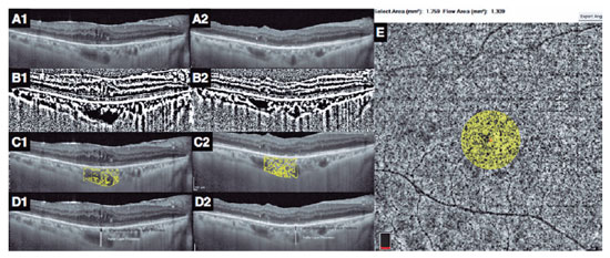
METHODS: The structure of the choroid was studied in 33 patients with unilateral idiopathic epiretinal membrane using optical coherence tomography with enhanced depth imaging and optical coherence tomography angiography. Eyes with epiretinal membrane underwent 25-gauge vitrectomy with epiretinal membrane and internal limiting membrane peeling. The choroidal vascularity index, Haller layer/choroidal thickness ratio, and choriocapillaris flow density were used to evaluate changes in choroidal structure after surgery and compare with the healthy fellow eyes.
RESULTS: The choroidal vascularity index and Haller layer/choroidal thickness ratio of the eyes with epiretinal membrane were higher than those of the fellow eyes at baseline (p=0.009 and p=0.04, respectively) and decreased postoperatively compared with preoperative values (p=0.009 and p=0.001, respectively). The choriocapillaris flow of eyes with epiretinal membrane was lower than that of the fellow eyes at baseline (p=0.001) and increased after surgery compared with the preoperative value (p=0.04). The choroidal vascularity index, Haller layer/choroidal thickness ratio, and choriocapillaris flow values of the healthy fellow eyes were comparable at baseline and final visit. In eyes with epiretinal membrane, the final choroidal vascularity index correlated with the final choriocapillaris flow (r=-0.749, p=0.008) in the multivariate analysis.
CONCLUSION: Idiopathic epiretinal membrane appears to affect the choroidal structure with increased choroidal vascularity index and Haller layer/choroidal thickness ratio and decreased choriocapillaris flow. These macrovascular (choroidal vascularity index and Haller layer/choroidal thickness) and microvascular (choriocapillaris flow) alterations appear to be relieved by surgical treatment of the epiretinal membranes.
Keywords: Epiretinal membrane/surgery; Vitrectomy; Choroid/pathology; Choroid/blood supply; Tomography, optical coherence/methods; Optical coherence tomography angiography; Humans


 ABO is licensed under a Creative Commons Attribution-NonComercial 4.0 Internacional.