Arq. Bras. Oftalmol. 2023;86 (6 )
:1-5
| DOI: 10.5935/0004-2749.2021-0481
Abstract
Objetivos: A pandemia de COVID-19 foi iniciada em março de 2020 e mudou o sistema de saúde. Mudanças na alocação de recursos, sobrecarga de unidades de terapia intensiva, apreensão dos pacientes em procurar atendimento médico não relacionado ao COVID-19 e redução abrupta de todas as consultas e cirurgias não urgentes. Este estudo avalia o impacto em um pronto-socorro oftalmológico após 1 ano de pandemia, avaliando a correlação entre as fases de lockdown, a mortalidade do COVID-19 e as visitas ao pronto-socorro.
Métodos: Estudo observacional retrospectivo que incluiu todos os pacientes admitidos no serviço de emergência oftalmológica do Hospital São Paulo, vinculado a UNIFESP/EPM, entre 1º de janeiro de 2019 e 28 de março de 2021. As visitas foram classificadas e comparadas em um grupo pré-pandemia e pandemia.
Resultados: No período pré-pandemia, o hospital registrou um total de 71.485 atendimentos com média de 194,78 ± 49,74 atendimentos diários, e no grupo pandemia, um total de 41.791 com média de 114,18 ± 43,12 atendimentos diários, redução de 41,4%. Uma diminuição significativa de 16,4% (p<0,001) foi observada na prevalência de conjuntivite aguda e um aumento significativo de 6,4% (p<0,01) na prevalência de corpo estranho da córnea. Foi identificada uma correlação negativa entre a taxa de mortalidade do COVID-19 e as taxas de visita ao pronto-socorro.
Conclusão: Esta análise de um ano mostrou uma redução de 41,4% nas visitas ao pronto-socorro, e uma diminuição significativa nas conjuntivites agudas. A mudança nos hábitos de higiene e o distanciamento social poderiam explicar essa redução, e o aumento da prevalência de traumas corneanos. Achados destacam a necessidade de medidas preventivas e educativas durante os períodos restritivos.
Keywords: SARS-CoV-2; COVID-19; Infecções por coronavírus; Pandemias; Serviços médicos de emergência; Trauma ocular.
Arq. Bras. Oftalmol. 2023;86 (6 )
:1-8
| DOI: 10.5935/0004-2749.2021-0419
Abstract
Objetivo: Investigar sintomas oculares subjetivos e medir a secreção lacrimal objetivamente em pacientes com diagnóstico confirmado da doença coronavírus 2019 (COVID-19).
Métodos: Vinte e quatro pacientes que sobreviveram à infecção pela COVID-19 e 27 controles saudáveis foram incluídos neste estudo transversal prospectivo. Citologia de impressão da conjuntiva, teste de Schirmer, tempo de separação do filme lacrimal, pontuações de coloração da córnea foram aplicados a todos os participantes.
Resultados: Concluiu-se que não houve diferença significativa em relação ao gênero e idade média entre os dois grupos (p=0,484 e p=0,599, respectivamente). A análise dos resultados da citologia de impressão da conjuntiva revelou que a densidade das células do cálice diminuiu, enquanto os linfócitos e neutrófilos aumentaram nos pacientes do grupo COVID-19 quando comparados com os do grupo controle. Quando a classificação de Nelson foi aplicada às amostras de citologia de impressão da conjuntiva, determinou-se que 25% dos pacientes do grupo COVID-19 e 14,8% dos pacientes do grupo controle apresentaram alterações consistentes com grau 2 ou superior. O tempo médio de separação do filme lacrimal, teste de Schirmer e os resultados das pontuações de coloração da córnea foram determinados, diferindo entre o grupo COVID-19 e o grupo controle (p=0,02, p<0,001, and p=0,003, respectivamente).
Conclusões: As análises realizadas neste estudo revelaram as alterações conjuntivais patológicas de pacientes com diagnóstico confirmado de COVID-19 e mostraram que é possível que alterações patológicas da superfície ocular ocorram mesmo no final da infecção pela COVID-19, sem a ocorrência de manifestações oculares clínicas significativas.
Keywords: Infecções por coronavirus; COVID-19; SARS-CoV-2; Manifestações oculares; Lágrimas.
Arq. Bras. Oftalmol. 2021;84 (4 )
:374-379
| DOI: 10.5935/0004-2749.20210065
Abstract
Objetivo: Sincinesias são resultado de inervações anômalas e ocasionam movimentos aberrantes dos músculos envolvidos. Apresentamos uma série com casos raros de sincinesias oculares congênitas dos músculos extraoculares e do levantador da pálpebra superior e especulamos a possibilidade de classificá-las dentro do espectro das desordens congênitas da desnervação cranianana.
Métodos: Prontuários de pacientes com diagnóstico de sincinesia ocular congênita foram estudados retrospectivamente. Analisamos sexo, lateralidade e as características completas do exame de motilidade de cada paciente.
Resultados: Nove pacientes com sincinesias oculares congênitas foram incluídos. Houve discreta predominância no sexo feminino. Em termos de lateralidade, o olho direito foi o único envolvido em 4 casos, o olho esquerdo também em 4 casos e 1 caso apresentou acometimento bilateral. 55,5% dos pacientes eram ortotrópicos na posição primária. Os III, VI e IV nervos participaram da sincinesia em 100%, 44,4% e 11,1% dos casos, respectivamente.
Conclusões: Sincinesias oculares congênitas podem se apresentar de modo bastante eclético e incomum. A inervação aberrante presente em cada um desses casos os coloca na lista de candidatos a integrar o grupo das desordens congênitas da desenervação craniana.
Keywords: Sincinesia; Nervo troclear; Nervos cranianos/ anormalidades; Músculos oculomotores; Transtornos da motilidade ocular/congênito
Arq. Bras. Oftalmol. 2026;89 (1 )
:1-6
| DOI: 10.5935/0004-2749.2025-0071
Abstract
PURPOSE: This study aimed to evaluate the outcomes of strabismus surgical correction in patients with Down syndrome.
METHODS: We conducted a retrospective chart review of patients with Down syndrome who underwent strabismus surgery between January 1997 and May 2024 at an Ophthalmology Outpatient Clinic in São Paulo, Brazil. The data collected included age, sex, medical and ocular history, surgical details, and follow-up outcomes. The patients were categorized by strabismus type into esotropia, fourth nerve palsy, and mixed groups. Surgical success was defined as final alignment within 10Δ of orthotropia and, where applicable, whether there was resolution of abnormal head posture of ocular origin. Patients with postoperative follow-up <6 months were excluded.
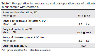
RESULTS: A total of 37 patients (21 females) were included. Of these, 22 (59.5%) were in the esotropia group, 10 (27.0%) in the fourth nerve palsy group, and 5 (13.5%) in the mixed group. The surgical success rate in the esotropia group was 86.4%, with a mean preoperative deviation of 35.2 (± 6.5)Δ, and mean surgical correction of 30.1 (± 10.4)Δ. The success rate in the fourth nerve palsy group was 40.0%, with a mean preoperative deviation of 10.4 (± 4.3)Δ. Overall, success was achieved with a single surgical procedure in 73.0% of the sample. No significant associations were found between surgical success and the clinical and demographic variables, including sex, age at surgery, oblique muscle overaction, pattern strabismus, visual acuity, amblyopia, preoperative deviation, or postoperative follow-up duration (p>0.05).
CONCLUSIONS: When standard surgical tables are applied, strabismus surgery in patients with Down syndrome appears to be safe and effective. We found high success rates, particularly among patients with esotropia. We observed no tendencies toward over- or under-correction. These findings support the use of conventional surgical protocols with this patient population.
Keywords: Down Syndrome/complications; Strabismus/surgery; Esotropia/surgery; Oculomotor nerve diseases/physiopathology; Vision disorders; Humans; Brazil.
Arq. Bras. Oftalmol. 2025;88 (4 )
:1-6
| DOI: 10.5935/0004-2749.2024-0236
Abstract
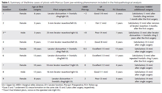
PURPOSE: This study was conducted to report the histopathological and clinical features of the Marcus Gunn phenomenon and other similar conditions of upper eyelid misfiring.
METHODS: This was a retrospective study of patients with congenital ptosis with Marcus Gunn phenomenon who have undergone surgical repair over a period of 12 years and another two patients with upper eyelid misfiring in association with extraocular movements to identify their histopathological findings as subtypes representing ocular congenital cranial dysinnervation disorder.
RESULTS: Among 136 patients with congenital ptosis, 11 (8%) patients with Marcus Gunn phenomenon or misfiring were identified, of whom 9 (6.6%) had typical known Marcus Gunn phenomenon and 2 (1.4%) had eyelid misfiring similar to Marcus Gunn phenomenon. In all patients, the histopathological changes of the excised levator muscle included overall loss and/or atrophy of muscle fibers and irregular-modified Gomori trichrome staining.
CONCLUSION: The Marcus Gunn phenomenon and similar misfiring conditions with synkinetic extraocular muscle movements share findings that are consistent with the neurogenic type of muscle atrophy. This result suggests a common underlying etiology with variable clinical findings, representing the ocular counterpart of congenital cranial dysinnervation disorder, which has been reported as ocular congenital cranial dysinnervation disorder.
Keywords: Eyelid diseases; Ocular motility disorders/surgery; Ophthalmologic surgical procedures
Arq. Bras. Oftalmol. 2025;88 (3 )
:1-6
| DOI: 10.5935/0004-2749.2024-0112
Abstract
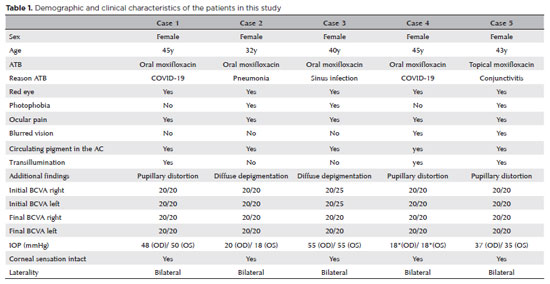
PURPOSE: To describe the ophthalmological findings of dry eye disease and its relation to the quality of life of COVID-19 survivors.
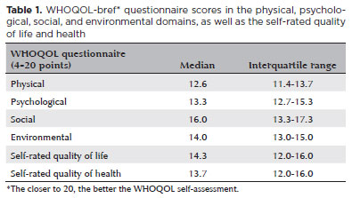
METHODS: COVID-19 survivors who had previously been hospitalized at Hospital das Clínicas de Ribeirão Preto complex underwent an ophthalmological evaluation, which included a dry eye disease questionnaire, break-up time, fluorescein staining, and Schirmer test. We collected the presenting and best-corrected visual acuity, sociodemographic data, personal medical history, and scores from a self-reported quality of life questionnaire (WHOQOL-bref). According to the severity of the acute phase of the disease, the patients were classified into mild-to-moderate, severe, and critical groups.
RESULTS: Ninety-five patients (190 eyes) were evaluated 100 ± 44 days after the onset of COVID-19 symptoms. Of these, 83 patients (87.3%) completed the WHOQOL-bref questionnaire. Ten patients (12.0%) had mild-to-moderate COVID-19, 41 (49.4%) had severe COVID-19, and 32 (38.6%) had critical COVID-19. The median best-corrected visual acuity was logMAR 0 (0-1). Approximately 26.3% patients had a history of dry eye disease or severe dry eye symptoms (frequent or constant ocular dryness and irritation). There was an association between the proportion of patients with dry eye disease and the quality of life (p=0.014) and health (p=0.001). Furthermore, there was a significant trend between the proportion of patients with dry eye disease and how they rated their health and quality of life (p=0.0004 and 0.0027, respectively.
CONCLUSIONS: There is a significant negative correlation between the proportion of patients with dry eye disease and their self-reported quality of life.
Keywords: COVID-19; Coronavirus infections; SARS-CoV-2; Eye diseases; Epidemiology; Ocular surface; Public health
 ABO is licensed under a Creative Commons Attribution-NonComercial 4.0 Internacional.
05-tab01tb.jpg)
14-fig01.jpg)
15-tab01tb.jpg)
12-fig01.jpg)
14-fig01.jpg)
11-fig01tb.jpg)
01-fig01.jpg)
03-fig01.jpg)