Arq. Bras. Oftalmol. 2023;86 (2 )
:121-126
| DOI: 10.5935/0004-2749.20230038
Abstract
Objetivo: Avaliar a pressão intraocular e as alterações da amplitude do pulso ocular em crianças e adolescentes obesos, usando tonometria de contorno dinâmico.
Métodos: Um total de 137 casos, sendo 64 crianças obesas e 73 crianças saudáveis, pareadas por idade e sexo, compôs a população estudada neste estudo transversal. Crianças com valores de índice de massa corporal superior ao percentil de 95% para seu sexo e idade foram consideradas obesas. Os participantes foram submetidos a exames oftalmológicos detalhados, incluindo a medição da pressão intraocular com um tonômetro de contorno dinâmico Pascal. As relações entre a pressão intraocular e as medidas da amplitude do pulso ocular com a idade, sexo, obesidade, estado puberal e resistência à insulina foram investigadas.
Resultados: A amplitude do pulso ocular bilateral foi menor no grupo obeso do que no grupo controle saudável (p<0,001), enquanto a pressão intraocular foi maior (p<0,001). Não foi observada nenhuma relação significativa entre a resistência à insulina e a pressão intraocular ou a amplitude de pulso ocular (p>0,005). Não foi determinada nenhuma correlação entre a pressão arterial sistólica e diastólica, a avaliação do modelo de homeostase para resistência à insulina ou os níveis de lipídios sanguíneos e a pressão intraocular e a amplitude de pulso ocular.
Conclusão: Os resultados mostraram que a obesidade causou um aumento da pressão intraocular e uma diminuição da amplitude do pulso ocular em crianças e adolescentes, independentemente da resistência à insulina. São necessários agora estudos prospectivos envolvendo o seguimento de longo prazo dos casos, para avaliar os prováveis efeitos adversos desses achados oculares observados em crianças obesas.
Keywords: Tanometria ocular; Pressão intraocular; Obesidade; Criança; Adolescente.
Arq. Bras. Oftalmol. 2025;88 (4 )
:1-7
| DOI: 10.5935/0004-2749.2023-0356
Abstract
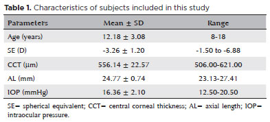
PURPOSE: Although the orthokeratology effects on corneal biomechanics have been proven with clinical trials, reports of stiffness parameter change are scarce. This study investigated the short-term orthokeratology effects in pediatric myopia and compared stiffness parameter changes to those published in recent clinical investigations. This prospective study aimed to investigate corneal biomechanics changes induced by short-term overnight orthokeratology treatment, focusing on stiffness parameter at A1 and stress-strain index
METHODS: Twenty-six children aged 8 to 18 were included in this study using orthokeratology lenses for two different durations: 1 day and 1 week. Corneal biomechanics were assessed using corneal visualization (Corvis) Scheimpflug technology. Measurements were taken at baseline and after each wearing session. Changes in corneal stiffness parameters and corneal curvature were analyzed.
RESULTS: All parameters changed significantly after 1 week of lens wear (p<0.05), except for velocity of corneal apex at the first and second applanation times highest concavity time, radius, stiffness parameter at A1 and stress-strain index. After 1 day, central corneal thickness, first applanation time, second applanation time, deformation amplitude ratio (2 mm), and Corvis biomechanical index (CBI) remained stable (p>0.05). After 1 week, central corneal thickness and first applanation time decreased, whereas second applanation time, deformation amplitude ratio, and Corvis Biomechanical Index significantly increased. With intraocular pressure and central corneal thickness as control variables, no significant correlation was found between stress-strain index and curvature changes (p>0.05). With age as the control variable, no significant correlation was found between stress-strain index and curvature changes (p>0.05).
CONCLUSIONS: Short-term orthokeratology treatment induced notable changes in several corneal biomechanical parameters. Stiffness parameter at A1 and stress-strain index are unaffected by increasing lens wear duration and do not influence the orthokeratology effect.
Keywords: Orthokeratologic procedures; Epithelium, corneal; Corneal topography; Myopia/therapy; Diagnostic techniques, ophthalmological; Biomechanical phenomena; Refraction, ocular; Visual acuity; Humans; Children; Adolescent
Arq. Bras. Oftalmol. 2022;85 (1 )
:19-24
| DOI: 10.5935/0004-2749.20220004
Abstract
Objetivo: Comparar os efeitos da ciclosporina tópica 0,1% e do bevacizumabe na neovascularização da córnea produzida experimentalmente em um modelo com ratos.
Métodos: Trinta ratos Sprague-Dawley adultos foram usados neste estudo experimental. A córnea central dos ratos foi cauterizada quimicamente. Os ratos foram distribuídos aleatoriamente em três grupos. O grupo 1 recebeu bevacizumabe a 1%, o grupo 2 recebeu ciclosporina tópica a 0,1% e o grupo 3 recebeu solução salina isotônica duas vezes ao dia durante 28 dias. O exame de lâmpada de fenda de todos os ratos foi realizado no terceiro e no vigésimo oitavo dias. Os ratos foram então sacrificados e as córneas excisadas. Nos cortes da córnea, o número de vasos sanguíneos, o estado de inflamação e a formação de colágeno foram avaliados em uma análise anatomopatológica.
Resultados: No Grupo 2, os graus de opacidade e de edema da córnea foram significativamente menores que no Grupo 3 (p=0,04 e 0,00, respectivamente). No exame histopatológico, o Grupo 2 apresentou um número significativamente menor de vasos sanguíneos do que o Grupo 3 (p=0,001). Em relação à avaliação da formação de colágeno, esta mostrou-se mais regular no Grupo 2 que no Grupo 1 e no Grupo 3 (p=0,03). Os graus de inflamação foram significativamente menores no Grupo 1 e no Grupo 2 em comparação com o Grupo 3 (p=0,014 e 0,001, respectivamente).
Conclusão: O bevacizumabe tópico é eficaz na inibição da neovascularização da córnea recém-formada. O tratamento tópico com ciclosporina a 0,1% parece ser mais eficaz em comparação ao tratamento tópico com bevacizumabe.
Keywords: Neovascularização da córnea; Bevacizumabe; Cyclosporina A; Ratos
Arq. Bras. Oftalmol. 2021;84 (6 )
:569-575
| DOI: 10.5935/0004-2749.20210101
Abstract
Objetivo: Utilizar aprendizado de máquina para predizer o risco de picos de pressão intraocular às 6 AM em pacientes com glaucoma primário de ângulo aberto e suspeitos.
Métodos: Esse estudo observacional transversal incluiu 98 olhos de 98 pacientes submetidos à curva de 24 horas de pressão intraocular (incluindo as medidas às 6 AM). A curva diurna de pressão intraocular foi definida como uma série de três medidas da curva de 24 horas de pressão intraocular às 8 AM, às 9 AM e às 11 AM. Duas novas variáveis foram apresentadas: inclinação e concavidade. A inclinação da curva às 8 AM foi calculada como a diferença entre pressão intraocular às 9 AM e 8 AM e reflete a variação da pressão intraocular na primeira hora. A concavidade da curva foi calculada como a diferença entre as inclinações às 9 AM e às 8 AM e pode ser para cima ou para baixo. Uma árvore de classificação foi usada para determinar um algoritmo multivariado a partir das medidas da curva diurna para prever o risco de pressão intraocular elevada às 6 AM.
Resultados: Quarenta e nove (50%) olhos apresentaram pressão intraocular às 6 AM >21 mmHg e a mediana do pico de pressão intraocularPIO foi 26 mmHg. Os melhores preditores de pressão intraocular às 6 AM >21 mmHg foram a pressão intraocular às 8 AM e a concavidade. O modelo proposto apresentou uma sensibilidade de 100% e uma especificidade de 86%, com uma acurácia de 93%.
Conclusões: A abordagem de aprendizado de máquina foi capaz de prever o risco de picos de pressão intraocular às 6 AM com uma boa acurácia. Essa nova abordagem para a curva diurna de pressão intraocular pode se tornar uma ferramenta amplamente utilizada na prática clínica e a indicação da curva de 24 horas de pressão intraocular pode ser racionalizada de acordo com a estratificação de risco.
Keywords: Glaucoma; Glaucoma de ângulo aberto; Suspeita de glaucoma; Pressão intraocular; Aprendizado de máquina
Arq. Bras. Oftalmol. 2022;85 (5 )
:490-497
| DOI: 10.5935/0004-2749.20220072
Abstract
Objetivo: Investigar a utilidade de quatro algoritmos diferentes para corrigir erros de medição sem contato da pressão intraocular em pacientes saudáveis e com ceratocone.
Métodos: A pressão intraocular não corrigida e as pressões intraoculares corrigidas foram medidas em um olho de 34 pacientes com ceratocone e 34 pacientes do grupo controle saudável pareados por idade e gênero usando a tecnologia Corvis Scheimpflug. Foi calculada a correlação da pressão intraocular não corrigida e das pressões intraoculares corrigidas com idade, comprimento axial e formato, espessura e biomecânica da córnea. As pressões intraoculares corrigidas foram comparadas com a pressão intraocular não corrigida usando o teste t pareado, e gráficos de Bland-Altman (limites de concordância de 95%).
Resultados: A pressão intraocular não corrigida correlacionou-se com a espessura da córnea e com os parâmetros biomecânicos em ambos os grupos (todos p≤0,047) e a ceratometria média frontal e posterior no grupo com ceratocone (r=-0,39, p=0,02, r=0,39, p=0,02, respectivamente). Após o ajuste com diferentes algoritmos de correção da pressão intraocular, a pressão intraocular corrigida biomecanicamente revelou uma correlação mínima com as características da córnea e uma diferença não significativa com a pressão intraocular não corrigida no grupo saudável (-0,1 ± 1,1 mmHg, p=0,58; limites de concordância de 95%: -2,3 a 2,1 mmHg).
Conclusões: A medição da pressão intraocular usando tonometria sem contato e suas formas corrigidas usando fórmulas lineares, simples, baseadas na espessura da córnea em pacientes com ceratocone estão associadas a muitos erros. O uso de fórmulas mais complexas que consideram mais parâmetros de rigidez da córnea além da espessura da córnea, como fórmula de pressão intraocular corrigida biomecanicamente, pode ser mais confiável e benéfico neste grupo de pacientes.
Keywords: Pressão intraocular; Tonometria sem contato; Córnea; Paquimetria corneana; Ceratocone
Arq. Bras. Oftalmol. 2026;89 (1 )
:1-9
| DOI: 10.5935/0004-2749.2025-0097
Abstract
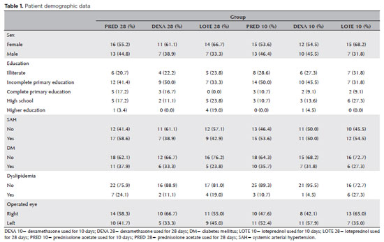
PURPOSE: To evaluate the efficacy of different corticosteroid eye drop formulations (prednisolone acetate 1.0%, dexamethasone 1.0%, and loteprednol etabonate 0.5%) administered for different treatment durations (10 vs. 28 days) in controlling postoperative inflammation following uncomplicated cataract surgery.
METHODS: This randomized, masked clinical trial was conducted at the Instituto Cearense de Oftalmologia. Eligible participants were aged ≥50 yr and scheduled for routine cataract surgery. Exclusion criteria included preexisting ocular disease (elevated intraocular pressure, retinopathy, maculopathy, or uveitis) or concurrent medication use that could confound results. Patients were randomized to receive prednisolone acetate (1.0%), dexamethasone (1.0%), or loteprednol etabonate (0.5%) four times daily for 28 days (with tapering) or for 10 days. Medication bottles, prescriptions, and examiners were masked. Postoperative assessments included ocular symptoms, visual acuity, intraocular pressure, anterior chamber cell count and flare, pachymetry, endothelial cell density, and macular thickness over a 30-day follow-up.
RESULTS: A total of 140 eyes from 140 patients were analyzed (29 prednisolone acetate 1.0%, 18 dexamethasone 1.0%, and 21 loteprednol etabonate 0.5% for 28 days; 28 prednisolone acetate 1.0%, 22 dexamethasone 1.0%, and 22 loteprednol etabonate 0.5% for 10 days). No significant differences were found among the six groups during follow-up. However, eyes treated with dexamethasone (1.0%) showed greater intraocular pressure fluctuations, particularly on Days 7 and 30, and a higher incidence of rebound inflammation in the 28-day regimen. Structural cystoid macular edema without visual impact was observed in 5.9% of eyes in the 28-day groups and 14.2% of eyes in the 10-day groups, as detected by optical coherence tomography at 30 days.
CONCLUSION: Equivalent postoperative inflammation control can be achieved using different corticosteroid eye drop formulations at varying treatment durations following cataract surgery. Brazilian Registry of Clinical Trials (ReBEC): RBR-2frpntv
Keywords: Adrenal cortex hormones; Cataract; Cystoid macular edema; Corticosteroids; Inflammation; Loteprednol etabonate; Ophthalmic solutions; Postoperative period; Intraocular pressure; Visual acuity
Arq. Bras. Oftalmol. 2025;88 (6 )
:1-5
| DOI: 10.5935/0004-2749.2024-0340
Abstract
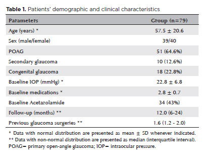
PURPOSE: This study aimed to report the surgical outcomes and success predictors of micropulse transscleral cyclophotocoagulation in eyes with refractory glaucoma.
METHODS: This was a noncomparative, interventional case series. Patients with refractory glaucomas, defined as eyes with prior incisional glaucoma surgery failure and uncontrolled intraocular pressure, who underwent micropulse transscleral cyclophotocoagulation between March 2017 and June 2021 were enrolled. A minimum follow-up period of 6 months was required. Preoperative and postoperative intraocular pressure, number of hypotensive medications, surgical complications, and any subsequent related events were recorded. Success criteria were as follows: 1) intraocular pressure reduction ≥20% and intraocular pressure ≤18 mmHg; 2) intraocular pressure reduction ≥30% and intraocular pressure ≤15 mmHg. The need for topical hypotensive medications was not considered a failure.
RESULTS: Seventy-nine (79) eyes (79 patients; mean age, 57.5 ± 20.6 years) were included. Overall, the median follow-up duration was 12.0 (interquartile interval, 6–24) months, and the mean intraocular pressure was reduced from 22.8 ± 6.8 mmHg to 15.5 ± 5.6 mmHg at the last follow-up visit (p<0.001). The mean number of medications was reduced from 2.8 ± 0.7 to 2.0 ± 1.0 (p<0.01). At 12 months postoperatively, the success rates for criteria 1 and 2 were 54.9% and 49.7%, respectively. Aside from one case of corneal ulcer, which fully resolved with clinical treatment, and two cases of persistent hypotony (with no visual acuity loss during follow-up), no other vision-threatening complications were observed during the postoperative period. The magnitude of intraocular pressure reduction at 1 month (adjusted to preoperative intraocular pressure; HR=1.01; p=0.002).
CONCLUSION: Our findings suggest that micropulse transscleral cyclophotocoagulation is a relatively effective alternative for managing refractory glaucomas, with minor postoperative complications. In addition, the initial intraocular pressure reduction was a statistically significant predictor of 1-year success in patients undergoing micropulse transscleral cyclophotocoagulation.
Keywords: Intraocular pressure/physiology; Glaucoma, open-angle/surgery; Trabeculectomy; Laser coagulation/methods; Tonometry, ocular/methods; Postoperative complications; Antihypertensive agents/therapeutic use.
Arq. Bras. Oftalmol. 2025;88 (3 )
:1-5
| DOI: 10.5935/0004-2749.2023-0174
Abstract
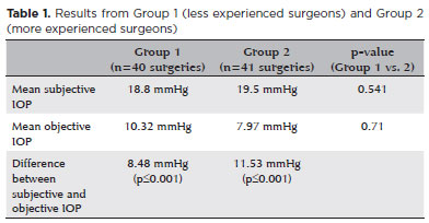
PURPOSE: To compare objective and subjective intraocular pressure measurements immediately after cataract surgery and intraocular pressure measurements between less experienced surgeons (Group 1) and experienced surgeons (Group 2).
METHODS: Surgeons were asked to estimate the IOP after corneal sealing after surgery based on their tactile perception of eye tension (subjective intraocular pressure) Objective intraocular pressure was measured using a Perkins tonometer while patients were still in the surgical field. Objective intraocular pressure was compared to subjective intraocular pressure. Results from less experienced surgeons were compared to more experienced surgeons.
RESULTS: The study comprised 81 surgeries (81 eyes) performed by 27 surgeons. The mean objective intraocular pressure (9.14 mmHg; SD=5.86) was statistically significantly lower (p<0.001) than the mean subjective intraocular pressure (19.21 mmHg; SD=4.82). Hypotony (intraocular pressure <6mmHg) was observed in 25 eyes (30.86%). The mean subjective intraocular pressure was 18.8 mmHg (SD=5.19) for less experienced surgeons and 19.5 mmHg (SD=4.46) for more experienced, without statistically significant difference (p=0.541). No statistically significant difference (p=0.71) was observed when comparing objective intraocular pressure in Group 1 (10.32 mmHg; SD=6.65) and Group 2 (7.97 mmHg; SD=4.7).
CONCLUSION: Objective intraocular pressure was significantly lower than subjective intraocular pressure, regardless of surgeons' experience. This study showed that the subjective method is unreliable compared to the gold standard (Perkins tonometer) and does not improve with surgeons' experience. Establishing standard training methods is paramount to developing surgeons' skills.
Keywords: Cataract; Intraocular pressure; Hypotony, Tonometry; Eye diseases; Training
Arq. Bras. Oftalmol. 2025;88 (5 )
:1-7
| DOI: 10.5935/0004-2749.2024-0217
Abstract
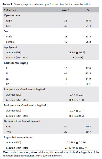
PURPOSE: This study aimed to evaluate the influence of intrastromal corneal ring segment implants on the intraocular pressure measurements using Goldmann applanation tonometry, rebound tonometry, and noncontact tonometry in keratoconic corneas and analyze the intertonometer agreement.
METHODS: We enrolled 74 eyes in this observational and prospective study. Each participant had a complete eye examination, corneal analysis with Scheimpflug Tomography (Pentacam®), and intraocular pressure evaluation with Goldmann applanation tonometry, rebound tonometry, and noncontact tonometry, before and after intrastromal corneal ring segment implantation (on postoperative days 1, 7, 45, and 90). Intertonometer agreement was assessed using Bland-Altman analysis.
RESULTS: The mean age was 29.9 ± 10.2 years, and 47 (63.5%) eyes had keratoconus grade II. Intraocular pressures were higher for noncontact tonometry preoperatively and on 90 postoperative day (mean ± SD: 12.4 ± and 12.1 ± 2.2 mmHg, respectively), followed by Goldmann applanation tonometry (11.1 ± 3.0 and 11.2 ± 2.7 mmHg, respectively), and were lower for rebound tonometry (9.7 ± and 9.4 ± 3.2 mmHg, respectively). The variation from the Goldmann tonometry on 7 postoperative day to the baseline (p=0.022) and that of noncontact tonometry on 90 postoperative day to the baseline (p=0.021) were statistically significant. The rebound tonometry underestimated intraocular pressure when compared with the Goldmann applanation tonometry by a mean of 1.47 ± 5.19 mmHg. Noncontact tonometry, when compared with Goldmann applanation tonometry, overesti-mated intraocular pressure by a mean of 1.23 ± 4.15 mmHg.
CONCLUSION: Despite statistically significant differences between some postoperative periods, the intraocular pressure measurement differences may not be clinically relevant.
Keywords: Keratoconus; Intraocular pressure; Cornea; Corneal stroma; Postoperative period; Tonometry ocular; Prostheses and implants
Arq. Bras. Oftalmol. 2024;87 (4 )
:1-5
| DOI: 10.5935/0004-2749.2022-0085
Abstract
Objetivo: Avaliar a influência das alterações da pressão atmosférica no comportamento da pressão intraocular de indivíduos militares saudáveis-alunos e instrutores da Escola de Mergulho e Resgate da Marinha Nacional na base naval “ARC BOLÍVAR”-durante uma imersão simulada na câmara hiperbárica do Hospital da Marinha de Cartagena.
Métodos: Realizamos um estudo exploratório descritivo. A pressão intraocular foi medida em diferentes pressões atmosféricas durante sessões de 60 minutos na câmara hiperbárica respirando ar comprimido. A profundidade máxima simulada foi de 60 pés. Os participantes eram alunos e instrutores do Departamento de Mergulho e Resgate da Base Naval.
Resultados: Quarenta e oito olhos de 24 mergulhadores foram estudados. Vinte e dois participantes (91,7%) eram do sexo masculino. A média de idade dos participantes foi de 30,6 (DP=5,5) anos, variando de 23 a 40. Nenhum participante tinha histórico de glaucoma ou hipertensão ocular. A média de base da pressão intraocular ao nível do mar foi de 14 mmHg, diminuindo para 13,1 mmHg (queda de 1,2 mmHg) a 60 pés de profundidade (p=0,0012). Entretanto, durante a parada de segurança a 30 pés, a pressão intraocular média continuou diminuindo até atingir 11,9 mmHg (p<0,001). Ao final da sessão, a pressão intraocular média atingiu 13,1 mmHg, valor inferior e estatisticamente significativo quando comparada à média de base da pressão intraocular (p=0,012).
Conclusões: Em indivíduos saudáveis, a pressão intraocular diminui ao atingir uma profundidade de 60 pés (2,8 de pressão atmosférica absoluta) e diminui ainda mais durante a ascensão a 30 pés. As medidas em ambos os pontos foram significativamente diferentes quando comparadas à pressão intraocular de base. A pressão intraocular final foi menor do que a pressão intraocular de base, sugerindo um efeito residual e prolongado da pressão atmosférica sobre a pressão intraocular.
Keywords: Pressão atmosférica; Tanometria; Pressão intraocular; Hipertensão ocular; Glaucoma; Militares
Arq. Bras. Oftalmol. 2025;88 (5 )
:1-7
| DOI: 10.5935/0004-2749.2024-0319
Abstract
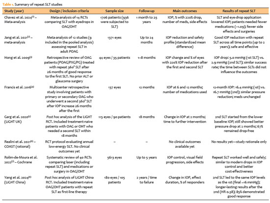
PURPOSE: This study evaluated rates of thyroid eye disease-related eyelid surgeries, strabismus surgeries, and orbital decompressions in active thyroid eye disease patients treated with teprotumumab compared to those who were not.
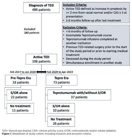
METHODS: In this single-center longitudinal study, we compared patients with active thyroid eye disease evaluated from 02/01/2017 to 01/31/2020 (pre-teprotumumab era) with those seen from 02/01/2020 to 04/30/2023 (teprotumumab era). Patients from the pre-teprotumumab era who received corticosteroids and/or orbital radiation were compared with those in the teprotumumab era treated with teprotumumab, with or without corticosteroids and/or orbital radiation. The primary outcomes were rates of orbital decompressions, strabismus surgery, and eyelid surgery among patients with at least 6 months of follow-up. Orbital decompressions involving two or more walls were classified as severe.
RESULTS: Of 486 records reviewed, 106 patients had active thyroid eye disease. Among them, 33 were from the pre-teprotumumab era; 22 received corticosteroids and/or orbital radiation, and 11 received no treatment. Seventy three patients were from the teprotumumab era; 37 received teprotumumab (with or without corticosteroids and/or orbital radiation), 10 received corticosteroids and/or orbital radiation alone, and 26 received no treatment. Demographics were comparable between groups. Orbital decompression was performed in 11 of 44 eyes (25.0%) in the pre-teprotumumab era treated with corticosteroids and/or orbital radiation (8 one-wall, 3 ≥two-wall), compared to 3 of 74 eyes (4.1%) in the teprotumumab era treated with teprotumumab with or without corticosteroids and/ or orbital radiation (all one-wall). The overall rate of orbital decompressions and the rate of ≥two-wall decompressions were significantly lower in the teprotumumab era (p=0.02 and p=0.0496, respectively). There was no significant difference in one-wall decompressions between era (p=0.07). Rates of strabismus surgeries (27.3% vs. 13.5%, p=0.19) and eyelid surgeries (22.7% vs. 21.6%, p=0.92) did not significantly differ between the era.
CONCLUSIONS: In patients with active thyroid eye disease, treatment with teprotumumab was associated with a significantly lower rate and severity of orbital decompressions compared to treatment with corticosteroids and/or orbital radiation alone. However, the rates of strabismus and eyelid surgeries remained similar between groups.
Keywords: Teprotumumab; Adrenal cortex hormone; Decompression; Graves ophthalmopathy; Strabismus
 ABO is licensed under a Creative Commons Attribution-NonComercial 4.0 Internacional.
15-tab01.jpg)
07-fig01.jpg)
07-fig01.jpg)
04-tab01tb.jpg)
11-tab01.jpg)
15-fig01tb.jpg)