Arq. Bras. Oftalmol. 2023;86 (5 )
:1-6
| DOI: 10.5935/0004-2749.20230064
Abstract
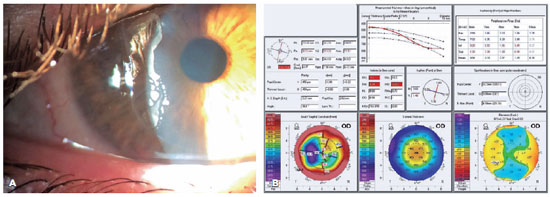
Objetivo: Avaliar a resposta tecidual e clínica a um implante orbitário de polimetilmetacrilato, oco e multiperfurado em sua porção posterior em modelo animal após evisceração.
Métodos: Dezesseis coelhos da raça Nova Zelândia foram submetidos à evisceração do globo ocular direito. Todos receberam implante oco de polimetilmetacrilato de 12 mm de diâmetro, multiperfurado em sua semiesfera posterior. O estudo foi dividido em avaliação clínica e histopatológica. A avaliação clínica foi diária até 14 dias pós-evisceração e, a cada sete dias, até completar 180 dias. Os animais foram divididos em grupos de quatro animais e cada um foi submetido à exenteração com 07, 30, 90 e 180 dias e depois à eutanásia. A análise histopatológica teve por fim caracterizar o padrão inflamatório, a estrutura do colágeno e o grau de neovascularização. Para isso, além da tradicional coloração pela hematoxilina-eosina, utilizou-se o corante Picrosirius Red (PSR) e imuno-histoquímica com o marcador CD 34.
Resultados: Não houve sinais de infecção, afinamento conjuntival ou escleral, exposição ou extrusão do implante em nenhum animal durante o estudo. Já no sétimo dia, o tecido neoformado migrou para dentro do implante formando uma rede fibrovascular através dos canais posteriores. A resposta inflamatória diminuiu ao longo do tempo avaliado e não foram encontradas células gigantes multinucleadas.
Conclusão: O implante analisado permite a sua integração aos tecidos orbitários pelo crescimento fibrovascular em seu interior. Os autores acreditam que este modelo de implante orbital pode fazer parte de testes com humanos.
Keywords: Implantes orbitários; Polimetilmetacrilato; Evisceração ocular; Anoftalmia; Procedimentos cirúrgicos oftalmológicos; Coelhos.
Arq. Bras. Oftalmol. 2025;88 (4 )
:1-6
| DOI: 10.5935/0004-2749.2024-0278
Abstract
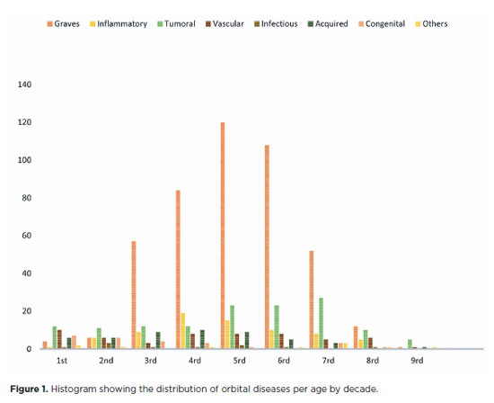
PURPOSE: This study aimed to evaluate the prevalence of orbital conditions in a tertiary ophthalmic outpatient hospital in Sao Paulo, Brazil, with a focus on the main diagnoses and their distribution.
METHODS: A retrospective chart review was conducted involving patients registered and admitted to the orbital disease unit at the Department of Ophthalmology, University of São Paulo Medical School, from January 2004 to March 2018. A total of 838 medical charts were analyzed, of which 37 were excluded due to incomplete data. The remaining charts were categorized into eight diagnostic groups: Graves’ orbitopathy , inflammatory disorders, tumors, vascular lesions, acquired structural abnormalities, congenital structural abnormalities, infectious diseases, and others.
RESULTS: Of the 837,300 ophthalmological appointments, 3,372 (0.4%) were related to orbital diseases. The study included 801 patients, of whom 63.45% were women. The patients’ mean age was 42.86 years. Graves’ orbitopathy was the most common (55%), followed by tumor (17%), inflammatory disorders (9%), vascular lesions (7%), acquired structural abnormalities (5%), congenital structural abnormalities (4%), others (2%), and infectious diseases (1%). The study found significant differences in the incidence and types of orbital diseases, indicating the specialized nature of tertiary care and referral biases.
CONCLUSION: Published data on epidemiological orbital diseases is scarce. Therefore, this study focused on the diverse nature of orbital diseases and their low incidence among ophthalmology appointments. The major trends align with other epidemiological studies, demonstrating a preponderance of Graves’ orbitopathy in middle-aged adults and a bimodal distribution of tumors. These findings are essential in shaping resident training programs and healthcare policies, particularly in tertiary settings. Understanding the epidemiology of orbital diseases can improve diagnostic accuracy, treatment approaches, and patient outcomes as well as support future systemic prospective studies.
Keywords: Orbital diseases; Orbital tumors; Neoplasms; Inflammation; Graves’ ophthalmopathy; Outpatients
 ABO is licensed under a Creative Commons Attribution-NonComercial 4.0 Internacional.
06-fig01.jpg)
14-fig01.jpg)
01-fig01.jpg)
12-fig01.jpg)
14-fig01.jpg)
02-fig01.jpg)
01-fig01tb.jpg)
03-fig01.jpg)
02-fig01.jpg)
03-fig01.jpg)
02-fig01.jpg)