Arq. Bras. Oftalmol. 2025;88 (6 )
:1-5
| DOI: 10.5935/0004-2749.2024-0321
Abstract
PURPOSE: To report the ophthalmological signs, symptoms, and clinical management observed during an unprecedented outbreak of chemical ocular injuries related to cosmetic hair ointments in Brazil.
METHODS: This descriptive, cross-sectional study reviewed medical records of patients treated at the emergency center of Fundação Altino Ventura for chemical ocular trauma associated with cosmetic hair ointment use between February 2022 and February 2023. Records with incomplete medical information were excluded.
RESULTS: The study included 168 patients (95.2% [n=160] female), with a mean age of 30.8 ± 9.7 years. The most frequently reported symptoms at presentation were pain (167/168, 99.4%) and photophobia (92/168, 54.8%). Severe pain was reported by 137 patients (80%). Keratitis was present in 280 of 336 eyes (83.3%), conjunctival hyperemia in 256 eyes (76.4%), and corneal abrasions in 174 eyes (51.8%). A decrease in visual acuity (worse than 20/25) was documented in 18.5% (31/168) of cases. Lubricants, antibiotics, and re-epithelialization
ointments were prescribed to 64.8% (109/168) of the patients. Topical corticosteroids and oral vitamin C were administered to 34% (57/168) and 1.2% (2/168) of patients, respectively. Followup visits were required in 19% (33/168) of cases.
CONCLUSION: The outbreak of chemical ocular injuries linked to cosmetic ointments used for braiding and hair modeling in Brazil was marked by intense ocular pain, conjunctival hyperemia, keratitis, and corneal abrasions. Most patients were treated with lubricants, antibiotics, and re-epithelialization ointments, although approximately one-fifth required followup care, and one-third received additional treatment with either topical corticosteroids and/or oral vitamin C.
Keywords: Cosmetics; Hair preparations; Eye injuries; Burns, chemical; Eye burns; Keratitis; Cornea; Corneal diseases; Visual low.
Arq. Bras. Oftalmol. 2026;89 (1 )
:1-4
| DOI: 10.5935/0004-2749.2024-0402
Abstract
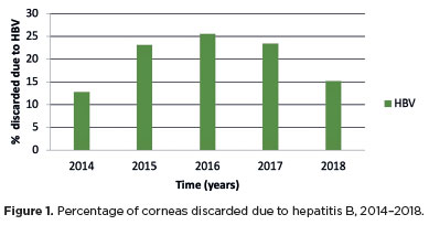
PURPOSE: This study evaluated the proportion of corneas discarded by the Eye Bank of Londrina, Paraná, due to positive serology over a 5-year period and its impact on transplant availability.
METHODS: A cross-sectional study was conducted, analyzing 1,968 corneas from 1,056 donors collected between January 2014 and December 2018 at the Eye Bank of Londrina. Serological tests for hepatitis B (HBsAg and anti-HBc), hepatitis C (anti-HCV), and HIV (anti-HIV 1 and 2) were performed using chemiluminescent microparticle immunoassays. Data were analyzed descriptively and presented in tables and graphs.
RESULTS: Of the 1,968 corneas processed, 897 (45.57%) were discarded. Among these, 333 (37.12%) tested positive for serological markers. Hepatitis B accounted for 34.67% of positive cases (15% of total donations), hepatitis C for 1.11% (0.50% of total), and HIV for 0.89% (0.4% of total). Hepatitis cases remained stable between 2014 and 2016, with a marked decline in 2017 and 2018. Most discarded corneas were positive for anti-HBc (31.88%) and negative for HBsAg; however, the anti-HBs test was not performed to confirm immunity to the hepatitis B virus.
CONCLUSION: The findings highlight the importance of serological testing to identify and eliminate contaminated corneas, thereby preventing the transmission of infectious diseases to recipients. Positive serology for hepatitis, particularly hepatitis B, was the leading cause of corneal disposal.
Keywords: Cornea; Corneal transplantation; Corneal donation; Eye banks; Hepatitis B virus; Hepatitis C virus; HIV infections; Seropositivity; Serologic tests
Arq. Bras. Oftalmol. 2022;85 (3 )
:277-285
| DOI: 10.5935/0004-2749.20230074
Abstract
Objetivos: Dimensionar o impacto da pandemia da COVID-19 nas doações e transplantes de córnea no Brasil e obter indicadores confiáveis para o embasamento de proposições de medidas efetivas para a manutenção e o aperfeiçoamento do sistema de obtenção, processamento, distribuição, utilização e controle dos tecidos oculares doados.
Métodos: Um questionário foi enviado, pelo escritório Brasil da Associação Pan-Americana de Bancos de Olhos (APABO), aos Bancos de Olhos brasileiros. Dados de janeiro a agosto de 2020 foram coletados para gerar indicadores confiáveis sobre o impacto da pandemia da COVID-19 nas doações e transplantes de córnea no Brasil.
Resultados: Dados de 37 Bancos de Olhos mostraram que 76,1% das 3.060 doações e 74,5% dos 3.167 transplantes aconteceram no período pré-pandemia. Das 6.052 córneas processadas 71,8% foram disponibilizadas para fins terapêuticos: 72,9% foram transplantadas, 26,1% acabaram sendo inviabilizadas (45% destas, classificadas para indicações ópticas) e 1%, em glicerina, permanecia em estoque. Das 1.706 córneas que não puderam ser disponibilizadas para uso terapêutico, 47,9% foram excluídas por fatores relacionados às condições dos tecidos, 43,6% por fatores sorológicos, 6,7% por contraindicações constatadas em histórico clínico após a captação e 1,8% por outros fatores.
Conclusões: O impacto negativo da pandemia nas doações e transplantes de córnea no Brasil se deveu à recomendação do Ministério da Saúde de suspender, por quase seis meses, as captações de doadores em parada cardiorrespiratória. Os indicadores tornam evidente a necessidade de atualização dos critérios de classificação e disponibilização das córneas pelos Bancos de Olhos e do sistema nacional de distribuição destes tecidos.
Keywords: Bancos de Olhos; Córnea; Doação de tecidos; Transplante de Córnea; COVID-19; Política pública; Brasil.
Arq. Bras. Oftalmol. 2022;85 (5 )
:506-512
| DOI: 10.5935/0004-2749.20220074
Abstract
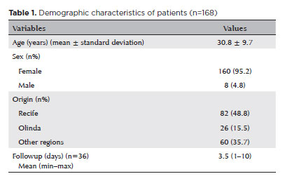
Objetivo: Avaliar o perfil clínico e epidemiológico dos transplantes de córnea realizados em um centro de referência oftalmológica de Recife no estado de Pernambuco, localizado no nordeste do Brasil.
Métodos: Esse estudo transversal coletou através de prontuários médicos dados clínicos e epidemiológicos de pacientes submetidos a ceratoplastia na Fundação Altino Ventura, de janeiro a dezembro de 2017.
Resultados: Um total de 356 procedimentos foram realizados em 327 pacientes dos quais 165 (50.5%) eram mulheres. A média de idade na cirurgia foi de 50.9 ± 22.6 anos (variação, 10 - 89 anos). A maioria dos pacientes (n=152 [46.5%]) era da capital e região metropolitana. A média de tempo de espera na fila para o transplante de córnea foi de 52.4 ± 58.9 dias (variação, 0 - 460 dias). As principais indicações de transplante foram ceratite infecciosa (n=88 [24.7%]), ceratocone (n=80 [22.5%]) e falência de transplante prévio (n=75 [21.1%]). Transplante penetrante foi a técnica mais realizada (n=213 [59.9%]) e foi mais comum em homens (n=132 [76.7%]), enquanto os transplantes lamelares posteriores (n=143 [41.1%]) foram mais realizados nas mulheres (p<0.001).
Conclusão: Ceratites infecciosas foram a causa mais comum de transplante, com prevalência similar em adultos economicamente ativos de ambos os sexos. Transplante penetrantes foram os prevalentes em homens e os transplantes lamelares em mulheres.
Keywords: Doença da córnea/epidemiologia; Transplante de córnea; Ceratoplastia penetrante; Brasil/epidemiologia
Arq. Bras. Oftalmol. 2025;88 (4 )
:1-8
| DOI: 10.5935/0004-2749.2024-0151
Abstract
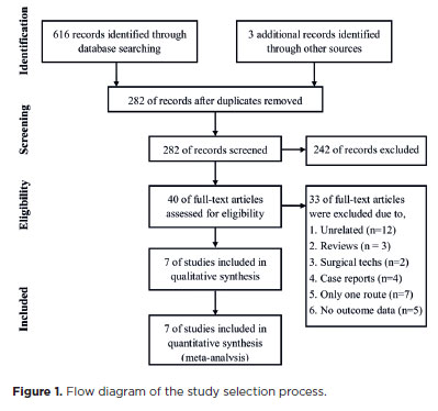
PURPOSE: To compare the incidence rates of complications following pediatric cataract surgery between the limbal and pars plana approaches.
METHODS: PubMed, EMBASE, Web of Science, Scopus, Cochrane Library, and ClinicalTrials.gov were systematically searched for studies comparing the two surgical approaches. We pooled the incidence rates of postoperative complications using a random-effects model.
RESULTS: Seven studies comprising 375 eyes from 260 patients were included. No significant differences in complication rates were observed between the limbal and pars plana approaches. The pooled incidence rates (95% confidence Interval) of postoperative visual axis opacity (VAO), VAO treated with laser or surgery, secondary glaucoma, wound leakage, corneal edema, anterior chamber reaction, posterior iris synechiae, capsular phimosis, intraocular lens dislocation, posterior capsular rupture, and intravitreal lens fragmentation were 4.7% (0.8%10.8%), 3.9% (1.0%-8.1%) , 2.8% (0%-11.4%), 0 (0%-1.3%), 2.9% (0%-11.8%), 5.6% (0.1%-16.5%), 2.4% (0%-8.5%), 3.8% (0.6%-8.9%), 2.2% (0%-6.4%), 9.2% (4.1%-15.8%) and 1.3% (0%-6.3%), respectively. Both surgical approaches demonstrated improved visual acuity postoperatively.
CONCLUSIONS: Pediatric cataract surgery, performed via the limbal or pars plana approach, is effective and safe, with a low incidence of complications when conducted by trained surgeons. Neither method demonstrated a significant difference in the visual acuity improvement or complication rates.
Keywords: Pediatric cataract surgery; Postoperative complications; Limbal route; Pars plana routes; Meta-analysis
Arq. Bras. Oftalmol. 2024;87 (3 )
:1-8
| DOI: 10.5935/0004-2749.2022-0076
Abstract
MÉTODOS: Córneas humanas de treinamento disponibilizadas foram randomizadas em quatro grupos: Pachy-100 (profundidade de incisão = espessura corneana central - margem de segurança de 100 µm), Pachy-50 (margem de segurança de 50 µm), Pachy-0 (sem margem de segurança) e Pachy+50 (profundidade de incisão = espessura corneana central + 50 µm). Todas as lamelas foram dissecadas através um método padronizado e já publicado (Pachy-DSEK). As espessuras das lamelas (centro, zona de 3,0mm e zona de 6,0mm) foram medidas com tomografia de coerência óptica. A razão de espessura centro-periferia foi calculada aos 3,0 e 6,0 mm de diâmetro.
RESULTADOS: Perfuração endotelial ocorreu apenas no grupo Pachy+50 (n=3, 30%). A espessura central da lamela nos grupos Pachy-100, Pachy-50, Pachy-0 e Pachy+50 foi de 185 ± 42 µm, 122 ± 29 µm, 114 ± 29 µm, e 58 ± 31 µm, respectivamente (p<0,001). As razões C/P aos 3,0 e 6,0 mm foram de 0,97 ± 0,06 e 0,92 ± 0,14, respectivamente. Os parâmetros de características do doador não se correlacionaram com os resultados de espessura de lamela. A profundidade planejada de incisão se correlacionou com a maioria dos parâmetros de espessura de lamela (p<0,001). A espessura de lamela se correlacionou negativamente com a profundidade planejada da incisão (p<0.001, r=-0,580). O melhor resultado foi observado no grupo Pachy-0, em que 75% das lamelas mediram abaixo de 130 µm e não houve perfuração endotelial.
CONCLUSÃO: Através de um método padronizado de dissecção, a maioria das lamelas endoteliais apresentou uma configuração planar. O planejamento de profundidade de incisão igual à espessura corneana central resultou em alta porcentagem de lamelas ultrafinas sem ocorrência de perfuração.
Keywords: Transplante de córnea; Ceratoplastia lamelar; Endotélio corneano; Dissecção; Tomografia de coerência óptica
Arq. Bras. Oftalmol. 2024;87 (6 )
:1-6
| DOI: 10.5935/0004-2749.2022-0205
Abstract
Objetivos: Descrever as características demográficas e clínicas das vítimas de trauma ocular por fogos de artifício atendidas nas emergências oftalmológicas de dois centros de referência em Pernambuco e identificar fatores relacionados a mau prognóstico visual.
Métodos: Avaliação retrospectiva dos prontuários de pacientes admitidos na emergência oftalmológica com história de trauma por fogos de artifício entre janeiro de 2012 e dezembro de 2018. A coleta de dados incluiu idade, gênero, procedência, mês e ano do acidente, estruturas oculares acometidas e características das lesões, além do tipo de tratamento a que os pacientes foram submetidos. Naqueles pacientes acompanhados por mais de 30 dias, analisou-se a acuidade visual final e a associação com sua procedência.
Resultados: Foram incluídos 370 olhos de 314 pacientes. Destes, 248 (79,0%) vítimas eram do sexo masculino e 160 (51,0%) da região metropolitana do Recife, com uma média de idade de 25.6 ± 18.8 anos. Em 56 (17,8%) dos casos o trauma foi bilateral. No mês de junho ocorreu um total de 152 (48,4%) casos. Os sítios mais acometidos foram pálpebras em 91 (24,6%) olhos e superfície ocular em 252 (68,1%). O tratamento cirúrgico foi necessário em 87 (23,5%) olhos. Após manejo clínico-cirúrgico, 37 (10.0%) olhos desenvolveram visão pior do que 20/400. Destes, 34 (91,9%) olhos eram de pacientes do interior do estado de Pernambuco ou de outro estado. Os pacientes provenientes do interior do estado apresentaram maior chance de desenvolver cegueira quando comparados aos que eram provenientes da região metropolitana (Odds Ratio de 5,46).
Conclusões: As vítimas de trauma ocular por fogos de artificio foram em sua maioria do sexo masculino, procedentes da região metropolitana do estado e das faixas etárias pediátrica e economicamente ativa. Aqueles provenientes do interior ou de outros estados apresentaram maior chance de desenvolver cegueira.
Keywords: Emergências; Queimaduras oculares/epidemiologia; Incêndios;Traumatismos por explosões; Substâncias explosivas
Arq. Bras. Oftalmol. 2025;88 (3 )
:1-6
| DOI: 10.5935/0004-2749.2024-0207
Abstract
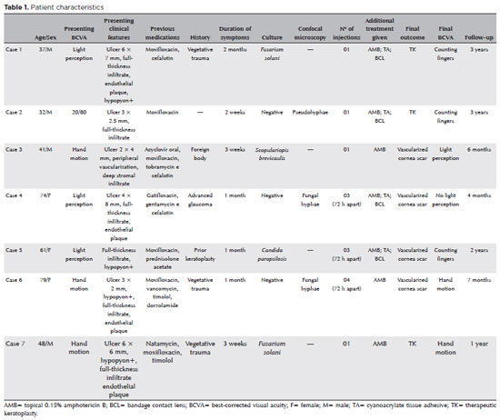
PURPOSE: This study aimed to report the use, efficacy, and safety of intracameral voriconazole as an adjuvant treatment for deep fungal keratitis.
METHODS: This was a prospective case series of seven eyes with fungal keratitis with anterior chamber involvement or a corneal ulcer refractory to conventional topical treatment. In addition to topical treatment with 0.15% amphotericin B eye drops, voriconazole 50 μg/ 0.1 mL
was administered to the anterior chamber of each affected eye up to four times within 72 h. The primary outcome measures were healing (fungal eradication) and the need for therapeutic keratoplasty. Best-corrected visual acuity was a secondary outcome measure.
RESULTS: Three cases were confirmed by confocal microscopy, and four were diagnosed from positive culture tests. At presentation, one patient had a best-corrected visual acuity of 20/80, while all others had hand motion or worse. Four cases received one intracameral injection, two cases received three, and one case received four injections. There were no complications after any of the intracameral voriconazole injections. Four patients had imminent corneal perforations and were treated with cyanoacrylate adhesive and bandage contact lenses. Four patients recovered from the infection, and three underwent therapeutic keratoplasty. The final best-corrected visual acuity was improved in two cases but all patients had a final visual acuity of counting fingers or worse.
CONCLUSION: As an adjuvant treatment for deep fungal keratitis, intracameral voriconazole injection is a feasible option. Although fungal eradication was achieved in all patients, three required therapeutic keratoplasty and all patients had unsatisfactory visual acuity outcomes.
Keywords: Antifungal agents; Fungi; Corneal transplantation; Keratitis; Eye infections, fungal; Voriconazole
Arq. Bras. Oftalmol. 2024;87 (3 )
:1-8
| DOI: 10.5935/0004-2749.2023-0109
Abstract
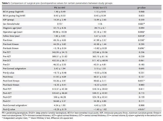
PURPOSES: This study aims to assess and compare the postoperative visual and topographic outcomes, complications, and graft survival rates following deep anterior lamellar keratoplasty and penetrating keratoplasty in patients with macular corneal dystrophy.
METHODS: In this study we enrolled 59 patients (23 male; and 36 female) with macular corneal dystrophy comprising 81 eyes. Out of these, 64 eyes underwent penetrating keratoplasty, while 17 eyes underwent deep anterior lamellar keratoplasty. The two groups were analyzed and compared based on best-corrected visual acuity, corneal tomography parameters, pachymetry, complication rates, and graft survival rates.
RESULTS: After 12 months, 70.6% of the patients who underwent deep anterior lamellar keratoplasty (DALK) and 75% of those who had penetrating keratoplasty (PK) achieved a best-corrected visual acuity of 20/40 or better (p=0.712). Following surgery, DALK group showed lower front Kmean (p=0.037), and Q values (p<0.01) compared to the PK group. Postoperative interface opacity was observed in seven eyes (41.2%) in the DALK group. Other topography values and other complications (graft rejection, graft failure, cataract, glaucoma, microbial keratitis, optic atrophy) did not show significant differences between the two groups. The need for regrafting was 9.4% and 11.8% in the PK and DALK groups, respectively (p=0.769). Graft survival rates were 87.5% and 88.2% for PK and DALK; respectively (p=0.88 by Log-rank test).
CONCLUSION: Both PK and DALK are equally effective in treating macular corneal dystrophy, showing similar visual, topographic, and survival outcomes. Although interface opacity occurs more frequently after DALK the visual results were comparable in both groups. Therefore, DALK emerges as a viable surgical choice for patients with macular corneal dystrophy without Descemet membrane involvement is absent.
Keywords: Macular corneal dystrophy; Corneal dystrophies; Hereditary; Keratoplasty; Penetrating; Corneal transplantation
Arq. Bras. Oftalmol. 2024;87 (2 )
:1-8
| DOI: 10.5935/0004-2749.2022-0328
Abstract
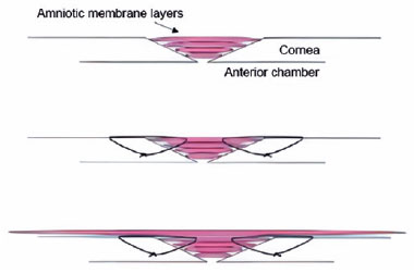
PURPOSE: Wet bio-amniotic membrane plugging combined with transplantation is a novel option that combined amniotic membrane plugging with amniotic membrane transplantation for the treatment of small corneal perforations. This study aimed to evaluate the efficacy of wet bio-amniotic membrane plugging in the treatment of small corneal perforations and compared it with that of the penetrating keratoplasty procedure.
METHODS: Forty patients (41 eyes) with small corneal perforations <3 mm in diameter treated at our hospital between July 2018 and January 2021 were retrospectively included. Among them, 21 eyes were treated with wet bio-amniotic membrane plugging (wet bio-amniotic membrane plugging group), and 20 eyes were treated with penetrating keratoplasty procedure (penetrating keratoplasty procedure group). The best-corrected visual acuity, anterior chamber formation, corneal thickness, primary disease control, postoperative complications, and graft survival rate were assessed.
RESULTS: No significant difference in baseline characteristics was found between the wet bio-amniotic membrane plugging and penetrating keratoplasty procedure groups (p>0.05). The postoperative control rates of primary diseases in the wet bio-amniotic membrane plugging and penetrating keratoplasty procedure groups were 95.2% and 90.0%, respectively (p=0.481). Visual acuity was improved 6 months after the operation in the wet bio-amniotic membrane plugging group and was improved at postoperative 1 month in the penetrating keratoplasty procedure group. The formation time of the anterior chamber in the wet bio-amniotic membrane plugging group was significantly shorter than that in the penetrating keratoplasty procedure group (p=0.023). The corneal thickness of the two groups significantly increased 12 months after the operation; however, the degree of thickening in the penetrating keratoplasty procedure group was higher than that in the wet bio-amniotic membrane plugging group (p<0.001). During the follow-up, postoperative complications were not different between the two groups (p>0.999).
CONCLUSION: The results suggest that wet bio-amniotic membrane plugging is effective and safe in the treatment of small corneal perforations. Thus, it can be used as an emergency treatment alternative to penetrating keratoplasty procedure for small corneal perforations.
Keywords: Amnion; Transplantation; Amniotic membrane; Keratoplasty, penetrating; Corneal perforation; Wet bio-amniotic membrane plugging; Wet bio-amniotic membrane transplantation
Arq. Bras. Oftalmol. 2024;87 (2 )
:1-6
| DOI: 10.5935/0004-2749.2023-2022-0341
Abstract
PURPOSE: To evaluate the clinical results of cryopreserved amniotic membrane transplantation as a treatment option for refractory neurotrophic corneal ulcers.
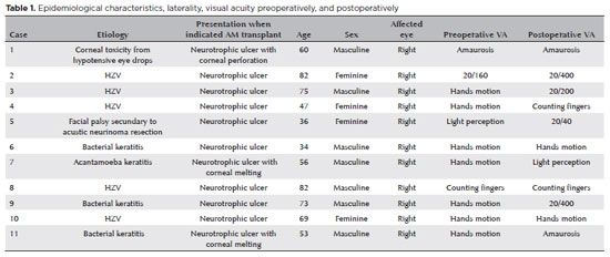
METHODS: This prospective study included 11 eyes of 11 patients who underwent amniotic membrane transplantation for the treatment of refractory neurotrophic corneal ulcers at Hospital de Clínicas da Universidade Federal do Paraná, in the city of Curitiba, from May 2015 to July 2021. Patients underwent different surgical techniques in which the amniotic membrane was applied with the epithelium facing upward to promote corneal re-epithelialization.
RESULTS: The median age of the patients was 60 years (range, 34-82 years), and 64% were men. The predominant etiology of corneal ulcers was herpes zoster (45% of cases). Approximately one-third of the patients (27%) were chronically using hypotensive eye drops, and more than half (54%) had previously undergone penetrating corneal transplantation. At the time of amniotic membrane transplantation, 18% of the eyes had corneal melting, 9% had corneal perforation, and the others had corneal ulceration without other associated complications (73%). The time between clinical diagnosis and surgical treatment ranged from 9 days to 2 years. The corrected visual acuity was worse than 20/400 in 90% of the patients preoperatively, with improvement in 36% after 3 months of the procedure, worsening in 18% and remaining stable in 36%. Of the patients, 81% complained of preoperative pain, and 66% of them reported total symptom relief after the surgical procedure. In one month, 54.6% of the patients presented a closure of epithelial defect, and half of the total group evolved with corneal thinning. The failure rate was 45.5% of the cases.
CONCLUSION: Cryopreserved amniotic membrane transplantation can be considered a good alternative for treating refractory neurotrophic corneal ulcers, as it resulted in significant improvement in pain (66%) and complete epithelial closure (60%) in many patients at 1 month postoperatively. Notably, the high failure rate highlights the need for further studies to identify patient- and ulcer-related factors that may influence the outcomes of this procedure.
Keywords: Amnion/transplantation; Corneal ulcer; Anterior eye segment; Keratitis
 ABO is licensed under a Creative Commons Attribution-NonComercial 4.0 Internacional.
03-qua01tb.jpg)
13-tab01.jpg)
06-tab01tb.jpg)
03-tab01.jpg)
15-fig01.jpg)
17-equ01.jpg)
09-fig01.jpg)