Arq. Bras. Oftalmol. 2026;89 (1 )
:1-6
| DOI: 10.5935/0004-2749.2025-0071
Abstract
PURPOSE: This study aimed to evaluate the outcomes of strabismus surgical correction in patients with Down syndrome.
METHODS: We conducted a retrospective chart review of patients with Down syndrome who underwent strabismus surgery between January 1997 and May 2024 at an Ophthalmology Outpatient Clinic in São Paulo, Brazil. The data collected included age, sex, medical and ocular history, surgical details, and follow-up outcomes. The patients were categorized by strabismus type into esotropia, fourth nerve palsy, and mixed groups. Surgical success was defined as final alignment within 10Δ of orthotropia and, where applicable, whether there was resolution of abnormal head posture of ocular origin. Patients with postoperative follow-up <6 months were excluded.
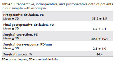
RESULTS: A total of 37 patients (21 females) were included. Of these, 22 (59.5%) were in the esotropia group, 10 (27.0%) in the fourth nerve palsy group, and 5 (13.5%) in the mixed group. The surgical success rate in the esotropia group was 86.4%, with a mean preoperative deviation of 35.2 (± 6.5)Δ, and mean surgical correction of 30.1 (± 10.4)Δ. The success rate in the fourth nerve palsy group was 40.0%, with a mean preoperative deviation of 10.4 (± 4.3)Δ. Overall, success was achieved with a single surgical procedure in 73.0% of the sample. No significant associations were found between surgical success and the clinical and demographic variables, including sex, age at surgery, oblique muscle overaction, pattern strabismus, visual acuity, amblyopia, preoperative deviation, or postoperative follow-up duration (p>0.05).
CONCLUSIONS: When standard surgical tables are applied, strabismus surgery in patients with Down syndrome appears to be safe and effective. We found high success rates, particularly among patients with esotropia. We observed no tendencies toward over- or under-correction. These findings support the use of conventional surgical protocols with this patient population.
Keywords: Down Syndrome/complications; Strabismus/surgery; Esotropia/surgery; Oculomotor nerve diseases/physiopathology; Vision disorders; Humans; Brazil.
Arq. Bras. Oftalmol. 2024;87 (2 )
:1-8
| DOI: 10.5935/0004-2749.2022-0319
Abstract
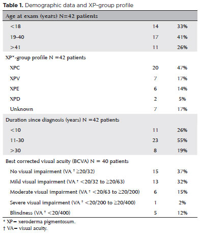
To assess Meibomian gland dysfunction using meibography in patients with xeroderma pigmentosum and correlate with ocular surface changes. This cross-sectional study evaluated patients with xeroderma pigmentosum. All patients underwent a comprehensive and standardized interview. The best-corrected visual acuity of each eye was determined. Detailed ophthalmic examination was conducted, including biomicroscopy examination of the ocular surface, Schirmer test type I, and meibography, and fundus examination was also performed when possible. Meibomian gland dysfunction was assessed by non-contact meibography using Oculus Keratograph® 5M (OCULUS Inc., Arlington, WA, USA). Saliva samples were collected using the Oragene DNA Self-collection kit (DNA Genotek Inc., Ottawa, Canada), and DNA was extracted as recommended by the manufacturer. Factors associated with abnormal meiboscores were assessed using generalized estimating equation models. A total of 42 participants were enrolled, and 27 patients underwent meibography. The meiboscore was abnormal in the upper eyelid in 8 (29.6%) patients and in the lower eyelid in 17 (62.9%). The likelihood of having abnormal meiboscores in the lower eyelid was 16.3 times greater than that in the upper eyelid.In the final multivariate model, age (p=0.001), mutation profile (p=0.006), and presence of ocular surface malignant tumor (OSMT) (p=0.014) remained significant for abnormal meiboscores. For a 1-year increase in age, the likelihood of abnormal meiboscores increased by 12%. Eyes with OSMT were 58.8 times more likely to have abnormal meiboscores than eyes without ocular surface malignant tumor.In the final model, age, xeroderma pigmentosum profile, previous cancer, and clinical alterations on the eyelid correlated with a meiboscore of ≥2.Meibomian gland dysfunction was common in patients with xeroderma pigmentosum, mainly in the lower eyelid. The severity of Meibomian gland dysfunction increases with age and is associated with severe eyelid changes.
Keywords: Meibomian glands/pathology; Meibomian glands/ diagnostic imaging; Photography; Xeroderma pigmentosum; Eyelid diseases/diagnostic imaging; Dry eye syndromes; DNA repair; Humans; Case report
 ABO is licensed under a Creative Commons Attribution-NonComercial 4.0 Internacional.
12-fig01.jpg)
09-fig01.jpg)
10-fig01.jpg)
08-fig01.jpg)
14-fig01.jpg)
11-fig01tb.jpg)
14-fig01.jpg)
01-fig01.jpg)
02-fig01.jpg)