Arq. Bras. Oftalmol. 2021;84 (4 )
:316-323
| DOI: 10.5935/0004-2749.20210045
Abstract
OBJETIVO: O objetivo deste estudo foi analisar a segurança do implante de lente intraocular primária em um grande número de olhos em crianças <24 meses.
MÉTODOS: Foram revisados os prontuários de pacientes com idade entre 5-24 meses, submetidos a implante primário de lente intraocular no saco capsular. Uma lente intraocular acrílica de três peças dobrável foi implantada pelo mesmo cirurgião usando uma única técnica cirúrgica. Pacientes que tiveram <1 ano de acompanhamento após a cirurgia foram excluídos. Os principais resultados incluíram medidas de acuidade visual, mudança miópica, complicações pós operatórias e cirurgias adicionais.
RESULTADOS: Foram analisados 68 pacientes (93 olhos). A média de idade dos pacientes no momento da cirurgia foi de 15,06 ± 6,19 (5 a 24) meses, e o equivalente esférico 1 mês após a cirurgia foi de 3,62 ± 2,32 D. Após 5,67 ± 3,10 anos, o equivalente esférico foi de -0,09 ± 3,22 D, e a acuidade visual corrigida à distância foi de 0,33 ± 0,33 e 0,64 ± 0,43 logMAR em casos bilaterais e casos unilaterais, respectivamente (p=0,000). A maior mudança míopica foi observado em bebês submetidos à cirurgia aos 5 e 6 meses de idade. As complicações mais frequentes incluíram opacificação do eixo visual e corectopia. Glaucoma e descolamento de retina não foram relatados.
CONCLUSÃO: O implante primário de lente intraocular no saco capsular em crianças de 5-24 meses é seguro e está associado à baixas taxas de eventos adversos e cirurgias adicional.
Keywords: Catarata pediátrica; Lente intraocular; Implante primário LIO; Mudança miópica; Catarata congênita
Arq. Bras. Oftalmol. 2023;86 (3 )
:1-7
| DOI: 10.5935/0004-2749.20230045
Abstract
Objetivo: Avaliar o implante de lente intraocular primária para tratamento da afacia pediátrica no Sistema Único de Saúde (SUS) e comparar os resultados em diferentes faixas etárias.
Métodos: Foram incluídas crianças com catarata congênita e do desenvolvimento unilateral ou bilateral de 0-12 anos de idade e submetidas a implante de lente intraocular primária.
Resultados: Cento e oito olhos de 68 crianças divididas em quatro grupos de idade (<7m; 7m-2a; 2-5a e > 5a) foram avaliados. Dezenove olhos (17,59%) apresentaram opacificação do eixo visual como complicação pós-operatória. Essa complicação foi mais frequente na faixa etária <7 meses (37,93%). A diferença foi significativa entre os grupos de idade <7 meses e > 5 anos (p=0,002). A opacificação do eixo visual foi dividida em duas categorias: membrana pupilar e proliferação de células do cristalino. Oito olhos apresentaram membrana pupilar e 14 proliferação de células do cristalino. Dos oito olhos com membrana pupilar, sete ocorreram na faixa etária <7 meses. A diferença entre o grupo de idade <7 meses e os grupos de 2-5 anos e > 5 anos foi significativa (p=0,01). A proliferação de células do cristalino foi mais frequente nos grupos de idade <7 meses e 2-5 anos, mas significativa apenas quando comparados o grupo de idade <7 meses com o grupo> 5 anos de idade (p=0,040). Glaucoma e suspeitos de glaucoma não foram observados durante o acompanhamento.
Conclusões: A principal complicação encontrada no estudo foi a opacificação do eixo visual. Sua incidência foi maior em crianças operadas antes dos 7 meses de idade.
Keywords: Extração de catarata; Lentes intraoculares; Complicações intraoperatórias; Glaucoma; Segmento anterior do olho; Criança.
Arq. Bras. Oftalmol. 2025;88 (1 )
:1-10
| DOI: 10.5935/0004-2749.2023-0073
Abstract
PURPOSE: To describe the epidemiological and clinical profile of hospitalized patients with retinoblastoma in Brazil.
METHODS: Using data from the Hospital Cancer Registry of the , patients with the morphological codes of retinoblastoma who were diagnosed between 2000 to 2018, aged 0–19 years, and followed up in registered hospitals (analytical cases) were selected. The relative and absolute frequencies of demographic, clinical, diagnostic, therapeutic, and outcome variables were described. Hospital performance indicators were calculated and compared between hospitals qualified and not qualified to treat pediatric oncology cases and between hospitals with different case volumes (<20, 20–75, >75 cases).
RESULTS: Of the 2,269 identified analytical cases from 86 institutions, 48% were from the Southeast, 54% were male, and 66% were aged <4 years. The proportion of missing data (NA) was too high for several variables. Approximately 84% of the patients were from the public health system, 40% had a positive family history, and 88% had unilateral involvement. The first treatment included surgery in 58.3% of the patients (NA=2), Approximately 36.6% of these patients achieved complete remission, 10.8% achieved partial remission, and 12.7% died (NA=59%). Hospital performance indicators were within the target in >90% of the patients. The median time between the first appointment and diagnosis (6 days, interquartile range [IQR] 1–14) was significantly lower and the median time to death was longer (343 days, IQR, 212-539) in high-volume hospitals (>75 cases) than in medium- and low-volume hospitals.
CONCLUSIONS: Despite the high proportion of missing data, we found that the delay in diagnosis is due to prehospital factors. Additionally, there is a need for educational programs for healthcare professionals and families that emphasize early identification and referral to specialized centers. Future studies should focus on the impact of Hospital Cancer Registry data completeness on outcomes, causes of delay in diagnosis, regional inequalities, and barriers to accessing specialized services.
Keywords: Retinoblastoma/diagnosis; Retinoblastoma/epidemiology; Patient care; Humans; Children; Adolescents; Brazil.
Arq. Bras. Oftalmol. 2025;88 (6 )
:1-5
| DOI: 10.5935/0004-2749.2025-0085
Abstract
PURPOSE: The purpose of this study was to assess visual outcomes and patient satisfaction following cataract surgery involving the implantation of quad-loop intraocular lenses, including trifocal, bifocal, and toric variants.
METHODS: Information was obtained from both physical and electronic medical records of patients who underwent phacoemulsification cataract surgery with implantation of different intraocular lenses between January 1, 2022, and December 31, 2023. The study included individuals aged over 18 who received bilateral implantation of bifocal, trifocal, or monofocal toric intraocular lenses. Visual acuity was assessed at various postoperative time points using the logMAR scale. Quantitative variables were analyzed using mean and standard deviation.
RESULTS: A total of 92 eyes received premium intraocular lenses: 4 bifocal, 32 trifocal, 52 toric monofocal, and 4 trifocal toric lenses. The average preoperative corrected visual acuity was logMAR 0.478 ± 0.259. On the first postoperative day, the average uncorrected visual acuity was logMAR 0.301 ± 0.207. By day 30, 67.4% of eyes achieved uncorrected distance visual acuity of logMAR 0.2 or better. Patient satisfaction was high, with few reports of glare or halos.
CONCLUSION: Quad-loop intraocular lenses-including trifocal, bifocal, and toric models-demonstrated effective improvement in visual acuity and high levels of patient satisfaction. These lenses represent a suitable option for enhancing visual outcomes after cataract surgery. Additional studies with larger cohorts are recommended to confirm these results.
Keywords: Cataract extraction; Aberrometry/methods; Lenses, intraocular; Lens implantation, intraocular; Prosthesis design
Arq. Bras. Oftalmol. 2026;89 (2 )
:1-8
| DOI: 10.5935/0004-2749.2025-0175
Abstract
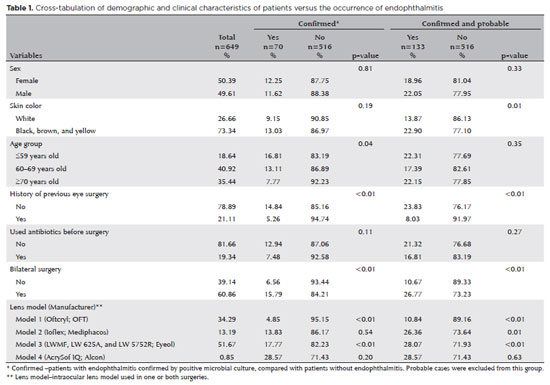
PURPOSE: Endophthalmitis is one of the most important adverse events after cataract surgery, as it can lead to total vision loss. This study aimed to describe the occurrence of endophthalmitis after phacoemulsification with intraocular lens implantation in patients treated in a community setting in Porto Velho, Rondônia, Brazil.
METHODS: This retrospective cohort study was conducted using a database of 649 medical records of patients who underwent surgery and were followed for three months. Poisson regression analysis was used to estimate relative risks and 95% confidence intervals (95% CIs).
RESULTS: The incidence of confirmed endophthalmitis was 11.94% (95% CI, 9.50-14.76), while the incidence of confirmed and probable cases was 20.50% (95% CI, 17.52-23.73). For confirmed cases, bilateral surgery and the use of lens model 3 were identified as risk factors for endophthalmitis, whereas age over 70 yr and preoperative antibiotic use were protective factors. For confirmed and probable cases, brown and yellow skin color, bilateral surgery, and the use of lens model 3 were also identified as risk factors. Gram-negative bacteria were the predominant etiological agents, and corneal edema was the main clinical manifestation. The mean duration of treatment was eight days, and 27.12% of patients used antibiotics.
CONCLUSION: The incidence observed was substantially higher than that reported in the literature, with a predominance of Gram-negative agents and an association with bilateral surgeries and the Eyeol intraocular lens model. These findings reinforce the need for continuous epidemiological surveillance and the implementation of specific biosafety and infection control protocols during cataract surgery campaigns.
Keywords: Endophthalmitis; Disease outbreaks; Phacoemulsification; Lens implantation, intraocular; Lenses, intraocular; Cataract; Risk factors; Anti-bacterial agents
Arq. Bras. Oftalmol. 2025;88 (2 )
:1-7
| DOI: 10.5935/0004-2749.2023-0265
Abstract
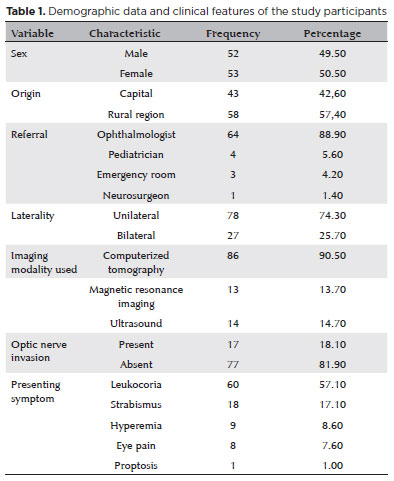
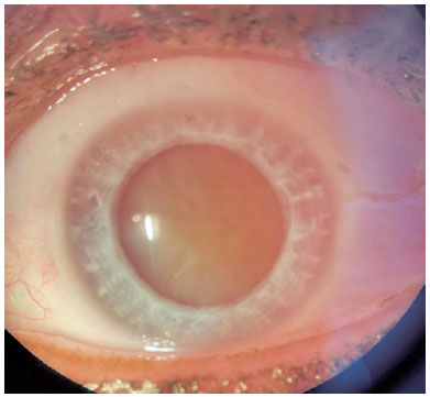
PURPOSE: Although Brazil has a high prevalence of retinoblastoma, there is a lack of epidemiological data on the disease. Thus, in this study, we aimed to evaluate the epidemiological profile of patients diagnosed with retinoblastoma in the ophthalmology department of a pediatric tertiary referral hospital in Ceara, Brazil.
METHODS: A descriptive and cross-sectional study was conducted by retrospectively analyzing the clinical and socioeconomic data from the medical records of pediatric patients followed-up at the hospital between 2007 and 2021. Retinoblastoma was diagnosed on the basis of a fundoscopic or histopathologic examination.
RESULTS: The data of 105 patients were included in the study, and the mean patient age at the time of diagnosis was 1.7 years. Most of the patients were women (50.5%) and hailed from rural areas (57.4%), which was associated with a higher tumor stage. Of the 150 patients, 57.1% initially presented with leukocoria. Ocular hyperemia was associated with more advanced stages of retinoblastoma (p=0.004). Bilateral involvement was observed in 25.7% of the patients and at a significantly younger age (p=0.009). The presence of retinal detachment, vascularized lesions, and vitreous seeds significantly increased the likelihood of requiring enucleation.
DISCUSSION: This study presents an epidemiological description of retinoblastoma in Brazil, which highlights the significance of early detection. Delayed diagnosis is associated with a poorer visual prognosis and higher mortality rate, particularly in patients with unilateral disease. Risk factors for a more severe disease were retinal detachment, vascularized lesions, and vitreous seeds. The correlation between histopathological features and clinical outcomes was limited.
CONCLUSION: Further studies are required to assess the influence of ocular hyperemia, fundoscopic assessment, and histopathologic findings on the prognosis of retinoblastoma. Moreover, it is critical to devise interventions to reduce the time-to-diagnosis in rural areas.
Keywords: Retinoblastoma; Retinal neoplasms; Epidemiology; Prevalence; Risk factors; Delayed diagnosis; Child
Arq. Bras. Oftalmol. 2025;88 (5 )
:1-8
| DOI: 10.5935/0004-2749.2024-0312
Abstract
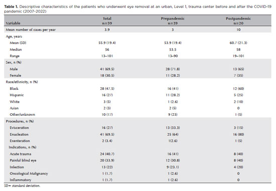
PURPOSE: To evaluate the changes in the rates and indications of eye removal procedures during the recent COVID-19 pandemic.
METHODS: The medical records of all patients who underwent eye removal from 2007 to 2022 were retrospectively reviewed. The patient demographic data and indications for surgery were collected. Data from two groups of patients (prepandemic surgery and postpandemic surgery) were compared. Statistical significance was set at p<0.05.
RESULTS: Fifty-nine patients underwent enucleation (69%), evisceration (27%), or exenteration (3%). The mean (SD) age of the patients was 55.9 (19.4) years, and most (69%) of the patients were males. Most (47%) of the study population were Black. The common indications for eye removal were trauma (41%), painful blind eye (34%), and infection/inflammation (24%). The types of trauma were assault (55%), accidental (39%), and self-inflicted (6%). The mean (SD) monthly rates of eye removal increased from 0.25 (0.50) in the prepandemic period to 0.77 (0.91) during the pandemic (p<0.001). These increases were noted in both males (p=0.003) and females (p=0.001) and were the highest among Black patients [0.42 (0.76); p<0.001]. Among the indications of eye removal, painful blind eyes [0.35 (0.75); p<0.001] and ocular trauma [0.31 (0.47); p=0.051] exhibited the greatest increases following the pandemic.
CONCLUSION: The rate of eye removal procedures increased during the recent pandemic. Although delayed care of chronic eye conditions may have contributed to the increased rates of painful blind eyes, the increased trauma-related eye removals may be attributed to the simultaneous spike in violent assaults in New York City.
Keywords: Eye injuries; Eye enucleation; COVID-19; Pandemics; Ethinicity; Inflammation, Trauma centers
Arq. Bras. Oftalmol. 2026;89 (4 )
:1-5
| DOI: 10.5935/0004-2749.2026-0010
Abstract
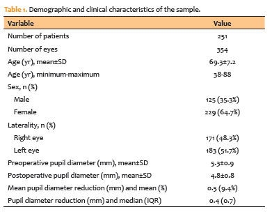
PURPOSE: To evaluate changes in scotopic pupil diameter before and after cataract surgery performed by phacoemulsification with intraocular lens implantation.
METHODS: This prospective longitudinal observational study included patients who underwent cataract surgery. Scotopic pupil diameter was measured preoperatively and 30-40 days postoperatively using an automated keratometer after a standardized dark-adaptation period under controlled ambient illumination. Each eye was considered an independent unit of observation. Because some participants contributed both eyes, intraindividual correlation was accounted for using a linear mixed-effects model with random patient intercepts. Time of assessment (preoperative versus postoperative), age, sex, and eye laterality were included as fixed effects.
RESULTS: A total of 354 eyes from 251 patients were analyzed. The mean patient age was 69.3±7.2 yr. Mean scotopic pupil diameter decreased from 5.3±0.9mm preoperatively to 4.8±0.8mm postoperatively, representing a mean reduction of 0.5mm (9.4%). In the linear mixed-effects model, cataract surgery was associated with a significant reduction in pupil diameter, with an adjusted mean difference of 0.45mm (95% confidence interval [95% CI], 0.39-0.51; p<0.001). Age (p=0.061), sex (p=0.920), and eye laterality (p=0.152) were not significantly associated with the magnitude of pupil diameter change.
CONCLUSION: Phacoemulsification with intraocular lens implantation was associated with a significant reduction in scotopic pupil diameter, independent of age, sex, and eye laterality. This finding should be considered during preoperative planning, particularly when selecting intraocular lenses whose optical performance depends on postoperative pupil size.
Keywords: Cataract; Pupil; Phacoemulsification; Lens implantation, intraocular; Lenses, intraocular; Pseudophakia
Arq. Bras. Oftalmol. 2024;87 (2 )
:1-6
| DOI: 10.5935/0004-2749.2021-0435
Abstract
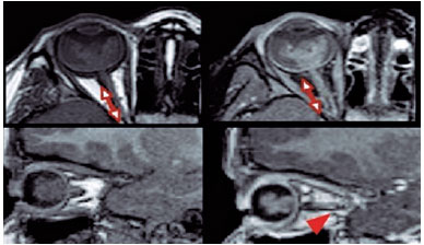
PURPOSE: This study aimed to analyze the association between magnetic resonance imaging apparent diffusion coefficient map value and histopathological differentiation in patients who underwent eye enucleation due to retinoblastomas.
METHODS: An observational chart review study of patients with retinoblastoma that had histopathology of the lesion and orbit magnetic resonance imaging with apparent diffusion coefficient analysis at Hospital de Clínicas de Porto Alegre between November 2013 and November 2016 was performed. The histopathology was reviewed after enucleation. To analyze the difference in apparent diffusion coefficient values between the two major histopathological prognostic groups, Student's t-test was used for the two groups. All statistical analyses were performed using SPSS version 19.0 for Microsoft Windows (SPSS, Inc., Chicago, IL, USA). Our institutional review board approved this retrospective study without obtaining informed consent.
RESULTS: Thirteen children were evaluated, and only eight underwent eye enucleation and were included in the analysis. The others were treated with photocoagulation, embolization, radiotherapy, and chemotherapy and were excluded due to the lack of histopathological results. When compared with histopathology, magnetic resonance imaging demonstrated 100% accuracy in retinoblastoma diagnosis. Optic nerve invasion detection on magnetic resonance imaging showed a 66.6% sensitivity and 80.0% specificity. Positive and negative predictive values were 66.6% and 80.0%, respectively, with an accuracy of 75%. In addition, the mean apparent diffusion coefficient of the eight eyes was 0.615 × 103 mm2/s. The mean apparent diffusion coefficient value of poorly or undifferentiated retinoblastoma and differentiated tumors were 0.520 × 103 mm2/s and 0.774 × 103 mm2/s, respectively.
CONCLUSION: This study revealed that magnetic resonance imaging is useful in the diagnosis of retinoblastoma and detection of optic nerve infiltration, with a sensitivity of 66.6% and specificity of 80%. Our results also showed lower apparent diffusion coefficient values in poorly differentiated retinoblastomas with a mean of 0.520 ×
103 mm2/s, whereas in well and moderately differentiated, the mean was 0.774 × 103 mm2/s.
Keywords: Retinoblastoma; Prognosis; Retinal neoplasms; Orbit; Diffusion magnetic resonance imaging
 ABO is licensed under a Creative Commons Attribution-NonComercial 4.0 Internacional.
02-tab01tb.jpg)
15-tab01tb.jpg)

02-fig01.jpg)
01-fig01.jpg)
13-fig01tb.jpg)