Arq. Bras. Oftalmol. 2023;86 (5 )
:1-6
| DOI: 10.5935/0004-2749.20230070
Abstract
Objetivo: A refração pós-operatória na cirurgia moderna de catarata por microincisão ganha ainda mais importância em pacientes com cirurgia prévia de ceratomileuse in situ assistida por laser (LASIK). As alterações astigmáticas induzidas cirurgicamente nesses olhos podem diferir não apenas em magnitude, mas também em direção em comparação com córneas virgens. O objetivo deste estudo foi comparar as alterações astigmáticas induzidas cirurgicamente após cirurgia de catarata por microincisão entre córneas pós-LASIK e olhos virgens.
Métodos: Foi revisada uma série de casos de cirurgia de catarata por microincisão em olhos com e sem cirurgia LASIK anterior. Os dados demográficos, o comprimento axial no momento da cirurgia de catarata, a espessura central da córnea, os valores esféricos e cilíndricos, as leituras da ceratometria e o astigmatismo corneano posterior pós-operatório foram avaliados retrospectivamente. O método Alpins modificado foi usado para análise vetorial astigmática e foram avaliados o astigmatismo basal, o astigmatismo induzido cirurgicamente, o vetor de diferença, o efeito de achatamento e o torque.
Resultados: Ao todo, 42 olhos de 24 indivíduos foram avaliados. O Grupo I consistiu em 14 olhos com LASIK prévio; o Grupo II incluiu 28 olhos sem qualquer cirurgia refrativa. A média da espessura corneana central pré-operatória no Grupo I foi significativamente mais fina (p=0,012). Não houve diferença significativa no astigmatismo basal entre os grupos em termos de magnitude e vetores de potência. Após a cirurgia de catarata por microincisão, não houve diferenças significativas nos valores médios esféricos, cilíndricos e leituras médias de ceratometria (todos com p>0,05). No entanto, o astigmatismo induzido cirurgicamente e o vetor de diferença foram significativamente maiores no componente do vetor J45 em olhos pós-LASIK, e o efeito de aumento da inclinação pela cirurgia de catarata por microincisão nas córneas pós-LASIK foi significativo em comparação com olhos virgens (p=0,001, p=0,002 e p=0,018, respectivamente).
Conclusões: A cirurgia de catarata aumentou a inclinação das córneas em ambos os grupos, sendo esse aumento significativamente maior nos olhos pós-LASIK. Certamente, a topografia da córnea antes da cirurgia de catarata é particularmente útil para fornecer interpretações mais precisas do astigmatismo induzido cirurgicamente.
Keywords: Cirurgia de catarata; Ceratomileuse; excimer laser in situ; Cirurgia refrativa; Astigmatismo induzido cirurgicamente; Análise vetorial.
Arq. Bras. Oftalmol. 2022;85 (6 )
:590-598
| DOI: 10.5935/0004-2749.20220081
Abstract
Objetivo: Identificar tendências no campo de pesquisa da orbitopatia de Graves nas últimas duas décadas e analisar os ramos de maior concentração de pesquisas nessa área.
Métodos: O banco de dados Web of Science foi usado para extrair artigos com “orbitopatia de Graves” ou seus sinônimos no título. Dados completos e referências foram exportados para o programa VOSviewer para serem analisados. Mapas e gráficos de visualização foram construídos a partir desses dados.
Resultados: Foram obtidos 1067 artigos sobre a orbitopatia de Graves a partir do banco de dados Web of Science. Os EUA ficaram em primeiro lugar em termos de número de publicações, seguidos pela Itália e pela República Popular da China. Dentre os autores, os artigos de Wiersinga WM tiveram o maior número de citações. Quanto às instituições, os artigos da Universidade de Amsterdã tiveram o maior número de citações, mas a Universidade de Pisa publicou o maior número de artigos. Dentre os periódicos, a revista Thyroid publicou o maior número de artigos. A análise de coautoria mostrou quatro agrupamentos de colaboração entre países. O primeiro agrupamento engloba países europeus; o segundo engloba os EUA, Brasil, Canadá, Coreia do Sul e Taiwan. A República Popular da China compreende um agrupamento por si só. O quarto agrupamento inclui Japão, Austrália e Polônia. A análise das palavras-chave revelou cinco agrupamentos de tópicos de palavras-chave: patogênese, gerenciamento, associação, qualidade de vida e cirurgia. A análise das referências citadas em conjunto revelou cinco agrupamentos: patogênese, manejo, fatores de risco, avaliação clínica e manejo cirúrgico.
Conclusão: A pesquisa no campo da orbitopatia de Graves cresceu nos últimos vinte anos. Os tópicos com a maior concentração de pesquisas são: patogênese, gerenciamento, fatores de risco, qualidade de vida e complicações. As tendências de pesquisa mudaram nas últimas duas décadas. Ficou evidente um aumento do interesse em explorar os mecanismos e associações da orbitopatia de Graves. Observou-se uma cooperação entre países europeus neste campo de pesquisa. Os EUA estabeleceram uma cooperação internacional mais ampla que outros países. Acreditamos que mais colaboração internacional envolvendo países em desenvolvimento seria recomendável.
Keywords: Oftalmopatia de Graves; Bibliometria; Oftalmopatia de Graves; Pesquisa
Arq. Bras. Oftalmol. 2023;86 (5 )
:1-5
| DOI: 10.5935/0004-2749.20230063
Abstract
Objetivo: A injeção peribulbar de triancinolona é um tratamento alternativo para doenças oculares da tireoide; no entanto, a segurança desse procedimento continua controversa. O objetivo deste artigo é descrever os efeitos adversos locais e sistêmicos de injeções peribulbares de triancinolona em pacientes com doença ocular da tireoide.
Métodos: Estudo retrospectivo de uma série de casos. Foram analisados os prontuários médicos dos pacientes com doença ocular da tireoide tratados com injeções de triancinolona peribulbar em uma única instituição acadêmica entre 2007 e 2019. Foram documentadas as complicações locais e sistêmicas.
Resultados: Um total de 123 pacientes foram tratados. Apenas 11 (8,9%) pacientes apresentaram complicações locais, sendo a mais frequente a presença de equimoses palpebrais superficiais (7,3%), enquanto 2 (1,6%) pacientes apresentaram complicações sistêmicas (hiperglicemia e inibição da suprarrenal após a interrupção do tratamento). Todas estas complicações foram transitórias e nenhum paciente apresentou sequelas de longo prazo.
Conclusões: As injeções peribulbares de triancinolona nas doenças oculares da tireoide têm uma taxa muito baixa de complicações, tanto locais quanto sistêmicas. São necessários estudos prospectivos para aprofundar este tópico.
Keywords: Órbita/diagnóstico por imagem; Imageamento por ressonância magnética; Oftalmopatia de Graves; Triancinolona/efeitos adversos; Injeções.
Arq. Bras. Oftalmol. 2025;88 (4 )
:1-8
| DOI: 10.5935/0004-2749.2024-0151
Abstract
PURPOSE: To compare the incidence rates of complications following pediatric cataract surgery between the limbal and pars plana approaches.
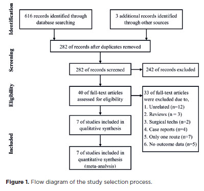
METHODS: PubMed, EMBASE, Web of Science, Scopus, Cochrane Library, and ClinicalTrials.gov were systematically searched for studies comparing the two surgical approaches. We pooled the incidence rates of postoperative complications using a random-effects model.
RESULTS: Seven studies comprising 375 eyes from 260 patients were included. No significant differences in complication rates were observed between the limbal and pars plana approaches. The pooled incidence rates (95% confidence Interval) of postoperative visual axis opacity (VAO), VAO treated with laser or surgery, secondary glaucoma, wound leakage, corneal edema, anterior chamber reaction, posterior iris synechiae, capsular phimosis, intraocular lens dislocation, posterior capsular rupture, and intravitreal lens fragmentation were 4.7% (0.8%10.8%), 3.9% (1.0%-8.1%) , 2.8% (0%-11.4%), 0 (0%-1.3%), 2.9% (0%-11.8%), 5.6% (0.1%-16.5%), 2.4% (0%-8.5%), 3.8% (0.6%-8.9%), 2.2% (0%-6.4%), 9.2% (4.1%-15.8%) and 1.3% (0%-6.3%), respectively. Both surgical approaches demonstrated improved visual acuity postoperatively.
CONCLUSIONS: Pediatric cataract surgery, performed via the limbal or pars plana approach, is effective and safe, with a low incidence of complications when conducted by trained surgeons. Neither method demonstrated a significant difference in the visual acuity improvement or complication rates.
Keywords: Pediatric cataract surgery; Postoperative complications; Limbal route; Pars plana routes; Meta-analysis
Arq. Bras. Oftalmol. 2025;88 (4 )
:1-6
| DOI: 10.5935/0004-2749.2024-0278
Abstract
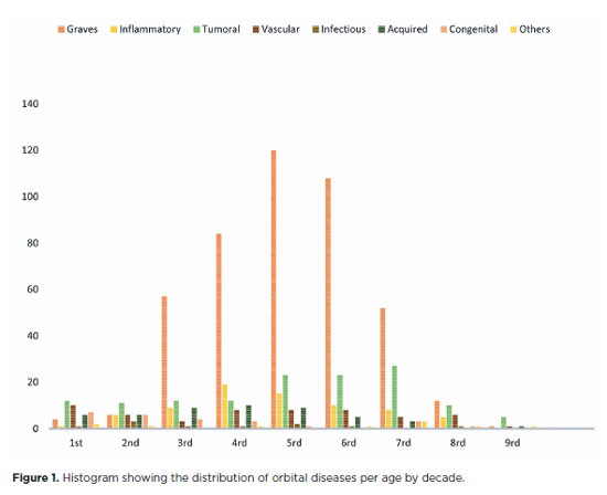
PURPOSE: This study aimed to evaluate the prevalence of orbital conditions in a tertiary ophthalmic outpatient hospital in Sao Paulo, Brazil, with a focus on the main diagnoses and their distribution.
METHODS: A retrospective chart review was conducted involving patients registered and admitted to the orbital disease unit at the Department of Ophthalmology, University of São Paulo Medical School, from January 2004 to March 2018. A total of 838 medical charts were analyzed, of which 37 were excluded due to incomplete data. The remaining charts were categorized into eight diagnostic groups: Graves’ orbitopathy , inflammatory disorders, tumors, vascular lesions, acquired structural abnormalities, congenital structural abnormalities, infectious diseases, and others.
RESULTS: Of the 837,300 ophthalmological appointments, 3,372 (0.4%) were related to orbital diseases. The study included 801 patients, of whom 63.45% were women. The patients’ mean age was 42.86 years. Graves’ orbitopathy was the most common (55%), followed by tumor (17%), inflammatory disorders (9%), vascular lesions (7%), acquired structural abnormalities (5%), congenital structural abnormalities (4%), others (2%), and infectious diseases (1%). The study found significant differences in the incidence and types of orbital diseases, indicating the specialized nature of tertiary care and referral biases.
CONCLUSION: Published data on epidemiological orbital diseases is scarce. Therefore, this study focused on the diverse nature of orbital diseases and their low incidence among ophthalmology appointments. The major trends align with other epidemiological studies, demonstrating a preponderance of Graves’ orbitopathy in middle-aged adults and a bimodal distribution of tumors. These findings are essential in shaping resident training programs and healthcare policies, particularly in tertiary settings. Understanding the epidemiology of orbital diseases can improve diagnostic accuracy, treatment approaches, and patient outcomes as well as support future systemic prospective studies.
Keywords: Orbital diseases; Orbital tumors; Neoplasms; Inflammation; Graves’ ophthalmopathy; Outpatients
Arq. Bras. Oftalmol. 2025;88 (5 )
:1-6
| DOI: 10.5935/0004-2749.2024-0103
Abstract
PURPOSE: This study aimed to evaluate abnormalities in the retinal nerve fiber layer and ganglion cell layer in patients with thyroid-associated orbitopathy using optical coherence tomography and to examine their relationship with disease severity.
METHODS: A cross-sectional study was conducted involving 74 participants, comprising 45 individuals with thyroid-associated orbitopathy and 29 healthy controls. All subjects underwent a comprehensive ophthalmological examination and optical coherence tomography using the Cirrus HD-OCT. The clinical activity score and the European Group on Graves’ Orbitopathy severity were also evaluated.
RESULTS: In the thyroid-associated orbitopathy group, the mean peripapillary retinal nerve fiber layer thickness was significantly reduced in the temporal quadrant (p<0.05). No significant differences were found in ganglion cell layer thickness across all sectors when compared with the control group. Besides, a significant correlation was observed between orbitopathy severity and decreased mean peripapillary retinal nerve fiber layer thickness (p<0.001).
CONCLUSION: Optical coherence tomography may serve as a useful tool for identifying changes in the retinal nerve fiber layer and ganglion cell layer in patients with thyroid-associated orbitopathy, including in the inactive phase and prior to the clinical manifestation of dysthyroid optic neuropathy. It may be a helpful adjunct in monitoring disease progression.
Keywords: Graves’ ophthalmopathy; Optic nerve disorders; Retinal nerve fiber layer; Retinal ganglion cells; Optical coherence tomography
Arq. Bras. Oftalmol. 2025;88 (5 )
:1-7
| DOI: 10.5935/0004-2749.2024-0319
Abstract
PURPOSE: This study evaluated rates of thyroid eye disease-related eyelid surgeries, strabismus surgeries, and orbital decompressions in active thyroid eye disease patients treated with teprotumumab compared to those who were not.
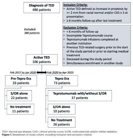
METHODS: In this single-center longitudinal study, we compared patients with active thyroid eye disease evaluated from 02/01/2017 to 01/31/2020 (pre-teprotumumab era) with those seen from 02/01/2020 to 04/30/2023 (teprotumumab era). Patients from the pre-teprotumumab era who received corticosteroids and/or orbital radiation were compared with those in the teprotumumab era treated with teprotumumab, with or without corticosteroids and/or orbital radiation. The primary outcomes were rates of orbital decompressions, strabismus surgery, and eyelid surgery among patients with at least 6 months of follow-up. Orbital decompressions involving two or more walls were classified as severe.
RESULTS: Of 486 records reviewed, 106 patients had active thyroid eye disease. Among them, 33 were from the pre-teprotumumab era; 22 received corticosteroids and/or orbital radiation, and 11 received no treatment. Seventy three patients were from the teprotumumab era; 37 received teprotumumab (with or without corticosteroids and/or orbital radiation), 10 received corticosteroids and/or orbital radiation alone, and 26 received no treatment. Demographics were comparable between groups. Orbital decompression was performed in 11 of 44 eyes (25.0%) in the pre-teprotumumab era treated with corticosteroids and/or orbital radiation (8 one-wall, 3 ≥two-wall), compared to 3 of 74 eyes (4.1%) in the teprotumumab era treated with teprotumumab with or without corticosteroids and/ or orbital radiation (all one-wall). The overall rate of orbital decompressions and the rate of ≥two-wall decompressions were significantly lower in the teprotumumab era (p=0.02 and p=0.0496, respectively). There was no significant difference in one-wall decompressions between era (p=0.07). Rates of strabismus surgeries (27.3% vs. 13.5%, p=0.19) and eyelid surgeries (22.7% vs. 21.6%, p=0.92) did not significantly differ between the era.
CONCLUSIONS: In patients with active thyroid eye disease, treatment with teprotumumab was associated with a significantly lower rate and severity of orbital decompressions compared to treatment with corticosteroids and/or orbital radiation alone. However, the rates of strabismus and eyelid surgeries remained similar between groups.
Keywords: Teprotumumab; Adrenal cortex hormone; Decompression; Graves ophthalmopathy; Strabismus
Arq. Bras. Oftalmol. 2025;88 (2 )
:1-5
| DOI: 10.5935/0004-2749.2024-0113
Abstract
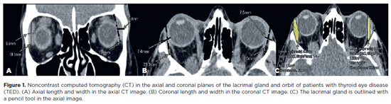
This study aimed to evaluate the morphometric and volumetric dimensions of the lacrimal gland in patients with inactive thyroid eye disease and compare them with the values reported in the literature. This case series evaluated consecutive patients with inactive thyroid eye disease treated at a tertiary eye hospital from 2015 to 2020. The patients' baseline demographics and clinical characteristics were obtained. The axial and coronal length, width, and volume of the lacrimal gland were measured on computed tomography scan images, and the results were statistically analyzed. A total of 21 patients (42 orbits) with inactive thyroid eye disease were evaluated. Their mean age was 49.0 ± 14.6 years, and 12 (57.1%) of them were men. The main complaint was dryness, and the majority of the patients had good vision and mild proptosis. The mean axial length and width of the lacrimal gland were 19.3 ± 3.9 mm and 7.5 ± 2.1 mm, respectively; coronal length and width, 20.4 ± 4.5 mm and 7.5 ± 2.1 mm, respectively; and lacrimal gland volume, 0.825 ± 0.326 mm3. Age, sex, or laterality were not found to be determinants of lacrimal gland enlargement. Patients with thyroid eye disease have enlarged lacrimal gland even in the nonactive phase of the disease multifactorial aspects influence the lacrimal gland in thyroid eye disease, making it difficult to establish a clear correlation with predisposing factors. Further studies are warranted to better understand the association between thyroid eye disease and the lacrimal gland.
Keywords: Graves' ophthalmology; Graves' disease; Lacrimal apparatus; Lacrimal apparatus diseases; X-ray computed tomography
Arq. Bras. Oftalmol. 2024;87 (5 )
:1-7
| DOI: 10.5935/0004-2749.2023-0296
Abstract
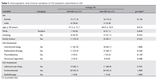
PURPOSE: To compare inferomedial wall orbital decompression to balanced medial plus lateral wall orbital decompression in patients with Graves’ orbitopathy in the inactive phase with regard to exophthalmos reduction and the effects on quality of life.
METHODS: Forty-two patients with inactive Graves’ orbitopathy were randomly divided into two groups and submitted to one of two orbital decompression techniques: inferomedial wall orbital decompression or medial plus lateral wall orbital decompression. Preoperative and postoperative assessments included Hertel’s exophthalmometry and a validated Graves’ orbitopathy quality of life questionnaire. The results of the two groups were compared.
RESULTS: Compared to preoperative measurement, exophthalmos reduction was statistically significant in both groups (p<0.001) but more so in patients undergoing medial plus lateral wall orbital decompression (p=0.010). Neither orbital decompression techniques increased the visual functioning subscale score on the Graves’ orbitopathy quality of life questionnaire (inferomedial wall orbital decompression p=0.362 and medial plus lateral wall orbital decompression p=0.727), but a statistically significant difference was observed in the score of the appearance subscale in patients submitted to medial plus lateral wall orbital decompression (p=0.006).
CONCLUSIONS: Inferomedial wall orbital decompression is a good alternative for patients who do not require large exophthalmos reduction. However, medial plus lateral wall orbital decompression offers greater exophthalmos reduction and greater improvement in appearance (higher Graves’ orbitopathy quality of life questionnaire scores), making it a suitable option for esthetic-functional rehabilitation.
Keywords: Graves’ ophthalmopathy; Quality of life; Exophthalmos; Strabismus; Diplopia; Decompression, surgical
Arq. Bras. Oftalmol. 2025;88 (3 )
:1-6
| DOI: 10.5935/0004-2749.2024-0215
Abstract
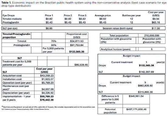
PURPOSE: To evaluate the economic impact of the following initial treatment scenarios for glaucoma on the Brazilian Public Health System (SUS): (1) traditional continuous instillation of hypotensive eye drops and (2) single session of selective laser trabeculoplasty.
METHODS: Economic impact was analyzed in three scenarios, from the least to the most conservative, for a hypothetical cohort of 5,000 individuals with open-angle glaucoma. Thereafter, projections were made on the basis of a glaucoma prevalence of 3% in the 2021 Brazilian population size.
RESULTS: All three scenarios demonstrated that selective laser trabeculoplasty exhibited a significantly lower economic impact than the eye drops on SUS over one and five years. Furthermore, the difference was more than United States Dollar 8 billion at five years when considering 3% of the Brazilian population aged >40 years in 2021.
CONCLUSION: As the initial treatment for primary open-angle glaucoma, selective laser trabeculoplasty exhibited a lower economic impact on SUS than latanoprost and timolol maleate eye drop instillation in all the studied scenarios over one and five-year periods.
Keywords: Glaucoma; Trabeculotomy; Laser therapy; Cost analysis; Health care cost Unified Health System; Brazil
Arq. Bras. Oftalmol. 2024;87 (2 )
:1-8
| DOI: 10.5935/0004-2749.2022-0241
Abstract
PURPOSE: We aimed to study reported cases of nasopharyngeal carcinoma presenting with ophthalmic manifestations with and without a prior diagnosis of nasopharyngeal carcinoma.
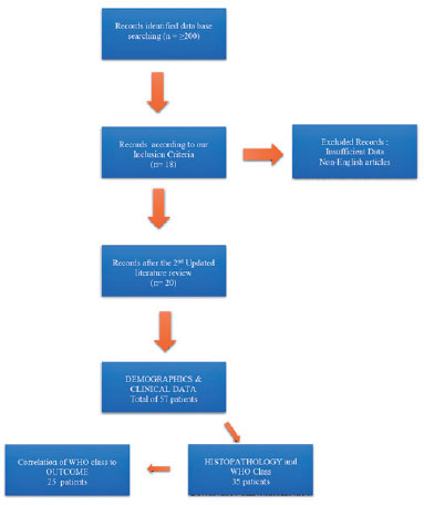
METHODS: We conducted a systematic review following the Preferred Reporting Items for Systematic Reviews and Meta-Analyses (PRISMA). A literature search was conducted using the MEDLINE database in PubMed and Google Scholar. We included patients with a previous diagnosis of nasopharyngeal carcinoma in Group I and those without a prior diagnosis of nasopharyngeal carcinoma in Group II. Data included demographics, clinical presentation, history of nasopharyngeal carcinoma, treatment, histopathological description, World Health Organization classification, and outcome.
RESULTS: Fifty-eight patients (26 in Group I and 32 in Group II) were included. The male-to-female ratio was 3:1. The mean age of the patients (53.3 ± 11.7 years and 54.8 ± 16.2 years, respectively) and gender did not differ significantly between the two groups. The most common ocular presentations were diplopia and proptosis in the first group (each in 34.6%), whereas visual disturbance was most common in the second group (46.9%). Treatment options and World Health Organization grading were comparable. The outcome in 38 patients (after a comparable follow-up period) was significantly better in group II (p=0.003). There was no statistically significant difference in the outcome of 23 patients in correlation with World Health Organization grades II versus III irrespective of group (p=0.094).
CONCLUSIONS: The demographics of patients with nasopharyngeal carcinoma presenting with ophthalmic manifestations were similar between the two study groups, with a wide age range and male predominance. Patients presenting initially to ophthalmologists with no history of nasopharyngeal carcinoma have a more favorable outcome. World Health Organization grading may have less value as a prognostic indicator.
Keywords: Nasopharyngeal carcinoma; Carcinoma; Eye manifestations; Exophthalmos; Diplopia; Systematic review
 ABO is licensed under a Creative Commons Attribution-NonComercial 4.0 Internacional.
12-tab01tb.jpg)
06-fig01.jpg)
05-tab01.jpg)

03-fig01.jpg)
01-fig01.jpg)
02-fig01.jpg)