Arq. Bras. Oftalmol. 2021;84 (3 )
:203-208
| DOI: 10.5935/0004-2749.20210034
Abstract
OBJETIVO: Avaliar comparativamente o limiar de sensibilidade macular da microperimetria e a estabilidade de fixação entre o primeiro (direito) e o segundo (esquerdo) olhos testados de indivíduos normais.
MÉTODOS: Trinta pacientes saudáveis foram divididos aleatoriamente em 2 grupos. Os participantes foram submetidos à microperimetria no “fast mode” e no “expert mode” no grupo I e II, respectivamente. Cada participante foi submetido a um único teste e o olho direito foi testado primeiro.
RESULTADOS: No grupo I, o limiar médio de sensibilidade macular (± DP) foi de 24,5 ± 2,3 dB e 25,7 ± 1,1 dB nos olhos direito e esquerdo, respectivamente (p=0,0415). No grupo II foi de 26,7 ± 4,5 dB e 27,3 ± 4,0 dB nos olhos direito e esquerdo, respectivamente (p=0,58). Não houve diferença estatisticamente significativa entre os olhos dos dois grupos (p=0,1512). Em relação à estabilidade de fixação (avaliada no grupo microperimetria no “expert mode”), a média das porcentagens dos pontos de fixação dentro do 1 grau central da mácula (P1) ± DP foi de 87,9 ± 11,5% no olho direito e de 93,8 ± 6,6% no olho esquerdo. O teste t pareado não mostrou diferença estatística entre os olhos (p=0,140). O valor médio de P2 ± DP foi de 95,5 ± 4,9% no olho direito e 98,5 ± 2,1% no olho esquerdo. Foi demonstrado um aumento na porcentagem de pontos de fixação no segundo olho testado quando comparado ao primeiro (teste t pareado= 2,364; p=0,034). Houve correlação negativa entre o limiar de sensibilidade macular do olho direito e a duração do exame nos dois grupos (microperimetria no “expert mode”: r=-0,717; p=0,0026; microperimetria no “fast mode”: r=-0,843; p <0,0001).
CONCLUSÃO: O limiar médio de sensibilidade macular foi maior no segundo olho testado no grupo microperimetria no “fast mode” e foi semelhante nos dois olhos no “expert mode”. Nossos dados sugerem que a compreensão do exame pelo indivíduo pode impactar nos resultados da microperimetria.
Keywords: Macula lutea; Fixação ocular; Viés; Campos visuais; Acuidade visual
Arq. Bras. Oftalmol. 2023;86 (2 )
:113-120
| DOI: 10.5935/0004-2749.20230022
Abstract
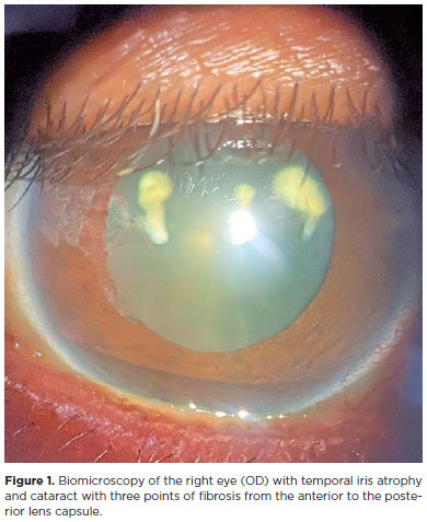
Objetivos: Avaliar a estabilidade e eficácia da técnica double-flanged com sutura de 5-0 polipropileno para fixação de cataratas subluxadas aos 18 meses e as possíveis complicações desta nova técnica.
Métodos: Esta técnica utiliza um monofilamento de polipropileno 5-0 para criar dois flanges com um termocautério para fixar um Segmento de Tensão Capsular na esclera a fim de estabilizar o saco capsular subluxado. Esta técnica foi implementada em 17 olhos que necessitavam do implante de lente intraocular em casos de diálise zonular devido a trauma, síndrome de Marfan, microesferofacia, subluxação idiopática ou pós-facoemulsificação que provocou subulxação do saco capsular intraoperatória.
Resultados: O seguimento dos pacientes foi de 18 meses. A acuidade visual corrigida melhorou significativamente de 0,85 para 0,39 (logMAR), enquanto os erros de refração esféricos e cilíndricos e a pressão intraocular permaneceram estáveis. Nenhuma fotodegradação de sutura ou pseudofacodonese foi encontrada.
Conclusão: A técnica double-flanged para fixação transescleral de saco capsular com sutura de 5-0 polipropileno mostrou resultados de estabilidade de longo prazo para o complexo lente/saco capsular. Então, aparenta ser uma opção segura para cirurgia de catarata, sem necessidade pontos, em olhos com fraqueza zonular ou diálise
Keywords: Catarata; Facoemulsificação; Lente intraocular; Técnica de sutura; Acuidade visual
Arq. Bras. Oftalmol. 2025;88 (6 )
:1-5
| DOI: 10.5935/0004-2749.2025-0085
Abstract
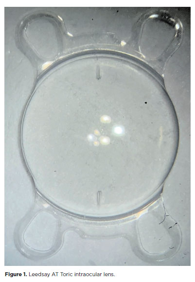
PURPOSE: The purpose of this study was to assess visual outcomes and patient satisfaction following cataract surgery involving the implantation of quad-loop intraocular lenses, including trifocal, bifocal, and toric variants.
METHODS: Information was obtained from both physical and electronic medical records of patients who underwent phacoemulsification cataract surgery with implantation of different intraocular lenses between January 1, 2022, and December 31, 2023. The study included individuals aged over 18 who received bilateral implantation of bifocal, trifocal, or monofocal toric intraocular lenses. Visual acuity was assessed at various postoperative time points using the logMAR scale. Quantitative variables were analyzed using mean and standard deviation.
RESULTS: A total of 92 eyes received premium intraocular lenses: 4 bifocal, 32 trifocal, 52 toric monofocal, and 4 trifocal toric lenses. The average preoperative corrected visual acuity was logMAR 0.478 ± 0.259. On the first postoperative day, the average uncorrected visual acuity was logMAR 0.301 ± 0.207. By day 30, 67.4% of eyes achieved uncorrected distance visual acuity of logMAR 0.2 or better. Patient satisfaction was high, with few reports of glare or halos.
CONCLUSION: Quad-loop intraocular lenses-including trifocal, bifocal, and toric models-demonstrated effective improvement in visual acuity and high levels of patient satisfaction. These lenses represent a suitable option for enhancing visual outcomes after cataract surgery. Additional studies with larger cohorts are recommended to confirm these results.
Keywords: Cataract extraction; Aberrometry/methods; Lenses, intraocular; Lens implantation, intraocular; Prosthesis design
Arq. Bras. Oftalmol. 2026;89 (2 )
:1-8
| DOI: 10.5935/0004-2749.2025-0175
Abstract
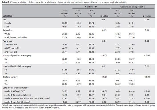
PURPOSE: Endophthalmitis is one of the most important adverse events after cataract surgery, as it can lead to total vision loss. This study aimed to describe the occurrence of endophthalmitis after phacoemulsification with intraocular lens implantation in patients treated in a community setting in Porto Velho, Rondônia, Brazil.
METHODS: This retrospective cohort study was conducted using a database of 649 medical records of patients who underwent surgery and were followed for three months. Poisson regression analysis was used to estimate relative risks and 95% confidence intervals (95% CIs).
RESULTS: The incidence of confirmed endophthalmitis was 11.94% (95% CI, 9.50-14.76), while the incidence of confirmed and probable cases was 20.50% (95% CI, 17.52-23.73). For confirmed cases, bilateral surgery and the use of lens model 3 were identified as risk factors for endophthalmitis, whereas age over 70 yr and preoperative antibiotic use were protective factors. For confirmed and probable cases, brown and yellow skin color, bilateral surgery, and the use of lens model 3 were also identified as risk factors. Gram-negative bacteria were the predominant etiological agents, and corneal edema was the main clinical manifestation. The mean duration of treatment was eight days, and 27.12% of patients used antibiotics.
CONCLUSION: The incidence observed was substantially higher than that reported in the literature, with a predominance of Gram-negative agents and an association with bilateral surgeries and the Eyeol intraocular lens model. These findings reinforce the need for continuous epidemiological surveillance and the implementation of specific biosafety and infection control protocols during cataract surgery campaigns.
Keywords: Endophthalmitis; Disease outbreaks; Phacoemulsification; Lens implantation, intraocular; Lenses, intraocular; Cataract; Risk factors; Anti-bacterial agents
Arq. Bras. Oftalmol. 2025;88 (6 )
:1-8
| DOI: 10.5935/0004-2749.2024-0394
Abstract
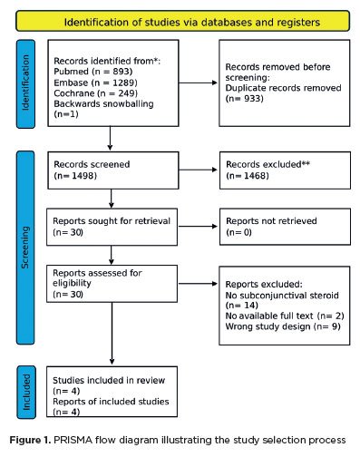
The advantages and disadvantages of using perioperative subconjunctival steroid injections in dropless cataract surgery continue to be debated. A systematic review of PubMed, EMBASE, and the Cochrane Central database identified five studies—two randomized controlled trials and three non-randomized studies—encompassing 70,751 eyes. Among these, 12,319 eyes (17.4%) received subconjunctival steroid injections, while 58,432 eyes (82.6%) were managed with topical steroids. The Cochrane Collaboration’s RoB 2 tool was applied for bias assessments in randomized controlled trials, and heterogeneity was assessed using the I² statistics. No statistically significant differences were found between the two groups regarding macular edema (p=0.249), visual acuity (p=0.73), or laser flare count (p=0.45). Both subconjunctival injections and topical steroids demonstrated comparable efficacy and safety in controlling postoperative inflammation after cataract surgery. Additional research is warranted to validate these conclusions.
Keywords: Cataract extraction; Phacoemulsification; Lens implantation, intraocular; Postoperative care; Intravitreal injections; Anti-inflammatory agents, non-steroidal/administration & dosage; Glucocorticoids; Triamcinolone acetonide; Research design; Randomiz
Arq. Bras. Oftalmol. 2024;87 (5 )
:1-7
| DOI: 10.5935/0004-2749.2021-0472
Abstract
Objetivos: A microperimetria tem sido usada há vários anos como uma forma de teste de função visual em pacientes com doenças da retina. Os valores normais de microperimetria obtidos com MP-3 ainda não foram totalmente publicados e os valores basais para sensibilidade macular topográfica e correlações com idade e sexo são necessários para estabelecer graus de comprometimento. O objetivo do trabalho é determinar valores para limiares de sensibilidade à luz e estabilidade de fixação usando o MP-3 em indivíduos normais.
Métodos: Trinta e sete voluntários saudáveis (idade: 28-68 anos), submetidos à microperimetria de limiar total usando uma estratégia de escada 4-2 (rápida) com o tamanho de estímulo padrão Goldmann III e 68 pontos de teste posicionados de forma idêntica aos do Humphrey Field Analyzer 10-2 grade de teste. A estabilidade da fixação foi registrada simultaneamente durante o teste de microperimetria. A relação entre a sensibilidade global e a idade foi calculada por meio de análise de regressão linear.
Resultados: A microperimetria foi realizada em 37 indivíduos (74 olhos). A sensibilidade média global foi de 29,01 ± 1,44 dB, intervalo: 26-31 dB. A mediana da sensibilidade central a 2° medida pelo MP-3 foi de 28,5 ± 1,77 dB (ER) e 28,75 ± 1,98 dB (OE). Os valores médios totais de estabilidade da fixação em 2° e 4° foram 80% e 96%, respectivamente. A análise de regressão linear também revelou um declínio de sensibilidade global relacionado à idade por ano de -0,051 dB ± 0,018 (ER) e -0,078 dB ± 0,021 (LE).
Conclusões: A microperimetria realizada com o MP-3 permite um exame automático, preciso e específico da topografia dos limiares de sensibilidade da retina. Os resultados deste estudo fornecem um banco de dados normal e de idade correspondente da microperimetria MP-3.
Keywords: Campos visual; Testes de campo visual; Retina; Microperímetro; Idade.
Arq. Bras. Oftalmol. 2025;88 (5 )
:1-6
| DOI: 10.5935/0004-2749.2024-0270
Abstract
PURPOSE: Standard automated perimetry has been the standard method for measuring visual field changes for several years. It can measure an individual’s ability to detect a light stimulus from a uniformly illuminated background. In the management of glaucoma, the primary objective of perimetry is the identification and quantification of visual field abnormalities. It also serves as a longitudinal evaluation for the detection of disease progression. The development of artificial intelligence-based models capable of interpreting tests could combine technological development with improved access to healthcare.
METHODS: In this observational, cross-sectional, descriptive study, we used an artificial intelligence-based model [Inception V3] to interpret gray-scale crops from standard automated perimetry that were performed in an ophthalmology clinic in the Brazilian Amazon rainforest between January 2018 and December 2022.
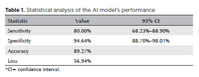
RESULTS: The study included 1,519 standard automated perimetry test results that were performed using Humphrey HFA-II-i-750 (Zeiss Meditech). The Subsequently, 70%, 10%, and 20% of the dataset were used for training, validation, and testing, respectively. The model achieved 80% (68.23%–88.9%) sensitivity and 94.64% (88.8%–98%) specificity for detecting altered perimetry results. Furthermore, the area under the receiver operating characteristic curve was 0.93.
CONCLUSIONS: The integration of artificial intelligence in the diagnosis, screening, and monitoring of pathologies represents a paradigm shift in ophthalmology, enabling significant improvements in safety, efficiency, availability, and accessibility of treatment.
Keywords: Glaucoma; Disease progression; Perimetry; Visual Fields; Visual field tests; Artificial intelligence; Neural networks, computers; Machine learning
Arq. Bras. Oftalmol. 2026;89 (3 )
:1-4
| DOI: 10.5935/0004-2749.2025-0263
Abstract
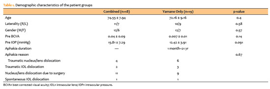
PURPOSE: To compare patients who underwent scleral fixation using the Yamane technique with and without simultaneous pars plana vitrectomy.
METHODS: A total of 37 patients were included in the study. Eighteen underwent simultaneous pars plana vitrectomy. The Yamane technique alone was performed only in patients with aphakia who had previously undergone pars plana vitrectomy for various reasons. Final lens position, best corrected visual acuity spherical equivalent, complication rates, and optical coherence tomography findings were recorded.
RESULTS: The duration of aphakia before intraocular lens implantation ranged from 1 month to 21 yr. Postoperative best corrected visual acuity improved in both groups, with no statistically significant difference (with pars plana vitrectomy: 0.42 ± 0.34; without pars plana vitrectomy: 0.32 ± 0.26; p=0.33). The spherical equivalent was also not significantly different between groups (with pars plana vitrectomy: 0.29 ± 1.08; without pars plana vitrectomy: 0.65 ± 2.23; p=0.53). There were no significant differences between the groups in complication rates, postoperative intraocular lens position or optical coherence tomography findings.
CONCLUSION: There was no difference in terms of safety or efficacy between the two approaches. Surgical decisions may be based on the surgeon’s experience and the patient’s systemic and ocular condition.
Keywords: Lens implantation, intraocular; Tomography, optical coherence; Vitrectomy; Intraocular lenses; Visual acuity; Aphakia; Yamane technique
Arq. Bras. Oftalmol. 2026;89 (4 )
:1-5
| DOI: 10.5935/0004-2749.2026-0010
Abstract
PURPOSE: To evaluate changes in scotopic pupil diameter before and after cataract surgery performed by phacoemulsification with intraocular lens implantation.
METHODS: This prospective longitudinal observational study included patients who underwent cataract surgery. Scotopic pupil diameter was measured preoperatively and 30-40 days postoperatively using an automated keratometer after a standardized dark-adaptation period under controlled ambient illumination. Each eye was considered an independent unit of observation. Because some participants contributed both eyes, intraindividual correlation was accounted for using a linear mixed-effects model with random patient intercepts. Time of assessment (preoperative versus postoperative), age, sex, and eye laterality were included as fixed effects.
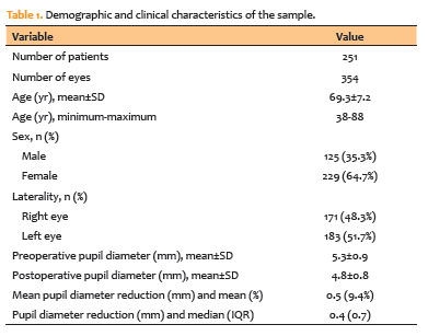
RESULTS: A total of 354 eyes from 251 patients were analyzed. The mean patient age was 69.3±7.2 yr. Mean scotopic pupil diameter decreased from 5.3±0.9mm preoperatively to 4.8±0.8mm postoperatively, representing a mean reduction of 0.5mm (9.4%). In the linear mixed-effects model, cataract surgery was associated with a significant reduction in pupil diameter, with an adjusted mean difference of 0.45mm (95% confidence interval [95% CI], 0.39-0.51; p<0.001). Age (p=0.061), sex (p=0.920), and eye laterality (p=0.152) were not significantly associated with the magnitude of pupil diameter change.
CONCLUSION: Phacoemulsification with intraocular lens implantation was associated with a significant reduction in scotopic pupil diameter, independent of age, sex, and eye laterality. This finding should be considered during preoperative planning, particularly when selecting intraocular lenses whose optical performance depends on postoperative pupil size.
Keywords: Cataract; Pupil; Phacoemulsification; Lens implantation, intraocular; Lenses, intraocular; Pseudophakia
Arq. Bras. Oftalmol. 2025;88 (5 )
:1-7
| DOI: 10.5935/0004-2749.2024-0368
Abstract
PURPOSE: To compare endothelial corneal cell changes following cataract surgery performed by phacoemulsification with intraocular lens implantation, conducted by surgeons with varying levels of experience.
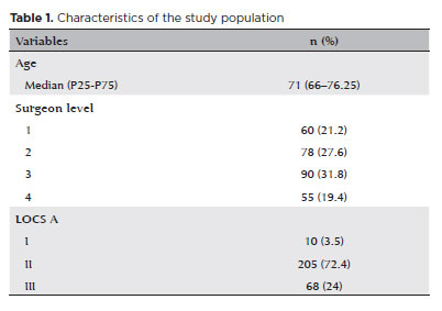
METHODS: Two hundred and eighty-three eyes diagnosed with cataract were included. Lens opacity was classified into three categories (I, II, and III). Surgeons were categorized into four experience levels (1, 2, 3, and 4), based on years of practice and lifetime surgeries performed. Corneal endothelial characteristics were assessed using non-contact specular microscopy, with measurements taken before surgery and 30-60 days post-surgery.
RESULTS: Pre- and postoperative endothelial analysis showed no significant differences between surgeon levels regarding visual acuity achieved, corneal thickness, and endothelial hexagonality. However, the central endothelial cell density index showed a significantly greater reduction among level 1 surgeons (p=0.026). Grade II cataracts exhibited significant variations in the central endothelial cell density (p=0.011) and average cell size, with level 1 surgeons showing the largest increases (p=0.024).
CONCLUSIONS: The analysis revealed significant differences in visual acuity and endothelial indices between surgeon experience levels, with less experienced surgeons showing greater variations and poorer performance. Clinical protocols should consider these data to establish safer training protocols.
Keywords: Cataract extraction; Phacoemulsification; Endothelium; corneal; Lens implantation, intraocular; Visual acuity; Internship and residency; Surgeons
Arq. Bras. Oftalmol. 2025;88 (5 )
:1-6
| DOI: 10.5935/0004-2749.2024-0015
Abstract
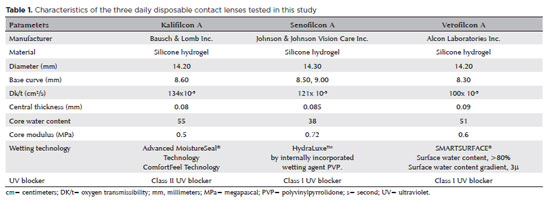
PURPOSE: This study aimed to compare the effects of three different daily disposable contact lens materials on contrast sensitivity.
METHODS: The participants were aged 18–45 years, with spherical equivalent refraction between -0.50 D and -6.00 D, astigmatism below 0.75 D, and best contact lens-corrected visual acuity of 0.0 logMAR or better. Each patient was fitted binocularly with three daily disposable contact lenses made of different materials on three separate examination days. These materials were kalifilcon A, senofilcon A, and verofilcon A. The contrast sensitivity of each patient was recorded at spatial frequencies of 3, 6, 12, and 18 cycles per degree (cpd) under photopic (85 cd/m2) and mesopic (3 cd/m2) conditions.
RESULTS: The current study comprised 72 eyes of 34 female and two male patients. The mean age of the participants was 25.63 (± 0.80) years. Under photopic conditions, the participants’ contrast sensitivity was significantly better with senofilcon A than with kalifilcon A at a frequency of 12 cpd (p=0.008). Under mesopic conditions, participants’ contrast sensitivity was significantly higher with kalifilcon A than verofilcon A at 3 cpd (p=0.001), and with senofilcon A than verofilcon A at 12 cpd (p=0.004). The pre-lens non-invasive break-up times did not differ significantly between the three daily disposable contact lenses (p>0.05).
CONCLUSION: In both photopic and mesopic lighting conditions, the participants in this study exhibited differences in contrast sensitivity when wearing three different daily disposable contact lens types, despite similar visual acuity and pre-lens tear film stability results in their clinical evaluations. These findings demonstrate the potential for subjective visual complaints arising from variations in the contrast sensitivity achieved by different daily disposable contact lenses.
Keywords: Contact lenses; Contrast sensitivity; Astigmatism; Lighting; Visual acuity
 ABO is licensed under a Creative Commons Attribution-NonComercial 4.0 Internacional.
01-tab01tb.jpg)
08-tab01.jpg)
05-fig01.jpg)
02-fig01.jpg)
01-fig01.jpg)