Arq. Bras. Oftalmol. 2022;85 (1 )
:7-12
| DOI: 10.5935/0004-2749.20220002
Abstract
Objetivo: A degeneração macular relacionada à idade é a causa mais comum de cegueira em países desenvolvidos e muitos fatores etiológicos têm-lhe sido atribuídos. O objetivo do presente estudo foi investigar a relação entre os níveis séricos de vitamina D e a degeneração macular relacionada à idade.
Métodos: Os dados de 114 pacientes com degeneração macular relacionada à idade foram analisados retrospectivamente. Foram alocados no Grupo Controle 102 pacientes sem registro de outras doenças além do erro refrativo. A acuidade visual melhor corrigida, os achados do exame de fundo de olho e os da tomografia de coerência óptica de domínio espectral foram analisados. Os pacientes foram alocados em grupos de acordo com a classificação do Age-Related Eye Disease Study (Estudo da Doença Ocular Relacionada à Idade). Os níveis séricos de vitamina D 25(OH) foram medidos. A espessura foveal central e a espessura da coroide subfoveal foram medidas com tomografia de coerência óptica.
Resultados: Os níveis de vitamina D 25(OH) em pacientes com degeneração macular relacionada à idade e em indivíduos saudáveis pareados por idade e sexo foram 14,6 ± 9,8 ng/mL e 29,14 ± 15,1 ng/mL, respectivamente. Os níveis de vitamina D foram significativamente menores no Grupo da Degeneração Macular relacionada à idade em comparação com o Grupo Controle (p>0,001). O valor da espessura da coroide subfoveal foi menor em pacientes com degeneração macular relacionada à idade (p>0,001). Foi encontrada uma fraca correlação positiva entre o nível de vitamina D 25(OH) e a espessura da coroide (r=0,357, p=0,01). O nível de vitamina D 25(OH), quando avaliado de acordo com os estágios da degeneração macular relacionada à idade, revelou ser menor na doença em estágio avançado (p=0,01). Constatou-se um risco aumentado de desenvolvimento de membrana neovascular da coroide e de fibrose sub-retiniana com a diminuição dos níveis de vitamina D.
Conclusões: A diminuição significativa dos níveis de vitamina D 25(OH) na degeneração macular relacionada à idade em estágio avançado sugere a presença de uma correlação significativa entre a deficiência de vitamina D e o desenvolvimento dessa patologia. Mais estudos são necessários para investigar se a suplementação de vitamina D tem ou não influência no desenvolvimento e progressão da degeneração macular relacionada à idade.
Keywords: Membrana neovascular da coroide; Degeneração macular; Tomografia de coerência óptica; Fibrose; Retina; Vitamina D; Deficiência de vitamina D
Arq. Bras. Oftalmol. 2022;85 (6 )
:584-589
| DOI: 10.5935/0004-2749.20220089
Abstract
Objetivo: Determinar os efeitos da deficiência de vitamina D nos microvasos da retina usando angiotomografia de coerência óptica.
Métodos: Este estudo foi planejado para ser do tipo caso-controle observacional. Foram avaliados 98 olhos de pacientes com deficiência de vitamina D e 96 olhos de participantes saudáveis com nível sérico de vitamina D superior a 30 ng/mL. Foram adquiridas imagens de varredura centralizadas na mácula, com um tamanho de 6,00 × 6,00 mm. Mediram-se a densidade dos vasos nos plexos capilares superficial e profundo da retina, a área da zona avascular foveal e a área do fluxo coriocapilar.
Resultados: Os grupos mostraram-se semelhantes em relação à melhor acuidade visual corrigida, ao gênero, ao comprimento axial, ao erro refrativo, à idade e à pressão intraocular ajustada. O nível médio de vitamina D foi significativamente menor no grupo de estudo (p=0,021). As densidades total, parafoveal e perifoveal do plexo capilar profundo foram significativamente maiores no grupo de estudo que no grupo controle (respectivamente, p=0,012, p=0,014 e p=0,023). As áreas da zona avascular foveal e do fluxo coriocapilar foram semelhantes nos dois grupos (respectivamente, p=0,37 e p=0,27). Além disso, houve uma forte correlação negativa do nível sérico de vitamina D com as densidades vasculares medidas em toda a imagem e nas regiões parafoveais e perifoveais do plexo capilar profundo no grupo de estudo (respectivamente, ρ de Spearman = -0,71, p=0,043; ρ de Spearman = -0,79, p=0,011; e ρ de Spearman = -0,74, p=0,032).
Conclusão: Pode ocorrer um aumento na densidade vascular da retina devido a alterações estruturais dos vasos causadas pela deficiência de vitamina D. O aumento da densidade vascular, especialmente no plexo capilar profundo, pode ser usado para o diagnóstico precoce da vasculopatia associada à deficiência de vitamina D.
Keywords: Deficiência de vitamina D; Vasos retinianos/fisiopatologia; Doenças vasculares/ prevenção & controle; Tomografia de coerência óptica
Arq. Bras. Oftalmol. 2020;83 (4 )
:312-317
| DOI: 10.5935/0004-2749.20200043
Abstract
Purpose: To analyze the clinical outcomes of the ocular surface in patients with vitamin D deficiency after oral replacement.
Methods: A total of 40 patients with vitamin D deficiency were enrolled in the study. The patients received 50,000 units of oral vitamin D weekly over a period of 8 weeks. After 8 weeks, 1,500-2,000 units/d were administered for 24 weeks. Eyelid margin score, meibomian gland expressibility score, Oxford grading, Schirmer I test, tear breakup time, tear osmolarity, and the Ocular Surface Disease Index score were evaluated at baseline, and at 8, 12, and 24 weeks.
Results: The meibomian gland expressibility score, Schirmer I, tear breakup time, tear osmolarity, and Ocular Surface Disease Index score showed improvement 8 weeks after vitamin D supplementation (p<0.05). Compared with the pretreatment values, the eyelid margin score and Oxford grading were decreased at week 12 (p<0.05).
Conclusion: Vitamin D replacement appears to improve ocular surface in individuals with vitamin D deficiency.
Keywords: Dry eye syndrome; Vitamin D deficiency; Dietary supplements
Arq. Bras. Oftalmol. 2025;88 (1 )
:1-63
| DOI: 10.5935/0004-2749.2023-0037
Abstract
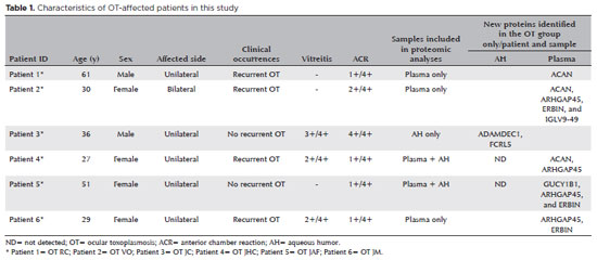
PURPOSE: To characterize the extracellular vesicle protein cargo in the aqueous humor and plasma of patients with ocular toxoplasmosis.
METHODS: Aqueous humor and plasma were collected from six patients with active ocular toxoplasmosis and six patients with cataract. Extracellular vesicles were isolated, and western blotting and mass spectrometry were performed for protein analysis.
RESULTS: All plasma samples from patients with ocular toxoplasmosis and cataract were positive for the tetraspanins CD63 and TSG101. However, the aqueous humor from patients with ocular toxoplasmosis was positive only for CD63. Sixty-seven new unreported proteins were identified in the aqueous humor and plasma of patients with the ocular toxoplasmosis and cataract. Of the 67 proteins, 10 and 7 were found only in the cataract and ocular toxoplasmosis groups, respectively. In general, these proteins were involved in immune system activation and retina homeostasis and were related to infections and retina-associated diseases. Conclusion: The distinct protein signatures between ocular toxoplasmosis and cataract may be helpful in the differential diagnosis of ocular toxoplasmosis. However, more studies are needed to better understand the role of these proteins in the pathogenesis of ocular toxoplasmosis.
Keywords: Extracellular vesicles; Proteomics; Toxoplasma gondii; Ocular toxoplasmosis, Aqueous humor; Plasma; Liquid biopsy
Arq. Bras. Oftalmol. 2024;87 (4 )
:1-7
| DOI: 10.5935/0004-2749.2023-0141
Abstract
PURPOSE: This study aimed to investigate the correlation between serum vitamin D levels and disease activity in patients with noninfectious uveitis.
METHODS: We conducted a prospective case-control study, assessing 51 patients with noninfectious uveitis, categorized into active (n=22) and inactive (n=29) groups, along with 51 healthy controls. Serum 25-hydroxy vitamin D [25(OH)D] levels were measured. The uveitis group also completed a questionnaire regarding sunlight exposure habits and vitamin D supplementation.
RESULTS: Patients with inflammation-related uveitis exhibited low serum 25(OH)D levels in 68% of cases. The median 25(OH)D level in patients with active uveitis was 17.8 ng/mL (interquartile range [IQR], 15-21 ng/mL), significantly lower compared to the 31.7 ng/mL (IQR, 25-39 ng/mL) in patients with inactive uveitis (p<0.001) and the 27 ng/mL (IQR, 23-31 ng/mL) in the Control Group (p<0.001). Significantly, nearly all patients with uveitis taking vitamin D supplementation were in the Inactive Group (p<0.005). Moreover, reduced sunlight exposure was associated with active uveitis (p<0.003). Furthermore, patients with 25(OH)D levels below 20 ng/mL had ten times higher odds of developing active uveitis (p=0.001).
CONCLUSIONS: This study revealed a prevalent 25(OH)D deficiency among patients with noninfectious uveitis and suggested a link between low 25(OH)D levels and disease activity. To prevent future episodes of intraocular inflammation, vitamin D supplementation and controlled sunlight exposure could be viable options.
Keywords: Vitamin D; 25-hydroxyvitamin D; Uveitis; Vitamin D deficiency; Immunity; Eye/immunology
Arq. Bras. Oftalmol. 2024;87 (2 )
:1-5
| DOI: 10.5935/0004-2749.2023-0001
Abstract
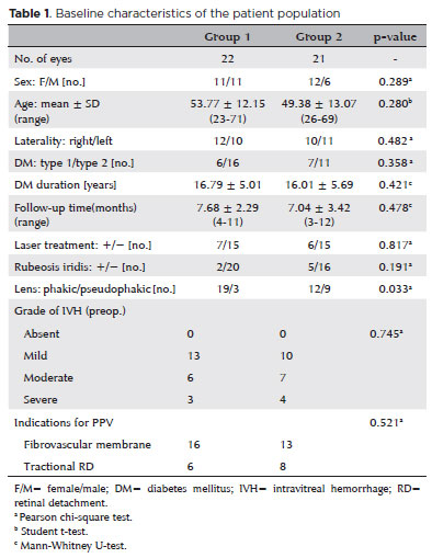
PURPOSE: To investigate the clinical benefits of the co-application of bevacizumab and tissue plasminogen activator as adjuncts in the surgical treatment of proliferative diabetic retinopathy.
METHODS: Patients who underwent vitrectomy for proliferative diabetic retinopathy complications were preoperatively given intravitreal injection with either bevacizumab and tissue plasminogen activator (Group 1) or bevacizumab alone (Group 2). Primary outcomes were surgery time and number of intraoperative iatrogenic retinal breaks. Secondary outcomes included changes in the best-corrected visual acuity and postoperative complications at 3 months postoperatively.
RESULTS: The mean surgery time in Group 1 (52.95 ± 5.90 min) was significantly shorter than that in Group 2 (79.61 ± 12.63 min) (p<0.001). The mean number of iatrogenic retinal breaks was 0.50 ± 0.59 (0-2) in Group 1 and 2.00 ± 0.83 (0-3) in Group 2 (p<0.001). The best-corrected visual acuity significantly improved in both groups (p<0.001). One eye in each group developed retinal detachment.
CONCLUSION: Preoperative co-application of bevacizumab and tissue plasminogen activator as adjuncts in the surgical treatment of proliferative diabetic retinopathy shortens the surgery time and reduces the number of intraoperative iatrogenic retinal breaks.
Keywords: Diabetic retinopathy; Bevacizumab; Plasminogen activators; Vitrectomy; Operative time


 ABO is licensed under a Creative Commons Attribution-NonComercial 4.0 Internacional.


04-tab01tb.jpg)
14-fig01.jpg)
08-fig01tb.jpg)

01-fig01.jpg)
04-fig01.jpg)
15-fig01tb.jpg)
10-tab01tb.jpg)