Arq. Bras. Oftalmol. 2023;86 (3 )
:1-5
| DOI: 10.5935/0004-2749.20230031
Abstract
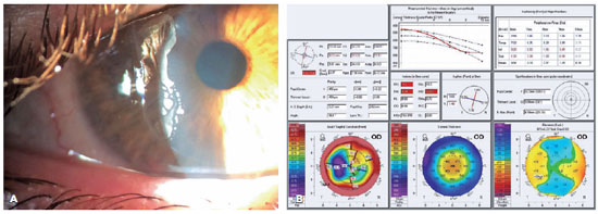
Objetivo: Descrever os resultados clínicos do tratamento do crescimento epitelial através da técnica de remoção manual seguido da utilização de um compressor de ar comprimido aquecido após a cirurgia de laser in situ keratomileusis (LASIK).
Métodos: Vinte olhos de 17 pacientes foram incluídos no estudo. Cada paciente havia sido submetido a cirurgia de LASIK com presença de crescimento epitelial e foi submetido a tratamento cirúrgico para sua retirada. O objetivo primário foi identificar a presença de crescimento epitelial recorrente ao final de 3 meses de seguimento. Os objetivos secundários foram as medidas de acuidade visual sem correção, acuidade visual com correção, e complicações pós-operatórias.
Resultados: Dez pacientes (58,8%) eram homens e 7 mulheres. Oito olhos de sete (41,2%) pacientes apresentavam cirurgia de LASIK primária e 12 olhos de 10 pacientes tinham cirurgia de LASIK com retratamento; dezesseis olhos (80%) utilizaram microcerátomo manual e quatro (20%) laser de femtosegundo. A média de idade no momento da cirurgia de remoção do epitélio era de 37,0 anos ± 9,3 (DP) (variando de 24 a 55 anos). Ocorreu recidiva do crescimento epithelial em dois olhos (10%) após 3 meses de seguimento. A acuidade visual sem correção antes da cirurgia era de 0,07 ± 0,09 logMAR, e após a cirurgia passou para 0,02 ± 0,04 logMAR (p=0,06). A chance (odds ration) de aparecimento do crescimento epithelial após uma reoperação de LASIK é 29,41 vezes maior do que no LASIK primário.
Conclusão: A técnica de remoção epitelial manual seguida da utilização de ar comprimido aquecido é segura e efetiva no tratamento do crescimento epitelial após LASIK. Ao final do último acompanhamento, nenhum olho apresentou perda de linhas de visão.
Keywords: Epitélio/crescimento & desenvolvimento; Endotélio corneano; Doenças da córnea; Ceratomileuse assistida por excimer laser in situ; Ceratectomia fotorrefrativa; Procedimentos cirúrgicos refrativos; Acuidade visual
Arq. Bras. Oftalmol. 2023;86 (5 )
:1-6
| DOI: 10.5935/0004-2749.20230070
Abstract
Objetivo: A refração pós-operatória na cirurgia moderna de catarata por microincisão ganha ainda mais importância em pacientes com cirurgia prévia de ceratomileuse in situ assistida por laser (LASIK). As alterações astigmáticas induzidas cirurgicamente nesses olhos podem diferir não apenas em magnitude, mas também em direção em comparação com córneas virgens. O objetivo deste estudo foi comparar as alterações astigmáticas induzidas cirurgicamente após cirurgia de catarata por microincisão entre córneas pós-LASIK e olhos virgens.
Métodos: Foi revisada uma série de casos de cirurgia de catarata por microincisão em olhos com e sem cirurgia LASIK anterior. Os dados demográficos, o comprimento axial no momento da cirurgia de catarata, a espessura central da córnea, os valores esféricos e cilíndricos, as leituras da ceratometria e o astigmatismo corneano posterior pós-operatório foram avaliados retrospectivamente. O método Alpins modificado foi usado para análise vetorial astigmática e foram avaliados o astigmatismo basal, o astigmatismo induzido cirurgicamente, o vetor de diferença, o efeito de achatamento e o torque.
Resultados: Ao todo, 42 olhos de 24 indivíduos foram avaliados. O Grupo I consistiu em 14 olhos com LASIK prévio; o Grupo II incluiu 28 olhos sem qualquer cirurgia refrativa. A média da espessura corneana central pré-operatória no Grupo I foi significativamente mais fina (p=0,012). Não houve diferença significativa no astigmatismo basal entre os grupos em termos de magnitude e vetores de potência. Após a cirurgia de catarata por microincisão, não houve diferenças significativas nos valores médios esféricos, cilíndricos e leituras médias de ceratometria (todos com p>0,05). No entanto, o astigmatismo induzido cirurgicamente e o vetor de diferença foram significativamente maiores no componente do vetor J45 em olhos pós-LASIK, e o efeito de aumento da inclinação pela cirurgia de catarata por microincisão nas córneas pós-LASIK foi significativo em comparação com olhos virgens (p=0,001, p=0,002 e p=0,018, respectivamente).
Conclusões: A cirurgia de catarata aumentou a inclinação das córneas em ambos os grupos, sendo esse aumento significativamente maior nos olhos pós-LASIK. Certamente, a topografia da córnea antes da cirurgia de catarata é particularmente útil para fornecer interpretações mais precisas do astigmatismo induzido cirurgicamente.
Keywords: Cirurgia de catarata; Ceratomileuse; excimer laser in situ; Cirurgia refrativa; Astigmatismo induzido cirurgicamente; Análise vetorial.
Arq. Bras. Oftalmol. 2026;89 (1 )
:1-8
| DOI: 10.5935/0004-2749.2024-0397
Abstract
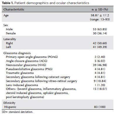
PURPOSE: Glaucoma is one of the leading causes of irreversible blindness worldwide. When topical hypotensive agents or laser trabeculoplasty fail to adequately control the disease, escalation of therapy becomes necessary, with transscleral cyclophotocoagulation being one of the available options. Several variations of transscleral cyclophotocoagulation exist, including traditional continuous wave, MicroPulse, and slow-coagulation techniques. We propose a novel variation – custom slow-coagulation transscleral cyclophotocoagulation – which combines elements of both continuous wave and slow-coagulation approaches. This study aimed to evaluate the outcomes of this technique in patients with refractory glaucoma.
METHODS: This retrospective, interventional study included 104 eyes of 83 patients with refractory glaucoma who underwent custom slow-coagulation transscleral cyclophotocoagulation. Changes in intraocular pressure, visual acuity, the number of glaucoma medications, and postoperative complications were analyzed. A paired t test was used to compare changes in intraocular pressure and visual acuity, while the Wilcoxon signed-rank test was applied to categorical variables. Success rates following custom slow-coagulation transscleral cyclophotocoagulation were estimated using Kaplan–Meier survival analysis.
RESULTS: Mean intraocular pressure decreased significantly from 38.9 ± 15.8 mmHg at baseline to 16.3 ± 9.9 mmHg at Month 12 (p<0.001). The mean number of glaucoma medications also decreased significantly from 3.6 ± 0.6 to 1.8 ± 1.4 (p<0.001). No significant reduction in mean visual acuity was observed during follow-up.
CONCLUSIONS: Custom slow-coagulation transscleral cyclophotocoagulation effectively reduced baseline intraocular pressure and the number of glaucoma medications, with a low rate of complications and no decline in visual acuity over a 12-month follow-up period. This novel technique demonstrated a high safety profile in a Hispanic population and represents a low-cost, minimally invasive procedure with rapid recovery and promising efficacy in intraocular pressure control.
Keywords: Glaucoma/surgery; Sclera; Filtering surgery; Laser coagulation/methods; Lasers, semiconductor/therapeutic use; Intraocular pressure; Blindness/prevention & control; Vision, low/epidemiology; Visual acuity
Arq. Bras. Oftalmol. 2025;88 (2 )
:1-9
| DOI: 10.5935/0004-2749.2023-0292
Abstract
PURPOSE: Myopia, or nearsightedness, is one of the most common eye conditions worldwide. However, a comparison of the effectiveness of different laser-assisted interventions is lacking. Thus, we aimed to compare the efficacy and safety of LASIK and IntraLASIK in addressing myopia.
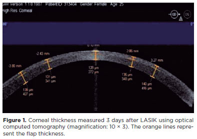
METHODS: The study was conducted in two ophthalmology clinics in Beijing, China, in 2022. A total of 84 patients (152 eyes) with different degrees of myopia were examined and underwent LASIK (n=46, 80 eyes) or IntraLASIK (n=38, 72 eyes). Keratometry, corneal topography, pachymetry, visual acuity evaluation, and corneal biomechanical analysis were performed before and after the intervention.
RESULTS: IntraLASIK produced more precise flaps than LASIK, with deviations of <8 mm and 0.1 mm from the intended thickness and diameter, respectively. LASIK resulted in nonuniform flaps, with thickness deviations of 5-86 mm. IntraLASIK demonstrated a superior efficacy for patients with severe myopia and thin corneas, with a mean spherical equivalent of 0.9 D at 6 months compared to the 1.4 D for LASIK. Approximately 91% and 83% of the patients with mild to moderate and severe myopia, respectively, achieved results within ± 0.49 D from the refractive target with IntraLASIK.
CONCLUSIONS: Corneal hysteresis and corneal resistance factor decreased with an increase in laser intensity, and they decreased faster with thinner corneas. Thus, IntraLASIK is more useful than LASIK in patients with thin corneas and severe myopia.
Keywords: Myopia; Lasers; Cornea; Keratomileusis; Laser in situ
Arq. Bras. Oftalmol. 2026;89 (3 )
:1-8
| DOI: 10.5935/0004-2749.2025-0330
Abstract
PURPOSE: To assess whether low-concentration brimonidine (0.025%) improves early postoperative signs and symptoms following femtosecond laser-assisted in situ keratomileusis and photorefractive keratectomy without affecting pupil diameter or flap safety.
METHODS: This prospective, randomized, double-masked, contralateral-eye, single-center study was conducted between January and September 2024. In each patient, one eye received 0.025% brimonidine 15–30 min before surgery (mean: 21.3 ± 2.4 min), whereas the fellow eye received 0.15% sodium hyaluronate (control). Primary outcomes on postoperative Day 1 included subconjunctival hemorrhage laser-assisted in situ keratomileusis and patient-reported symptoms (0–10 scale; composite score). Pupil diameter was measured pre-ablation. Statistical analyses included McNemar and paired t tests, with a significant threshold of α=0.05.
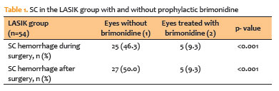
RESULTS: A total of 124 patients were included (54 laser-assisted in situ keratomileusis and 70 photorefractive keratectomy). Pupil diameter did not differ significantly between brimonidine-treated and control eyes (laser-assisted in situ keratomileusis: 2.63 ± 0.47 vs. 2.69 ± 0.42 mm, p=0.273; photorefractive keratectomy: 2.56 ± 0.44 vs. 2.61 ± 0.39 mm, p=0.116). In laser-assisted in situ keratomileusis, subconjunctival hemorrhage occurred less frequently in brimonidine-treated eyes both intraoperatively (9.3% vs. 46.3%, p<0.001) and on postoperative Day 1 (9.3% vs. 50.0%, p<0.001). Composite symptom scores were significantly lower in brimonidine-treated eyes in both laser-assisted in situ keratomileusis and photorefractive keratectomy groups (p=0.001 for both).
CONCLUSIONS: Preoperative administration of low-concentration brimonidine (0.025%) significantly reduced subconjunctival hemorrhage in laser-assisted in situ keratomileusis without comprising flap integrity. It also improved early postoperative symptoms in laser-assisted in situ keratomileusis and photorefractive keratectomy, without affecting pupil diameter. These findings support the use of dilute brimonidine as a safe and effective adjunct to enhance the immediate postoperative experience in refractive surgery.
Keywords: Brimonidine tartrate; Postoperative pain; Subconjunctival hemorrhage; Refractive surgery; Hemorrhage; Keratomileusis, laser in situ; Photorefractive keratectomy
Arq. Bras. Oftalmol. 2024;87 (3 )
:1-7
| DOI: 10.5935/0004-2749.2023-0049
Abstract
PURPOSE: To investigate the association of pre-photorefractive keratectomy Schirmer-1 test value with post-photorefractive keratectomy central corneal epithelial thickness, ocular surface disease index score, and uncorrected distance visual acuity.
METHODS: Patients were categorized according to preoperative Schirmer-1 value: the normal Schirmer Group (n=54; Schirmer-1 test value, >10 mm) and the low Schirmer Group (n=52; Schirmer-1 test value, between 6 and 10 mm). We analyzed ablation depth, visual acuity, result of Schirmer-1 test (with anesthesia), tear film break-up time, ocular surface disease index score, central corneal epithelial thickness, and spherical equivalent refraction.
RESULTS: We found significant differences between the groups in Schirmer-1 test value, tear film break-up time, and ocular surface disease index score, both preoperatively and postoperatively (p<0.001). The preoperative central corneal epithelial thicknesses of the two groups were similar (p>0.05). After photorefractive keratectomy, the Schirmer-1 test value and spherical equivalent refraction decreased in both groups (p<0.05), and ocular surface disease index scores and central corneal epithelial thickness values increased in the low Schirmer Group (p<0.001) but not in the normal Schirmer Group (p>0.05). The postoperative central corneal epithelial thicknesses of the low Schirmer Group were significantly higher than those of the normal Schirmer Group (p<0.001). Postoperative uncorrected distance visual acuity did not differ significantly between the two groups (p>0.05).
CONCLUSIONS: In patients with low Schirmer-1 test values before photorefractive keratectomy, the corneal epithelium thickened and ocular surface complaints increased during the postoperative period. However, changes in the corneal epithelium did not affect the postoperative uncorrected distance visual acuity. To reduce postoperative problems on the ocular surface in these patients, we recommend that dry eye be treated before photorefractive keratectomy.
Keywords: Epithelium, corneal; Cornea; Photorefractive keratectomy; Schirmer test; Visual acuity
Arq. Bras. Oftalmol. 2024;87 (2 )
:1-5
| DOI: 10.5935/0004-2749.2021-0395
Abstract
Objetivos: Avaliar a segurança e eficácia a longo prazo da vitreólise com Nd:YAG laser para moscas volantes sintomáticas, uma vez que permanece como um procedimento controverso devido a falta de evidência científica robusta sobre a manutenção dos resultados e ocorrência de efeitos adversos.
Métodos: Este estudo é uma extensão observacional de um ensaio clínico prospectivo, randomizado, duplo cego, previamente publicado. Oito de treze pacientes que foram submetidos a vitreólise com YAG laser foram acompanhados para uma reavaliação tardia, dezoito meses após o procedimento, para avaliar a eficácia e segurança do procedimento.
Resultados: Todos os pacientes mantiveram a melhora na sintomatologia notada ao final do procedimento original, com 25% dos casos apresentando melhora completa, e uma proporção semelhante (37,5%) demonstrando melhora significativa ou parcial. A melhora objetiva na opacidade foi similar ao achado no seguimento original de 6 meses. O questionário de qualidade de vida NEI-VFQ 25 não demonstrou diferença estatisticamente significativa nas respostas entre o sexto e o décimo oitavo mês de acompanhamento. Nenhum efeito adverso foi notado no exame clínico ou reportado pelos pacientes.
Conclusão: A eficácia da vitreólise observada ao sexto mês do acompanhamento foi mantida até o décimo oitavo mês, com todos os pacientes notando algum grau de melhora quando comparado ao estado pré procedimento. Nenhum efeito adverso tardio foi notado. Um ensaio clínico randomizado maior é necessário para confirmar a segurança do procedimento.
Keywords: Terapia a laser; Lasers de estado sólido; Vitrectomia; Corpo vítreo; Cirurgia vitreorretiniana; Acuidade visual; Doenças oculares; Qualidade de vida; Inquéritos e questionários
 ABO is licensed under a Creative Commons Attribution-NonComercial 4.0 Internacional.
08-fig01.jpg)
12-tab01tb.jpg)
08-fig01.jpg)
11-fig01.jpg)
01-tab01tb.jpg)
05-fig01tb.jpg)
04-fig01.jpg)