Arq. Bras. Oftalmol. 2023;86 (4 )
:359-364
| DOI: 10.5935/0004-2749.20230056
Abstract
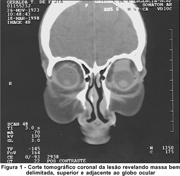
Objetivo: Comparar as características radiológicas e clínicas do adenoma pleomórfico primário e do carcinoma adenoide cístico da glândula lacrimal.
Métodos: Este estudo revisou retrospectivamente os achados de imagem e os prontuários médicos de casos de adenoma pleomórfico e carcinoma adenoide cístico da glândula lacrimal.
Resultados: Foram avaliados 11 pacientes com adenoma pleomórfico e 16 pacientes com carcinoma adenoide cístico. Não houve diferenças estatisticamente significativas em relação à idade e sexo. Proptose foi o sintoma de apresentação mais comum em ambos os grupos. Os carcinomas adenoides císticos foram mais propensos que os adenomas pleomórficos a apresentarem massas palpáveis, diplopia, dor e perda sensorial, mas essa diferença entre os grupos não foi estatisticamente significativa. Não houve diferenças estatísticas em termos de homogeneidade e indentação do globo ocular entre os dois tipos de tumores em imagens de tomografia computadorizada (p>0,05). Também à tomografia computadorizada, a invasão óssea, a calcificação do tumor e o sinal em cunha foram mais frequentes nos carcinomas adenoides císticos, enquanto a remodelação óssea foi mais frequente nos adenomas pleomórficos, com significância estatística para todas essas manifestações (p<0,05). À ressonância magnética, os adenomas pleomórficos foram significativamente mais propensos a terem margens bem definidas, contornos lobulados, realce heterogêneo pelo contraste e hiperintensidade na ressonância magnética ponderada em T2 (p<0,05).
Conclusão: Ao se diferenciar o adenoma pleomórfico e o carcinoma adenoide cístico da glândula lacrimal, é muito importante avaliar as características radiológicas juntamente com as características clínicas. Os contornos lobulados podem ser uma característica radiológica significativamente distinta em favor do adenoma pleomórfico.
Keywords: Aparelho lacrimal/patologia; Adenoma pleomorfo; Carcinoma adenoide cístico; Tomografia computadorizada por raios x; Imagem por ressonância magnética.
Arq. Bras. Oftalmol. 2025;88 (2 )
:1-5
| DOI: 10.5935/0004-2749.2024-0113
Abstract
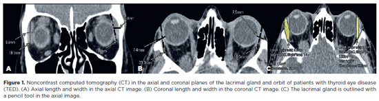
This study aimed to evaluate the morphometric and volumetric dimensions of the lacrimal gland in patients with inactive thyroid eye disease and compare them with the values reported in the literature. This case series evaluated consecutive patients with inactive thyroid eye disease treated at a tertiary eye hospital from 2015 to 2020. The patients' baseline demographics and clinical characteristics were obtained. The axial and coronal length, width, and volume of the lacrimal gland were measured on computed tomography scan images, and the results were statistically analyzed. A total of 21 patients (42 orbits) with inactive thyroid eye disease were evaluated. Their mean age was 49.0 ± 14.6 years, and 12 (57.1%) of them were men. The main complaint was dryness, and the majority of the patients had good vision and mild proptosis. The mean axial length and width of the lacrimal gland were 19.3 ± 3.9 mm and 7.5 ± 2.1 mm, respectively; coronal length and width, 20.4 ± 4.5 mm and 7.5 ± 2.1 mm, respectively; and lacrimal gland volume, 0.825 ± 0.326 mm3. Age, sex, or laterality were not found to be determinants of lacrimal gland enlargement. Patients with thyroid eye disease have enlarged lacrimal gland even in the nonactive phase of the disease multifactorial aspects influence the lacrimal gland in thyroid eye disease, making it difficult to establish a clear correlation with predisposing factors. Further studies are warranted to better understand the association between thyroid eye disease and the lacrimal gland.
Keywords: Graves' ophthalmology; Graves' disease; Lacrimal apparatus; Lacrimal apparatus diseases; X-ray computed tomography
 ABO is licensed under a Creative Commons Attribution-NonComercial 4.0 Internacional.
11-tab01.jpg)
01-fig01.jpg)
02-fig01.jpg)
02-fig01.jpg)
01-fig01tb.jpg)
02-fig01.jpg)
02-fig01.jpg)
09-fig01.jpg)
13-fig01.jpg)
11-fig01.jpg)
14-fig01.jpg)
14-fig01.jpg)