Arq. Bras. Oftalmol. 2021;84 (4 )
:316-323
| DOI: 10.5935/0004-2749.20210045
Abstract
OBJETIVO: O objetivo deste estudo foi analisar a segurança do implante de lente intraocular primária em um grande número de olhos em crianças <24 meses.
MÉTODOS: Foram revisados os prontuários de pacientes com idade entre 5-24 meses, submetidos a implante primário de lente intraocular no saco capsular. Uma lente intraocular acrílica de três peças dobrável foi implantada pelo mesmo cirurgião usando uma única técnica cirúrgica. Pacientes que tiveram <1 ano de acompanhamento após a cirurgia foram excluídos. Os principais resultados incluíram medidas de acuidade visual, mudança miópica, complicações pós operatórias e cirurgias adicionais.
RESULTADOS: Foram analisados 68 pacientes (93 olhos). A média de idade dos pacientes no momento da cirurgia foi de 15,06 ± 6,19 (5 a 24) meses, e o equivalente esférico 1 mês após a cirurgia foi de 3,62 ± 2,32 D. Após 5,67 ± 3,10 anos, o equivalente esférico foi de -0,09 ± 3,22 D, e a acuidade visual corrigida à distância foi de 0,33 ± 0,33 e 0,64 ± 0,43 logMAR em casos bilaterais e casos unilaterais, respectivamente (p=0,000). A maior mudança míopica foi observado em bebês submetidos à cirurgia aos 5 e 6 meses de idade. As complicações mais frequentes incluíram opacificação do eixo visual e corectopia. Glaucoma e descolamento de retina não foram relatados.
CONCLUSÃO: O implante primário de lente intraocular no saco capsular em crianças de 5-24 meses é seguro e está associado à baixas taxas de eventos adversos e cirurgias adicional.
Keywords: Catarata pediátrica; Lente intraocular; Implante primário LIO; Mudança miópica; Catarata congênita
Arq. Bras. Oftalmol. 2023;86 (3 )
:1-7
| DOI: 10.5935/0004-2749.20230045
Abstract
Objetivo: Avaliar o implante de lente intraocular primária para tratamento da afacia pediátrica no Sistema Único de Saúde (SUS) e comparar os resultados em diferentes faixas etárias.
Métodos: Foram incluídas crianças com catarata congênita e do desenvolvimento unilateral ou bilateral de 0-12 anos de idade e submetidas a implante de lente intraocular primária.
Resultados: Cento e oito olhos de 68 crianças divididas em quatro grupos de idade (<7m; 7m-2a; 2-5a e > 5a) foram avaliados. Dezenove olhos (17,59%) apresentaram opacificação do eixo visual como complicação pós-operatória. Essa complicação foi mais frequente na faixa etária <7 meses (37,93%). A diferença foi significativa entre os grupos de idade <7 meses e > 5 anos (p=0,002). A opacificação do eixo visual foi dividida em duas categorias: membrana pupilar e proliferação de células do cristalino. Oito olhos apresentaram membrana pupilar e 14 proliferação de células do cristalino. Dos oito olhos com membrana pupilar, sete ocorreram na faixa etária <7 meses. A diferença entre o grupo de idade <7 meses e os grupos de 2-5 anos e > 5 anos foi significativa (p=0,01). A proliferação de células do cristalino foi mais frequente nos grupos de idade <7 meses e 2-5 anos, mas significativa apenas quando comparados o grupo de idade <7 meses com o grupo> 5 anos de idade (p=0,040). Glaucoma e suspeitos de glaucoma não foram observados durante o acompanhamento.
Conclusões: A principal complicação encontrada no estudo foi a opacificação do eixo visual. Sua incidência foi maior em crianças operadas antes dos 7 meses de idade.
Keywords: Extração de catarata; Lentes intraoculares; Complicações intraoperatórias; Glaucoma; Segmento anterior do olho; Criança.
Arq. Bras. Oftalmol. 2021;84 (1 )
:31-36
| DOI: 10.5935/0004-2749.20210006
Abstract
Objetivo: Avaliar a prevalência de transtornos de depressão e ansiedade em pacientes com glaucoma e identificar fatores de riscos associados.
Métodos: Estudo transversal em pacientes com glaucoma, avaliados durante Agosto de 2016 e Agosto de 2017 no Hospital das Clínicas da Universidade de Campinas e no Hospital Oftalmológico de Brasília. Todos pacientes foram submetidos à exame oftalmológico completo para confirmar o diagnóstico de glaucoma. Todos pacientes preencheram o questionário “Hospital Anxiety and Depression Scale”.
Resultados: Foram incluídos 129 pacientes no estudo, sendo 74 homens (57.36%) e 55 (42.64%) mulheres, 90 pacientes eram brancos (69.77%) e 38 (29.46%) eram negros. A idade média foi de 70.14 ± 15.8 anos. O estudo demonstrou uma prevalência de 10.08% de transtornos depressivo e/ou ansiedade.
A regressão logística demonstrou que mulheres apresentam
maior risco de desenvolver transtornos depressivos e/ou ansiedade (Risco relativo: 5.25, p=0.015), assim como pacientes com maior número de co-morbidades clínicas (Risco relativo: 2.82, p=0.038).
Conclusão: Uma proporção significativa dos pacientes com glaucoma podem apresentar transtornos de depressão e/ou ansiedade. Pacientes com glaucoma do sexo feminino e que apresentem maiores co-morbidades clínicas apresentam maior risco de apresentar esses transtornos.
Keywords: Glaucoma; Depressão/epidemiologia; Ansiedade/epidemiologia; Estudos transversais
Arq. Bras. Oftalmol. 2025;88 (6 )
:1-9
| DOI: 10.5935/0004-2749.2024-0411
Abstract
PURPOSE: This study evaluated macular thickness using spectral-domain optical coherence tomography in healthy participants from a population-based eye survey.
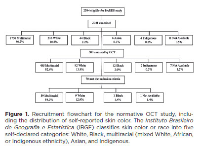
METHODS: The Brazilian Amazon Region Eye Survey was a population-based study assessing the prevalence and causes of visual impairment, blindness, and ocular diseases in adults aged ≥45 years from urban and rural areas of Parintins. A subgroup was selected based on inclusion criteria for both eyes: best-corrected visual acuity ≥20/32, normal eye examination results, and no prior ocular surgery. Scans were performed using the iVue optical coherence tomography device. Measurements were taken from the nine subfields defined by the Early Treatment Diabetic Retinopathy Study, examining the full retina as well as the inner and outer retinal layers. Associations of retinal thickness with age and sex were also analyzed. Statistical significance was set at p≤0.05.
RESULTS: In total, 70 healthy participants (25 males), aged 45–65 years (mean=52 ± 5), were included. Mean central foveal thickness was 248.71 ± 18.73 μm. A significant age-related reduction in macular thickness was observed, particularly in the inner superior parafovea (p=0.036), nasal perifovea (p=0.001), superior perifovea (p=0.028), outer layer of inferior parafovea (p=0.049), and the inferior perifovea of the full retina (p=0.029). Males showed significantly greater thickness in the outer layer, especially in the outer parafovea (p=0.004) and perifovea (p<0.0001).
CONCLUSIONS: This study established normative macular thickness values for healthy older adults in the Brazilian Amazon region using spectral-domain optical coherence tomography. Age and sex were found to significantly influence macular thickness and should be considered when interpreting measurements. These data will support future studies of retinal diseases in this population.
Keywords: Retinal diseases/diagnosis; Macula lutea/pathology; Macular degeneration/diagnosis; Diabetic retinopathy/diagnosis; Vision, low; Vision tests; Tomography, optical coherence/methods; Young adult; Cross-sectional studies; Brazil/epidemiology
Arq. Bras. Oftalmol. 2021;84 (3 )
:258-266
| DOI: 10.5935/0004-2749.20210040
Abstract
OBJETIVO: Investigar o efeito inibidor da fluorofenidona contra a proliferação e a transição epitélio-mesenquimal induzida pelo fator de transformação do crescimento β2 na linha HLEC FHL 124 e seu mecanismo potencial.
MÉTODOS: Avaliou-se o efeito in vitro da fluorofenidona na proliferação e na transição epitelial-mesenquimal da linha FHL 124 de células epiteliais do cristalino humano. Após tratamento com fluorofenidona nas concentrações de 0, 0,1, 0,2, 0,4, 0,6 e 1,0 mg/mL, a proliferação celular foi medida através de um ensaio de MTT. A viabilidade celular foi avaliada pela atividade da lactato-desidrogenase liberada por células danificadas. As células FHL 124 foram tratadas com diferentes concentrações do fator de transformação do crescimento β2 (0-10 ng/mL) por 24 horas e a expressão de CTGF, α-SMA, COL-I, E-caderina e Fn foram avaliadas por qPCR e Western blot. Após tratamento com 0, 0,2 e 0,4 mg/mL de fluorofenidona, as expressões do fator de transformação do crescimento β2 e de SMADs foram detectadas com PCR em tempo real e Western blot. As expressões do CTGF, α-SMA, COL-I e Fn foram analisadas através de um ensaio imunocitoquímico.
RESULTADOS: A viabilidade das células FHL 124 não foi inibida com concentrações de fluorofenidona menores ou iguais a 0,4 mg/mL após 24 horas de tratamento. Não foi detectada nenhuma citotoxicidade pelo ensaio da lactato-desidrogenase após 24 e 36 horas de tratamento com 0,2 e 0,4 mg/mL de fluorofenidona. O fator de transformação do crescimento β2 aumentou a expressão de mRNA e proteína do CTGF, α-SMA, COL-I e Fn. Porém, a fluorofenidona suprimiu significativamente a expressão de SMADs, CTGF, α-SMA, COL-I e Fn, tanto na ausência quanto na presença de estimulação pelo fator de transformação do crescimento β2.
CONCLUSÕES: A fluorofenidona inibiu significativamente a expressão de SMADs, CTGF, α-SMA, COL-I e Fn em células FHL 124. Devido à ausência de incompatibilidade em lactentes, a fluorofenidona pode vir a se tornar um novo medicamento contra a opacificação capsular posterior em lactentes.
Keywords: Fator de crescimento transformador beta2; Fluorofenidona; Lentes; Catarata; Lactente
Arq. Bras. Oftalmol. 2025;88 (6 )
:1-5
| DOI: 10.5935/0004-2749.2025-0085
Abstract
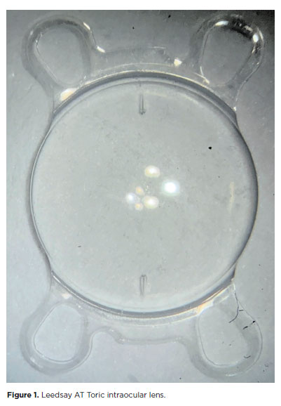
PURPOSE: The purpose of this study was to assess visual outcomes and patient satisfaction following cataract surgery involving the implantation of quad-loop intraocular lenses, including trifocal, bifocal, and toric variants.
METHODS: Information was obtained from both physical and electronic medical records of patients who underwent phacoemulsification cataract surgery with implantation of different intraocular lenses between January 1, 2022, and December 31, 2023. The study included individuals aged over 18 who received bilateral implantation of bifocal, trifocal, or monofocal toric intraocular lenses. Visual acuity was assessed at various postoperative time points using the logMAR scale. Quantitative variables were analyzed using mean and standard deviation.
RESULTS: A total of 92 eyes received premium intraocular lenses: 4 bifocal, 32 trifocal, 52 toric monofocal, and 4 trifocal toric lenses. The average preoperative corrected visual acuity was logMAR 0.478 ± 0.259. On the first postoperative day, the average uncorrected visual acuity was logMAR 0.301 ± 0.207. By day 30, 67.4% of eyes achieved uncorrected distance visual acuity of logMAR 0.2 or better. Patient satisfaction was high, with few reports of glare or halos.
CONCLUSION: Quad-loop intraocular lenses-including trifocal, bifocal, and toric models-demonstrated effective improvement in visual acuity and high levels of patient satisfaction. These lenses represent a suitable option for enhancing visual outcomes after cataract surgery. Additional studies with larger cohorts are recommended to confirm these results.
Keywords: Cataract extraction; Aberrometry/methods; Lenses, intraocular; Lens implantation, intraocular; Prosthesis design
Arq. Bras. Oftalmol. 2026;89 (2 )
:1-8
| DOI: 10.5935/0004-2749.2025-0175
Abstract
PURPOSE: Endophthalmitis is one of the most important adverse events after cataract surgery, as it can lead to total vision loss. This study aimed to describe the occurrence of endophthalmitis after phacoemulsification with intraocular lens implantation in patients treated in a community setting in Porto Velho, Rondônia, Brazil.
METHODS: This retrospective cohort study was conducted using a database of 649 medical records of patients who underwent surgery and were followed for three months. Poisson regression analysis was used to estimate relative risks and 95% confidence intervals (95% CIs).
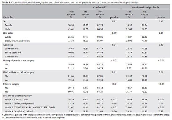
RESULTS: The incidence of confirmed endophthalmitis was 11.94% (95% CI, 9.50-14.76), while the incidence of confirmed and probable cases was 20.50% (95% CI, 17.52-23.73). For confirmed cases, bilateral surgery and the use of lens model 3 were identified as risk factors for endophthalmitis, whereas age over 70 yr and preoperative antibiotic use were protective factors. For confirmed and probable cases, brown and yellow skin color, bilateral surgery, and the use of lens model 3 were also identified as risk factors. Gram-negative bacteria were the predominant etiological agents, and corneal edema was the main clinical manifestation. The mean duration of treatment was eight days, and 27.12% of patients used antibiotics.
CONCLUSION: The incidence observed was substantially higher than that reported in the literature, with a predominance of Gram-negative agents and an association with bilateral surgeries and the Eyeol intraocular lens model. These findings reinforce the need for continuous epidemiological surveillance and the implementation of specific biosafety and infection control protocols during cataract surgery campaigns.
Keywords: Endophthalmitis; Disease outbreaks; Phacoemulsification; Lens implantation, intraocular; Lenses, intraocular; Cataract; Risk factors; Anti-bacterial agents
Arq. Bras. Oftalmol. 2025;88 (6 )
:1-8
| DOI: 10.5935/0004-2749.2025-0053
Abstract
PURPOSE: This pilot study evaluated the diagnostic accuracy of a deep learning model for detecting pterygium in anterior segment photographs taken using smartphones in the Brazilian Amazon. The model’s performance was benchmarked against assessments made by experienced ophthalmologists, considered the clinical gold standard.
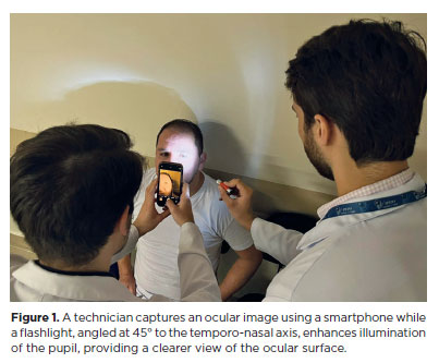
METHODS: In this cross-sectional study, 38 participants (76 eyes) from Barcelos, Brazil, were enrolled. Trained nonmedical health workers captured high-resolution anterior segment images using smartphones. These images were analyzed using a deep learning model based on the MobileNet-V2 convolutional neural network. Diagnostic metrics–including sensitivity, specificity, accuracy, positive predictive value, negative predictive value, and area under the receiver operating characteristic curve–were calculated and compared with the ophthalmologists’ evaluations.
RESULTS: The deep learning model achieved a sensitivity of 91.43%, specificity of 90.24%, positive predictive value of 88.46%, negative predictive value of 92.79%, and an area under the curve of 0.91. Logistic regression revealed no statistically significant association between pterygium and demographic variables such as age or gender.
CONCLUSIONS: The deep learning model demonstrated high diagnostic performance in identifying pterygium in a remote Amazonian population. These preliminary findings support the potential use of artificial intelligence–based tools to facilitate early detection and screening in underserved regions, thereby enhancing access to ophthalmic care.
Keywords: Pterygium/diagnostic imaging; Smartphone; Diagnostic techniques, ophthalmological; Deep learning; Telemedicine; Artificial intelligence; Cross-sectional studies; Brazil/epidemiology
Arq. Bras. Oftalmol. 2025;88 (4 )
:1-6
| DOI: 10.5935/0004-2749.2024-0083
Abstract
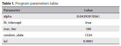
PURPOSE: We developed an artificial intelligence program for calculating intraocular lenses and analyzed its accuracy rate via ultrasonic biometry. This endeavor is aimed at enhancing precision and efficacy in the selection of intraocular lenses, particularly in cases where optical biometry is unavailable.
METHODS: Data was collected from the Hospital de Clínicas de Porto Alegre, which included cases of phacoemulsification with intraocular lens implantation, in which the lens selection was based on ultrasonic biometry. The program, implemented in Python, Java, and PHP, employs the ridge regression method. Two design options were developed: a basic model, which uses only keratometry variables (K1 and K2), axial size and final target refraction in the spherical equivalent, and an advanced model, which incorporates preoperative refraction and the patient's age. The Universal Barrett II formula was used to compare both models.
RESULTS: The sample consisted of 486 eyes from 313 patients, with 350 eyes used for program training and 136 for program validation. The spherical equivalent hit rates, with a variation of ±0.5 D, were 86% and 87.5% for the basic and advanced models, respectively, with no statistically significant difference between them. With the Barret Universal II formula, the success rate was 69%, which was significantly different from the values of the two aforementioned models (p<0.0001). The system was better for medium and long eyes but worse for short eyes (<=22.00 mm).
CONCLUSION: The developed artificial intelligence program was superior to the Barrett formula in terms of performance, in the general context and within the subgroup of patients with longer eyes. This innovation can considerably contribute to the selection of intraocular lenses, particularly in cases where optical biometry is unavailable.
Keywords: Biometry; Intraocular lens; Cataract; Artificial intelligence
Arq. Bras. Oftalmol. 2024;87 (6 )
:1-6
| DOI: 10.5935/0004-2749.2022-0257
Abstract
OBJETIVO: Descrever as características clínicas e os fatores associados à presença de ceratite em pacientes com corpos estranhos na córnea em uma população colombiana.
MÉTODOS: Trata-se de um estudo transversal baseado na revisão dos registros clínicos de pacientes com corpos estranhos na córnea admitidos em um departamento de emergência em Cali, Colômbia, entre junho de 2018 e junho de 2019. O desfecho primário foi a presença de ceratite diagnosticada através de critérios clínicos. Foram utilizados modelos de regressão logística univariada e multivariada para identificar os fatores associados.
RESULTADO: Neste estudo, foi analisado um total de 381 corpos estranhos na córnea em 372 pacientes (idade média: 40,0 anos, intervalo interquartil: 29,0-53,0; sexo masculino: 94,7% [352 casos]). Noventa e cinco casos desenvolveram ceratite (24,9%, intervalo de confiança de 95% — IC 95%: 20,8%-29,5%). Na análise multivariada, para idade ≤30 anos (razão de chances — RC: 2,15, IC 95%: 1,06-4,36), o achado de flare aquoso (RC: 2,81, IC 95%: 1,39-5,66]) e a presença de corpo estranho na periferia da córnea (RC: 2,05, IC 95%: 1,19-3,50) foram associados a um risco aumentado de ceratite. Sexo, tempo entre a lesão e a internação, e edema da córnea não foram relacionados à ceratite (p>0,05).
CONCLUSÃO: Há uma proporção elevada de ceratite em casos de corpos estranhos na córnea em Cali, Colômbia. Os três fatores associados à ceratite foram a idade, o achado de flare aquoso e a presença de corpo estranho na periferia da córnea.
Keywords: Corpos estranhos no olho; Lesões da córnea; Ceratite/epidemiologia; Estudos transversais; Colômbia.
Arq. Bras. Oftalmol. 2026;89 (4 )
:1-5
| DOI: 10.5935/0004-2749.2026-0010
Abstract
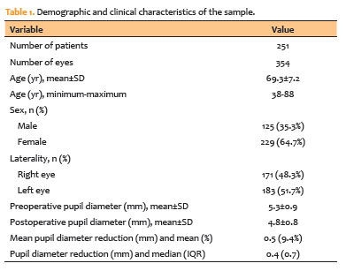
PURPOSE: To evaluate changes in scotopic pupil diameter before and after cataract surgery performed by phacoemulsification with intraocular lens implantation.
METHODS: This prospective longitudinal observational study included patients who underwent cataract surgery. Scotopic pupil diameter was measured preoperatively and 30-40 days postoperatively using an automated keratometer after a standardized dark-adaptation period under controlled ambient illumination. Each eye was considered an independent unit of observation. Because some participants contributed both eyes, intraindividual correlation was accounted for using a linear mixed-effects model with random patient intercepts. Time of assessment (preoperative versus postoperative), age, sex, and eye laterality were included as fixed effects.
RESULTS: A total of 354 eyes from 251 patients were analyzed. The mean patient age was 69.3±7.2 yr. Mean scotopic pupil diameter decreased from 5.3±0.9mm preoperatively to 4.8±0.8mm postoperatively, representing a mean reduction of 0.5mm (9.4%). In the linear mixed-effects model, cataract surgery was associated with a significant reduction in pupil diameter, with an adjusted mean difference of 0.45mm (95% confidence interval [95% CI], 0.39-0.51; p<0.001). Age (p=0.061), sex (p=0.920), and eye laterality (p=0.152) were not significantly associated with the magnitude of pupil diameter change.
CONCLUSION: Phacoemulsification with intraocular lens implantation was associated with a significant reduction in scotopic pupil diameter, independent of age, sex, and eye laterality. This finding should be considered during preoperative planning, particularly when selecting intraocular lenses whose optical performance depends on postoperative pupil size.
Keywords: Cataract; Pupil; Phacoemulsification; Lens implantation, intraocular; Lenses, intraocular; Pseudophakia
Arq. Bras. Oftalmol. 2025;88 (5 )
:1-7
| DOI: 10.5935/0004-2749.2024-0368
Abstract
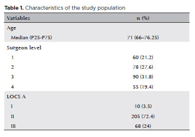
PURPOSE: To compare endothelial corneal cell changes following cataract surgery performed by phacoemulsification with intraocular lens implantation, conducted by surgeons with varying levels of experience.
METHODS: Two hundred and eighty-three eyes diagnosed with cataract were included. Lens opacity was classified into three categories (I, II, and III). Surgeons were categorized into four experience levels (1, 2, 3, and 4), based on years of practice and lifetime surgeries performed. Corneal endothelial characteristics were assessed using non-contact specular microscopy, with measurements taken before surgery and 30-60 days post-surgery.
RESULTS: Pre- and postoperative endothelial analysis showed no significant differences between surgeon levels regarding visual acuity achieved, corneal thickness, and endothelial hexagonality. However, the central endothelial cell density index showed a significantly greater reduction among level 1 surgeons (p=0.026). Grade II cataracts exhibited significant variations in the central endothelial cell density (p=0.011) and average cell size, with level 1 surgeons showing the largest increases (p=0.024).
CONCLUSIONS: The analysis revealed significant differences in visual acuity and endothelial indices between surgeon experience levels, with less experienced surgeons showing greater variations and poorer performance. Clinical protocols should consider these data to establish safer training protocols.
Keywords: Cataract extraction; Phacoemulsification; Endothelium; corneal; Lens implantation, intraocular; Visual acuity; Internship and residency; Surgeons


 ABO is licensed under a Creative Commons Attribution-NonComercial 4.0 Internacional.


02-tab01tb.jpg)
15-tab01tb.jpg)
05-tab01.jpg)
12-tab01.jpg)
11-tab01tb.jpg)
10-fig01.jpg)
01-fig01.jpg)