Arq. Bras. Oftalmol. 2026;89 (2 )
:1-8
| DOI: 10.5935/0004-2749.2025-0105
Abstract
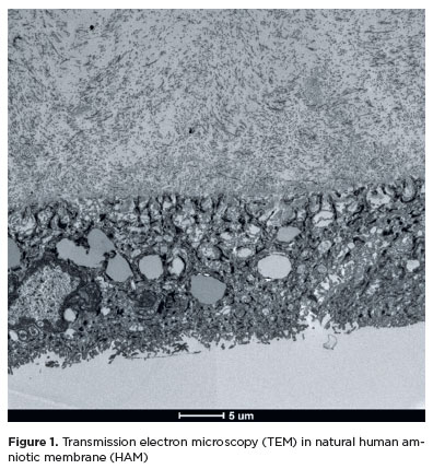
PURPOSE: To evaluate structural differences in amniotic membrane fragments subjected to different preservation techniques for potential ophthalmologic applications.
METHODS: Three placentas were collected from healthy donors, and four amniotic membrane fragments were prepared from each placenta. The fragments were divided into four groups with three samples each: cryopreserved, lyophilized, vacuum-dried using a vacuum concentrator, and fresh (control). After processing, the fragments were fixed, sectioned, and analyzed using scanning transmission electron microscopy to assess tissue morphology.
RESULTS: All samples met the established evaluation criteria. No morphological differences were observed among the groups. The structural characteristics of lyophilized and vacuum-dried membranes were comparable with those of cryopreserved and fresh membranes. However, vacuum drying demonstrated the greatest practicality for ophthalmologic use, as it allows membrane availability at any time and storage at room temperature.
CONCLUSION: Vacuum drying using a vacuum concentrator, lyophilization, and cryopreservation preserve the morphological characteristics of the human amniotic membrane similar to those of fresh tissue. A standardized protocol using a vacuum concentrator may be established owing to its advantages in storage convenience and accessibility.
Keywords: Amnion/transplantation; Cryopreservation/methods; Freeze drying; Lyophilization; Ophthalmologic surgical procedures; Regenerative medicine
Arq. Bras. Oftalmol. 2023;86 (4 )
:359-364
| DOI: 10.5935/0004-2749.20230056
Abstract
Objetivo: Comparar as características radiológicas e clínicas do adenoma pleomórfico primário e do carcinoma adenoide cístico da glândula lacrimal.
Métodos: Este estudo revisou retrospectivamente os achados de imagem e os prontuários médicos de casos de adenoma pleomórfico e carcinoma adenoide cístico da glândula lacrimal.
Resultados: Foram avaliados 11 pacientes com adenoma pleomórfico e 16 pacientes com carcinoma adenoide cístico. Não houve diferenças estatisticamente significativas em relação à idade e sexo. Proptose foi o sintoma de apresentação mais comum em ambos os grupos. Os carcinomas adenoides císticos foram mais propensos que os adenomas pleomórficos a apresentarem massas palpáveis, diplopia, dor e perda sensorial, mas essa diferença entre os grupos não foi estatisticamente significativa. Não houve diferenças estatísticas em termos de homogeneidade e indentação do globo ocular entre os dois tipos de tumores em imagens de tomografia computadorizada (p>0,05). Também à tomografia computadorizada, a invasão óssea, a calcificação do tumor e o sinal em cunha foram mais frequentes nos carcinomas adenoides císticos, enquanto a remodelação óssea foi mais frequente nos adenomas pleomórficos, com significância estatística para todas essas manifestações (p<0,05). À ressonância magnética, os adenomas pleomórficos foram significativamente mais propensos a terem margens bem definidas, contornos lobulados, realce heterogêneo pelo contraste e hiperintensidade na ressonância magnética ponderada em T2 (p<0,05).
Conclusão: Ao se diferenciar o adenoma pleomórfico e o carcinoma adenoide cístico da glândula lacrimal, é muito importante avaliar as características radiológicas juntamente com as características clínicas. Os contornos lobulados podem ser uma característica radiológica significativamente distinta em favor do adenoma pleomórfico.
Keywords: Aparelho lacrimal/patologia; Adenoma pleomorfo; Carcinoma adenoide cístico; Tomografia computadorizada por raios x; Imagem por ressonância magnética.
Arq. Bras. Oftalmol. 2024;87 (4 )
:1-7
| DOI: 10.5935/0004-2749.2022-0024
Abstract
Objetivo: A síndrome de opsoclonia-mioclonia é extremamente rara em adultos e tem uma fisiopatologia autoimune. Devido à raridade dessa síndrome, o reconhecimento da síndrome de opsoclonia-mioclonia-ataxia precisa melhorar urgentemente em todo o mundo. Assim sendo, este estudo visou aumentar a conscientização sobre a síndrome de opsoclonia-mioclonia-ataxia e ajudar os médicos para um melhor diagnóstico e o uso correto da imunoterapia.
Métodos: Este é o relato de um caso adulto de síndrome de opsoclonia-mioclonia idiopática com movimentos oculares conjugados, multidirecionais, arrítmicos e espontâneos, mioclonia, ataxia, distúrbios do sono e medo intenso. Além disso, foram pesquisadas as publicações recentes relevantes e resumiu-se a fisiopatologia, a apresentação clínica, o diagnóstico e o tratamento da síndrome de opsoclonia-mioclonia-ataxia.
Resultados: A paciente recuperou-se totalmente da opsoclonia, da mioclonia e da ataxia através de imunoterapia. O artigo também fornece um resumo atualizado sobre a síndrome de opsoclonia-mioclonia-ataxia.
Conclusão: Adultos com síndrome de opsoclonia-mioclonia-ataxia têm uma baixa frequência de sequelas residuais. O diagnóstico e o tratamento precoces podem levar a melhores prognósticos. Espera-se que a imunoterapia combinada reduza a incidência da síndrome de opsoclonia-mioclonia-ataxia refratária e recorrente.
Keywords: Síndrome de opsoclonia-mioclonia/diagnóstico; Síndrome de opsoclonia-mioclonia/tratamento farmacológico; Imunoterapia/ métodos; Humanos
Arq. Bras. Oftalmol. 2025;88 (2 )
:1-5
| DOI: 10.5935/0004-2749.2024-0113
Abstract
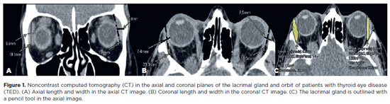
This study aimed to evaluate the morphometric and volumetric dimensions of the lacrimal gland in patients with inactive thyroid eye disease and compare them with the values reported in the literature. This case series evaluated consecutive patients with inactive thyroid eye disease treated at a tertiary eye hospital from 2015 to 2020. The patients' baseline demographics and clinical characteristics were obtained. The axial and coronal length, width, and volume of the lacrimal gland were measured on computed tomography scan images, and the results were statistically analyzed. A total of 21 patients (42 orbits) with inactive thyroid eye disease were evaluated. Their mean age was 49.0 ± 14.6 years, and 12 (57.1%) of them were men. The main complaint was dryness, and the majority of the patients had good vision and mild proptosis. The mean axial length and width of the lacrimal gland were 19.3 ± 3.9 mm and 7.5 ± 2.1 mm, respectively; coronal length and width, 20.4 ± 4.5 mm and 7.5 ± 2.1 mm, respectively; and lacrimal gland volume, 0.825 ± 0.326 mm3. Age, sex, or laterality were not found to be determinants of lacrimal gland enlargement. Patients with thyroid eye disease have enlarged lacrimal gland even in the nonactive phase of the disease multifactorial aspects influence the lacrimal gland in thyroid eye disease, making it difficult to establish a clear correlation with predisposing factors. Further studies are warranted to better understand the association between thyroid eye disease and the lacrimal gland.
Keywords: Graves' ophthalmology; Graves' disease; Lacrimal apparatus; Lacrimal apparatus diseases; X-ray computed tomography
Arq. Bras. Oftalmol. 2024;87 (2 )
:1-8
| DOI: 10.5935/0004-2749.2022-0328
Abstract
PURPOSE: Wet bio-amniotic membrane plugging combined with transplantation is a novel option that combined amniotic membrane plugging with amniotic membrane transplantation for the treatment of small corneal perforations. This study aimed to evaluate the efficacy of wet bio-amniotic membrane plugging in the treatment of small corneal perforations and compared it with that of the penetrating keratoplasty procedure.
METHODS: Forty patients (41 eyes) with small corneal perforations <3 mm in diameter treated at our hospital between July 2018 and January 2021 were retrospectively included. Among them, 21 eyes were treated with wet bio-amniotic membrane plugging (wet bio-amniotic membrane plugging group), and 20 eyes were treated with penetrating keratoplasty procedure (penetrating keratoplasty procedure group). The best-corrected visual acuity, anterior chamber formation, corneal thickness, primary disease control, postoperative complications, and graft survival rate were assessed.
RESULTS: No significant difference in baseline characteristics was found between the wet bio-amniotic membrane plugging and penetrating keratoplasty procedure groups (p>0.05). The postoperative control rates of primary diseases in the wet bio-amniotic membrane plugging and penetrating keratoplasty procedure groups were 95.2% and 90.0%, respectively (p=0.481). Visual acuity was improved 6 months after the operation in the wet bio-amniotic membrane plugging group and was improved at postoperative 1 month in the penetrating keratoplasty procedure group. The formation time of the anterior chamber in the wet bio-amniotic membrane plugging group was significantly shorter than that in the penetrating keratoplasty procedure group (p=0.023). The corneal thickness of the two groups significantly increased 12 months after the operation; however, the degree of thickening in the penetrating keratoplasty procedure group was higher than that in the wet bio-amniotic membrane plugging group (p<0.001). During the follow-up, postoperative complications were not different between the two groups (p>0.999).
CONCLUSION: The results suggest that wet bio-amniotic membrane plugging is effective and safe in the treatment of small corneal perforations. Thus, it can be used as an emergency treatment alternative to penetrating keratoplasty procedure for small corneal perforations.
Keywords: Amnion; Transplantation; Amniotic membrane; Keratoplasty, penetrating; Corneal perforation; Wet bio-amniotic membrane plugging; Wet bio-amniotic membrane transplantation
Arq. Bras. Oftalmol. 2024;87 (2 )
:1-6
| DOI: 10.5935/0004-2749.2023-2022-0341
Abstract
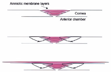
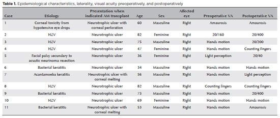
PURPOSE: To evaluate the clinical results of cryopreserved amniotic membrane transplantation as a treatment option for refractory neurotrophic corneal ulcers.
METHODS: This prospective study included 11 eyes of 11 patients who underwent amniotic membrane transplantation for the treatment of refractory neurotrophic corneal ulcers at Hospital de Clínicas da Universidade Federal do Paraná, in the city of Curitiba, from May 2015 to July 2021. Patients underwent different surgical techniques in which the amniotic membrane was applied with the epithelium facing upward to promote corneal re-epithelialization.
RESULTS: The median age of the patients was 60 years (range, 34-82 years), and 64% were men. The predominant etiology of corneal ulcers was herpes zoster (45% of cases). Approximately one-third of the patients (27%) were chronically using hypotensive eye drops, and more than half (54%) had previously undergone penetrating corneal transplantation. At the time of amniotic membrane transplantation, 18% of the eyes had corneal melting, 9% had corneal perforation, and the others had corneal ulceration without other associated complications (73%). The time between clinical diagnosis and surgical treatment ranged from 9 days to 2 years. The corrected visual acuity was worse than 20/400 in 90% of the patients preoperatively, with improvement in 36% after 3 months of the procedure, worsening in 18% and remaining stable in 36%. Of the patients, 81% complained of preoperative pain, and 66% of them reported total symptom relief after the surgical procedure. In one month, 54.6% of the patients presented a closure of epithelial defect, and half of the total group evolved with corneal thinning. The failure rate was 45.5% of the cases.
CONCLUSION: Cryopreserved amniotic membrane transplantation can be considered a good alternative for treating refractory neurotrophic corneal ulcers, as it resulted in significant improvement in pain (66%) and complete epithelial closure (60%) in many patients at 1 month postoperatively. Notably, the high failure rate highlights the need for further studies to identify patient- and ulcer-related factors that may influence the outcomes of this procedure.
Keywords: Amnion/transplantation; Corneal ulcer; Anterior eye segment; Keratitis
 ABO is licensed under a Creative Commons Attribution-NonComercial 4.0 Internacional.
11-tab01.jpg)
09-fig01.jpg)
08-fig01.jpg)
13-fig01.jpg)
04-fig01.jpg)
14-fig01.jpg)
03-fig01.jpg)
14-fig01.jpg)