Arq. Bras. Oftalmol. 1999;62 (6 )
:705-711
| DOI: 10.1590/S0004-27491999000600010
Abstract
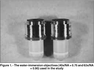
Objetivo: Este estudo prospectivo compara as imagens de microscopia confocal do epitélio corneano do coelho e do homem, obtidas através de 2 objetivas com aberturas numéricas (AN) diferentes. Métodos: Dez olhos de coelhos foram enucleados e fixados através de um suporte pneumático para garantir o melhor desempenho de cada objetiva. Cinco pacientes normais foram selecionados após consentimento. Os olhos de coelhos e dos pacientes foram previamente examinados na lâmpada de fenda. O exame de microscopia confocal (Tomey, Erlangen-Tennenlohe, Alemanha) foi realizado com as objetivas Achroplan 40x/AN = 0,75 e 63x/AN = 0,9 (Zeiss, Oberkochen, Alemanha). Imagens selecionadas do epitélio corneano foram avaliadas qualitativamente com relação ao tamanho, forma e refletividade das células. Resultados: As células no epitélio superficial dos coelhos e dos pacientes, previamente à descamação, tiveram uma refletividade maior que as células adjacentes. Este aspecto foi claramente observado somente com a objetiva 63x/AN = 0,9. As camadas basal e intermediária do epitélio em coelhos foram visualizadas somente através desta objetiva. Estas camadas nos pacientes tornaram-se mais nítidas com a objetiva de abertura numérica maior (63x/AN = 0,9). Conclusão: Uma objetiva de abertura numérica elevada produz melhor resolução dos cortes ópticos, facilitando a análise das camadas do epitélio no coelho e no homem.
Keywords: Corneal epithelium; Confocal microscopy; Epitélio corneano; Microscopia confocal
Arq. Bras. Oftalmol. 2023;86 (3 )
:1-5
| DOI: 10.5935/0004-2749.20230031
Abstract
Objetivo: Descrever os resultados clínicos do tratamento do crescimento epitelial através da técnica de remoção manual seguido da utilização de um compressor de ar comprimido aquecido após a cirurgia de laser in situ keratomileusis (LASIK).
Métodos: Vinte olhos de 17 pacientes foram incluídos no estudo. Cada paciente havia sido submetido a cirurgia de LASIK com presença de crescimento epitelial e foi submetido a tratamento cirúrgico para sua retirada. O objetivo primário foi identificar a presença de crescimento epitelial recorrente ao final de 3 meses de seguimento. Os objetivos secundários foram as medidas de acuidade visual sem correção, acuidade visual com correção, e complicações pós-operatórias.
Resultados: Dez pacientes (58,8%) eram homens e 7 mulheres. Oito olhos de sete (41,2%) pacientes apresentavam cirurgia de LASIK primária e 12 olhos de 10 pacientes tinham cirurgia de LASIK com retratamento; dezesseis olhos (80%) utilizaram microcerátomo manual e quatro (20%) laser de femtosegundo. A média de idade no momento da cirurgia de remoção do epitélio era de 37,0 anos ± 9,3 (DP) (variando de 24 a 55 anos). Ocorreu recidiva do crescimento epithelial em dois olhos (10%) após 3 meses de seguimento. A acuidade visual sem correção antes da cirurgia era de 0,07 ± 0,09 logMAR, e após a cirurgia passou para 0,02 ± 0,04 logMAR (p=0,06). A chance (odds ration) de aparecimento do crescimento epithelial após uma reoperação de LASIK é 29,41 vezes maior do que no LASIK primário.
Conclusão: A técnica de remoção epitelial manual seguida da utilização de ar comprimido aquecido é segura e efetiva no tratamento do crescimento epitelial após LASIK. Ao final do último acompanhamento, nenhum olho apresentou perda de linhas de visão.
Keywords: Epitélio/crescimento & desenvolvimento; Endotélio corneano; Doenças da córnea; Ceratomileuse assistida por excimer laser in situ; Ceratectomia fotorrefrativa; Procedimentos cirúrgicos refrativos; Acuidade visual
Arq. Bras. Oftalmol. 2021;84 (4 )
:324-329
| DOI: 10.5935/0004-2749.20210046
Abstract
OBJETIVO: O ceratocone na população pediátrica apresenta algumas particularidades em relação à população adulta. O maior desafio é devido à doença ser geralmente mais severa e rapidamente progressiva em crianças.
MÉTODOS: Este artigo utiliza uma análise retrospectiva para relatar o uso do crosslinking em jovens menores de 18 anos e sua evolução pelo menos 24 meses após o procedimento. Foram estudados 12 olhos de 10 pacientes, e dados como acuidade visual com e sem correção, ceratometria máxima, espessura corneana, espessura foveal e microscopia endotelial avaliados no pré e pós-operatórios. O crosslinking corneano foi realizado em todos os pacientes pelo mesmo cirurgião.
RESULTADOS: Observou-se uma tendência de redução do valor do Kmax e melhora da acuidade visual corrigida em todos os momentos de pós operatório. Com relação à paquimetria, observou-se afinamento corneano do ponto mais fino, nos primeiros quatro meses de pós-operatório.
CONCLUSÃO: Resultados encorajadores foram obtidos com relação à estabilização da doença, progressão e segurança do procedimento, corroborando com as conclusões de outros autores. A importância do diagnóstico precoce e do acompanhamento a curto prazo do paciente deve ser destacada.
Keywords: Ceratocone/diagnóstico; Ceratocone/tratamento farmacológico; Córnea; Doenças da córnea; Topografia da córnea; Colágeno/metabolismo; Raios ultravioleta; Reagentes para ligações cruzadas/uso terapêutico; Riboflavina/uso terapêutico; Acuidade visual; Adolesc
Arq. Bras. Oftalmol. 2025;88 (4 )
:1-7
| DOI: 10.5935/0004-2749.2023-0356
Abstract
PURPOSE: Although the orthokeratology effects on corneal biomechanics have been proven with clinical trials, reports of stiffness parameter change are scarce. This study investigated the short-term orthokeratology effects in pediatric myopia and compared stiffness parameter changes to those published in recent clinical investigations. This prospective study aimed to investigate corneal biomechanics changes induced by short-term overnight orthokeratology treatment, focusing on stiffness parameter at A1 and stress-strain index
METHODS: Twenty-six children aged 8 to 18 were included in this study using orthokeratology lenses for two different durations: 1 day and 1 week. Corneal biomechanics were assessed using corneal visualization (Corvis) Scheimpflug technology. Measurements were taken at baseline and after each wearing session. Changes in corneal stiffness parameters and corneal curvature were analyzed.
RESULTS: All parameters changed significantly after 1 week of lens wear (p<0.05), except for velocity of corneal apex at the first and second applanation times highest concavity time, radius, stiffness parameter at A1 and stress-strain index. After 1 day, central corneal thickness, first applanation time, second applanation time, deformation amplitude ratio (2 mm), and Corvis biomechanical index (CBI) remained stable (p>0.05). After 1 week, central corneal thickness and first applanation time decreased, whereas second applanation time, deformation amplitude ratio, and Corvis Biomechanical Index significantly increased. With intraocular pressure and central corneal thickness as control variables, no significant correlation was found between stress-strain index and curvature changes (p>0.05). With age as the control variable, no significant correlation was found between stress-strain index and curvature changes (p>0.05).
CONCLUSIONS: Short-term orthokeratology treatment induced notable changes in several corneal biomechanical parameters. Stiffness parameter at A1 and stress-strain index are unaffected by increasing lens wear duration and do not influence the orthokeratology effect.
Keywords: Orthokeratologic procedures; Epithelium, corneal; Corneal topography; Myopia/therapy; Diagnostic techniques, ophthalmological; Biomechanical phenomena; Refraction, ocular; Visual acuity; Humans; Children; Adolescent
Arq. Bras. Oftalmol. 2025;88 (6 )
:1-7
| DOI: 10.5935/0004-2749.2025-0120
Abstract
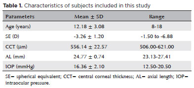
PURPOSE: To describe the technique and outcomes of intrastromal autologous blood injection in patients with severe corneal hydrops.
METHODS: Nineteen patients with corneal hydrops underwent intrastromal autologous blood injection. Postoperative assessments included best-corrected visual acuity and time to resolution of corneal edema
RESULTS: Corneal edema resolved within 1 week in 5 patients, within 1 month in 11, and within 3 months in 3. The mean duration of edema persistence was 37.94 ± 33.05 days (range, 6–124). Corneal thickness decreased from 2.06 ± 0.71-mm preoperatively to 1.34 ± 0.65-mm at day 7, 0.85 ± 0.56-mm at day 30, and 0.57 ± 0.13-mm at day 90 (p<0.001). Descemet’s membrane (DM) detachment decreased from 1.01 ± 0.75-mm to 0.44 ± 0.57-mm, 0.24 ± 0.36-mm, and 0.08 ± 0.11-mm on postoperative days 7, 30, and 90, respectively (p<0.001). DM break size decreased from 1.12 ± 1.19-mm to 0.62 ± 0.84-mm at 3 months (p<0.005). Three patients developed hematocornea; no other major complications were observed. At 3 months, mean best-corrected visual acuity improved from 2.37 ± 0.66 to 0.41 ± 0.17 logMAR with hard contact lenses (p<0.001).
CONCLUSIONS: Intrastromal autologous blood injection is an effective treatment for severe corneal hydrops, promoting faster edema resolution and visual improvement with minimal complications.
Keywords: Corneal edema; Corneal diseases; Edema; Visual acuity; keratoconus.
Arq. Bras. Oftalmol. 2025;88 (5 )
:1-9
| DOI: 10.5935/0004-2749.2024-0326
Abstract
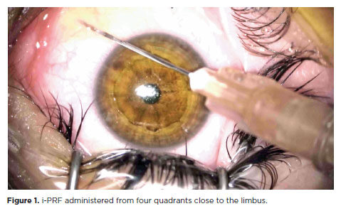
PURPOSE: This study was conducted to investigate the effect of injectable platelet-rich fibrin on the recovery of compromised epithelium due to crosslinking treatment.
METHODS: In this comparative study, the epithelial closure rates and in vivo confocal biomicroscopy results of 26 patients with keratoconus who underwent subconjunctival injection of injectable platelet-rich fibrin near the limbus after epithelium-off corneal crosslinking treatment were compared with those of 25 patients who did not receive the injection of injectable platelet-rich fibrin.
RESULTS: The average time to epithelial defect closure in the injectable platelet-rich fibrin group was 2.76 ± 0.90 days compared to 3.56 ± 0.86 days in the non-injectable platelet-rich fibrin group (p=0.003). At the end of the 1st month, the mean subbasal nerve plexus density was 1.26 ± 1.61 nerves/mm2 in the injectable platelet-rich fibrin group, whereas it was 0.72 ± 0.89 nerves/mm2 in the non-injectable platelet-rich fibrin group (p=0.016). By the 3rd month, the density increased to 3.42 ± 1.13 nerves/mm2 in the injectable platelet-rich fibrin group and 2.36 ± 1.15 nerves/mm2 in the non-injectable platelet-rich fibrin group (p=0.002). Similarly, the anterior stromal keratocyte density at the end of the 1st month was 93.6 ± 33.5 cells/mm2 in the injectable platelet-rich fibrin group compared to 67.3 ± 26.4 cells/mm2 in the non-injectable platelet-rich fibrin group (p=0.001). By the end of the 3rd month, the density increased to 255.2 ± 45.7 cells/mm2 in the injectable platelet-rich fibrin group and 222.1 ± 43.6 cells/mm2 in the non-injectable platelet-rich fibrin group (p=0.011). In the non-injectable platelet-rich fibrin group, one patient developed a sterile infiltrate at the end of the 1st week, whereas no complications were observed in the injectable platelet-rich fibrin group.
CONCLUSION: Subconjunctival injectable platelet-rich fibrin application is an effective and safe method for corneal epithelial healing after crosslinking treatment.
Keywords: Keratoconus; Platelet-rich fibrin; Epithelium; corneal; Corneal crosslinking; Wound healing
Arq. Bras. Oftalmol. 2024;87 (3 )
:1-7
| DOI: 10.5935/0004-2749.2023-0049
Abstract
PURPOSE: To investigate the association of pre-photorefractive keratectomy Schirmer-1 test value with post-photorefractive keratectomy central corneal epithelial thickness, ocular surface disease index score, and uncorrected distance visual acuity.
METHODS: Patients were categorized according to preoperative Schirmer-1 value: the normal Schirmer Group (n=54; Schirmer-1 test value, >10 mm) and the low Schirmer Group (n=52; Schirmer-1 test value, between 6 and 10 mm). We analyzed ablation depth, visual acuity, result of Schirmer-1 test (with anesthesia), tear film break-up time, ocular surface disease index score, central corneal epithelial thickness, and spherical equivalent refraction.
RESULTS: We found significant differences between the groups in Schirmer-1 test value, tear film break-up time, and ocular surface disease index score, both preoperatively and postoperatively (p<0.001). The preoperative central corneal epithelial thicknesses of the two groups were similar (p>0.05). After photorefractive keratectomy, the Schirmer-1 test value and spherical equivalent refraction decreased in both groups (p<0.05), and ocular surface disease index scores and central corneal epithelial thickness values increased in the low Schirmer Group (p<0.001) but not in the normal Schirmer Group (p>0.05). The postoperative central corneal epithelial thicknesses of the low Schirmer Group were significantly higher than those of the normal Schirmer Group (p<0.001). Postoperative uncorrected distance visual acuity did not differ significantly between the two groups (p>0.05).
CONCLUSIONS: In patients with low Schirmer-1 test values before photorefractive keratectomy, the corneal epithelium thickened and ocular surface complaints increased during the postoperative period. However, changes in the corneal epithelium did not affect the postoperative uncorrected distance visual acuity. To reduce postoperative problems on the ocular surface in these patients, we recommend that dry eye be treated before photorefractive keratectomy.
Keywords: Epithelium, corneal; Cornea; Photorefractive keratectomy; Schirmer test; Visual acuity
Arq. Bras. Oftalmol. 2024;87 (4 )
:1-8
| DOI: 10.5935/0004-2749.2023-0144
Abstract
PURPOSE: To assess the outcomes of deep anterior lamellar keratoplasty or penetrating keratoplasty at the scar and the edema stages.
METHODS: Forty-five patients (45 eyes) with keratoconus scar stage (scar group, n=26; penetrating keratoplasty a subgroup, n=7; deep anterior lamellar keratoplasty b subgroup, n=19) and keratoconus edema stage (edema group, n=19; penetrating keratoplasty c subgroup, n=12; deep anterior lamellar keratoplasty d group, n=7) who received penetrating keratoplasty or deep anterior lamellar keratoplasty from 2000 to 2022 were retrospectively studied. At 1, 6, and 12 months after surgery, the best-corrected visual acuity, astigmatism, spherical equivalent, corneal endothelial cell density, and complications were analyzed.
RESULTS: The best-corrected visual acuity and average corneal endothelial cell loss rate were not significantly different between the scar and edema groups (p>0.05). At 6 and 12 months after surgery, the astigmatism and spherical equivalent in the scar group were significantly lower than those in the edema group (p<0.05). The spherical equivalent of the deep anterior lamellar keratoplasty b subgroup was lower than that of the penetrating keratoplasty a subgroup in the scar group 6 months after surgery (p<0.05). In the edema group, there was no significant difference in spherical equivalent between subgroups (p>0.05). There were no significant differences in best-corrected visual acuity and astigmatism between subgroups within the two groups (p>0.05). In comparison to the scar group, the edema group experienced more complications. According to a survival analysis, there was no statistically significant difference between the scar group and the edema group regarding the progression of vision.
CONCLUSIONS: In terms of the outcomes and prognosis for vision after keratoplasty with edema stage and scar stage, deep anterior lamellar keratoplasty may be as effective as penetrating keratoplasty.
Keywords: keratoconus; Edema; Cicatrix; keratoplasty, penetrating; Corneal transplantation; Astigmatism; Corneal endothelial cell loss; Endothelial cells
Arq. Bras. Oftalmol. 2024;87 (2 )
:1-7
| DOI: 10.5935/0004-2749.2022-0084
Abstract
OBJETIVOS: Foram estudadas cinco técnicas de cultivo primário de células epiteliais de córnea humana para se determinar o melhor protocolo para a obtenção do maior rendimento de meio de cultivo condicionado para ser utilizado na diferenciação de células tronco mesenquimais para células epiteliais de córnea.
MÉTODOS: As técnicas de cultivo estudadas foram: explantes em frascos de cultivo com e sem tratamento hidrofílico de superfície, sobre membrana amniótica, com digestão enzimática e por raspado de córnea. O meio de cultivo condicionado foi coletado e as células tronco mesenquimais induzidas a se diferenciarem em células epiteliais da córnea utilizando o meio de cultivo condicionado. As células foram caracterizadas por citometria de fluxo com pan-citoqueratina e com os marcadores específicos da córnea, citoqueratina 3 e citoqueratina 12.
RESULTADOS: A técnica utilizando frascos com o tratamento de superfície apresentou o maior rendimento de meio de cultivo condicionado. Os frascos sem tratamento de superfície levaram a uma taxa de sucesso muito baixa. A digestão enzimática e a raspagem da córnea mostraram contaminação das culturas com fibroblastos de córnea. A cultura sobre membranas amnióticas só permitiu a coleta do meio de cultivo condicionado durante a 1ª confluência celular. A análise de citometria de fluxo confirmou o sucesso da diferenciação celular utilizando o meio de cultivo condicionado coletado, demonstrada pela expressão de citoqueratina 3 (95,3%), citoqueratina 12 (93,4%) e pan-citoqueratina (95,3%).
CONCLUSÃO: O cultivo de explantes de células tronco mesenquimais em frascos com tratamento hidrofílico de superfície é a melhor técnica para a obtenção de um alto rendimento de meio de cultivo condicionado.
Keywords: Cultivo de células; Células tronco mesenquimais; Diferenciação celular; Células epiteliais; Córnea; Meio de cultivo condicionado; Técnicas de cultivo
 ABO is licensed under a Creative Commons Attribution-NonComercial 4.0 Internacional.
08-fig01.jpg)
03-tab01.jpg)


14-fig01.jpg)
02-fig01.jpg)
01-fig01.jpg)