Arq. Bras. Oftalmol. 2023;86 (6 )
:1-8
| DOI: 10.5935/0004-2749.2021-0105
Abstract
Objetivo: Avaliar a prática e tratamento da ptose da pálpebra superior por membros das sociedades latino-americanas e espanhola de Cirurgia Plástica Ocular.
Métodos: Os membros das referidas sociedades foram convidados por e-mail para responder a um questionário eletrônico garantindo o anonimato. O questionário constou de dados demográficos do cirurgião e outras quatro seções: avaliação pré-operatória da ptose da pálpebra superior, preferências cirúrgicas, conduta pós-operatória e complicações. Estatística descritiva foi utilizada para análise da frequência e proporções percentuais.
Resultados: Trezentos e cinquenta e quatro experientes cirurgiões oculoplásticos dos quais 47,7% realizam mais de 20 cirurgias de ptose da pálpebra superior por ano responderam ao questionário. Na avaliação pré-operatória, 68,9% realizam testes para olho seco, mas o teste da fenilefrina é feito por menos da metade dos entrevistados (47,4%). A ptose da pálpebra superior leve geralmente é corrigida por conjuntivo-mullerectomia (43,6%), a ptose da pálpebra superior grave por cirurgia do músculo frontal (57%) ou ressecção da aponeurose do levantador via anterior, principalmente usando a supramáxima (17,5%). O principal motivo para operar a ptose congênita grave é o risco de ambliopia (37,3%). A ptose involucional associada à dermatocálase costuma ser corrigida pela via anterior (63,3%). Hipocorreção é complicação comum após a ressecção da aponeurose do levantador (40%) ou suspensão ao frontal (27,5%).
Conclusões: As práticas atuais dos cirurgiões oculoplásticos espanhóis e latino-americanos para diagnóstico e tratamento de ptose da pálpebra superior foram relatadas. Os dados apresentados podem ser usados para comparar a abordagem dos cirurgiões com a de seus pares.
Keywords: Blefaroptose/diagnóstico; Ambliopia; Fenillefrina; Inquéritos e questionários; Demografia; Cirurgiões.
Arq. Bras. Oftalmol. 2022;85 (6 )
:599-605
| DOI: 10.5935/0004-2749.20220082
Abstract
Objetivo: Avaliar as características clínicas de pacientes pediátricos com blefaroptose adquirida unilateral, transitória e de início agudo.
Métodos: Neste estudo retrospectivo, foram revisados prontuários clínicos entre abril de 2015 e junho de 2020. Os pacientes foram avaliados em termos de características demográficas, manifestações neurológicas e oftalmológicas associadas, duração dos sintomas, etiologia e achados de imagem. Foram excluídos pacientes com blefaroptose congênita e com blefaroptose adquirida de etiologia crônica.
Resultados: Foram incluídos neste estudo 16 pacientes pediátricos (10 masculinos e 6 femininos) com blefaroptose adquirida transitória unilateral de início agudo. A média de idade dos pacientes foi de 6,93 ± 3,16 anos. As causas etiológicas mais comumente identificadas foram trauma em 7 pacientes (43,75%) e infecção (casos parainfecciosos) em 5 pacientes (31,25%). Além disso, a síndrome de Miller-Fisher, a síndrome de Horner secundária a neuroblastoma, a síndrome de Brown adquirida e pseudotumor cerebral foram determinados como causas etiológicas em um paciente cada uma. Achados oculares adicionais estavam associados à blefaroptose em 7 pacientes (58,33%). Foi observada a resolução espontânea da blefaroptose, sem tratamento, em todos os pacientes, exceto nos pacientes com síndrome de Miller-Fisher, neuroblastoma e pseudotumor cerebral. Nenhum paciente precisou de tratamento cirúrgico. Morbidades oculares, como ambliopia, não foram encontradas em nenhum paciente.
Conclusão: Este estudo demonstrou que a blefaroptose transitória unilateral de início agudo, rara na infância, pode regredir sem a necessidade de tratamento cirúrgico na população pediátrica. No entanto, também não deve ser esquecido que patologias graves que requerem tratamento podem se apresentar com blefaroptose.
Keywords: Blefaroptose; Trauma craniocerebral; Síndrome de Miller Fisher; Síndrome de Horner; Criança
Arq. Bras. Oftalmol. 2023;86 (3 )
:1-7
| DOI: 10.5935/0004-2749.20230020
Abstract
Objetivos: Blefaroptose e estrabismo podem ser coexistentes em adultos e ambos afetam a aparência estética e o domínio psicossocial. Ambos também geralmente requerem cirurgia, realizada tradicionalmente em uma abordagem sequencial. O objetivo do presente estudo foi avaliar a eficácia da execução simultânea da ressecção musculoconjuntival de Müller, com ou sem cirurgia de tarsectomia, e da cirurgia de estrabismo em pacientes adultos com ptose e estrabismo coexistentes.
Métodos: Foram retrospectivamente avaliados pacientes com ptose e estrabismo coexistentes submetidos simultaneamente à ressecção musculoconjuntival de Müller, com ou sem tarsectomia, e à cirurgia de estrabismo horizontal. A análise incluiu a mensuração do ângulo de desvio das dioptrias de prisma, a distância do reflexo à margem, a assimetria da altura palpebral e quaisquer complicações após a cirurgia. A ressecção musculoconjuntival de Müller, com ou sem sucesso na tarsectomia, foi considerada bem-sucedida com uma distância reflexo-margem medindo entre 3,5 e 5 mm, e uma diferença entre as duas pálpebras superiores menor que 1 mm. O sucesso da cirurgia de estrabismo foi definido como um alinhamento com ± 10 dioptrias prismáticas de ortotropia.
Resultados: Os pacientes foram 3 mulheres e 5 homens, com média de idade de 37,12 anos (faixa de 22 a 62 anos). A parte de estrabismo da cirurgia foi realizada primeiro em todos os pacientes. Os resultados da simetria palpebral superior foram avaliados como perfeitos (<0,5 mm) em 4 pacientes, bons (≥0,5 mm, <1 mm) em 4 pacientes e regulares (≥1 mm) em nenhum. A ressecção musculoconjuntival de Müller, com ou sem sucesso na tarsectomia, teve sucesso em 6 dos 8 pacientes (75%) e a intervenção para o estrabismo foi bem-sucedida em todos os pacientes. Não foi necessária cirurgia de revisão da pálpebra ou do estrabismo após a cirurgia simultânea em nenhum paciente.
Conclusões: A ressecção musculoconjuntival de Müller, com ou sem tarsectomia, pode ser combinada com a cirurgia de estrabismo em uma abordagem alternativa para pacientes com ptose e estrabismo coexistentes.
Keywords: Blefaroptose/cirurgia; Ambliopia; Estrabismo/cirurgia; Músculos oculomotores/cirurgia; Pálpebras; Procedimentos cirúrgicos oftalmológicos/métodos
Arq. Bras. Oftalmol. 2025;88 (4 )
:1-6
| DOI: 10.5935/0004-2749.2024-0278
Abstract
PURPOSE: This study aimed to evaluate the prevalence of orbital conditions in a tertiary ophthalmic outpatient hospital in Sao Paulo, Brazil, with a focus on the main diagnoses and their distribution.
METHODS: A retrospective chart review was conducted involving patients registered and admitted to the orbital disease unit at the Department of Ophthalmology, University of São Paulo Medical School, from January 2004 to March 2018. A total of 838 medical charts were analyzed, of which 37 were excluded due to incomplete data. The remaining charts were categorized into eight diagnostic groups: Graves’ orbitopathy , inflammatory disorders, tumors, vascular lesions, acquired structural abnormalities, congenital structural abnormalities, infectious diseases, and others.
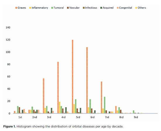
RESULTS: Of the 837,300 ophthalmological appointments, 3,372 (0.4%) were related to orbital diseases. The study included 801 patients, of whom 63.45% were women. The patients’ mean age was 42.86 years. Graves’ orbitopathy was the most common (55%), followed by tumor (17%), inflammatory disorders (9%), vascular lesions (7%), acquired structural abnormalities (5%), congenital structural abnormalities (4%), others (2%), and infectious diseases (1%). The study found significant differences in the incidence and types of orbital diseases, indicating the specialized nature of tertiary care and referral biases.
CONCLUSION: Published data on epidemiological orbital diseases is scarce. Therefore, this study focused on the diverse nature of orbital diseases and their low incidence among ophthalmology appointments. The major trends align with other epidemiological studies, demonstrating a preponderance of Graves’ orbitopathy in middle-aged adults and a bimodal distribution of tumors. These findings are essential in shaping resident training programs and healthcare policies, particularly in tertiary settings. Understanding the epidemiology of orbital diseases can improve diagnostic accuracy, treatment approaches, and patient outcomes as well as support future systemic prospective studies.
Keywords: Orbital diseases; Orbital tumors; Neoplasms; Inflammation; Graves’ ophthalmopathy; Outpatients
Arq. Bras. Oftalmol. 2026;89 (5 )
:1-8
| DOI: 10.5935/0004-2749.2026-0041
Abstract
PURPOSE: To evaluate the efficacy of autologous fat grafting for treating enophthalmos in anophthalmic sockets.
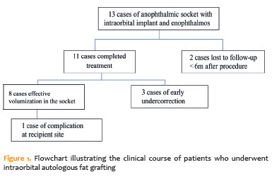
METHODS: In this prospective interventional case series, consecutive patients with anophthalmic sockets and enophthalmos were enrolled between August 2013 and July 2025. Clinical ophthalmologic examination, Hertel exophthalmometry, and orbital computed tomography scans were performed. Enophthalmos was assessed clinically and radiologically before and after orbital lipofilling with autologous fat. Fat was harvested from the inner thigh and injected into the inferolateral orbital compartment. The volume of injected fat was determined according to the degree of enophthalmos measured by Hertel exophthalmometry. Follow-up ranged from 2 to 60 months (median, 7 months).
RESULTS: Thirteen patients (13 sockets) underwent orbital autologous fat grafting using Coleman's technique. The injected fat volume ranged from 3.7 to 6 mL per procedure (median, 4.2 mL). Clinically, a reduction in enophthalmos of 1–5 mm was observed in 12 cases (median, 2 mm), which was associated with a decrease in prosthesis thickness. One patient developed a fat cyst due to anterior fat migration, and three patients required repeat grafting because of undercorrection. Computed tomography scans, available for nine patients, demonstrated a 6.8%–10.8% increase in intraorbital volume, particularly in the inferolateral quadrant. Conclusions: Clinical and radiological assessments demonstrated that lipofilling is an effective technique for restoring orbital volume in anophthalmic sockets with enophthalmos.
Keywords: Anophthalmos; Enophthalmos; Orbital volumetry; Fat grafting; Orbital reconstruction
Arq. Bras. Oftalmol. 2025;88 (2 )
:1-7
| DOI: 10.5935/0004-2749.2024-0029
Abstract
PURPOSE: To evaluate the effect of upper eyelid ptosis repair with Muller muscle-conjunctival resection on meibomian gland function and ocular surface parameters.
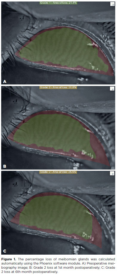
METHODS: Thirty-eight patients who underwent ptosis repair with Muller muscle-conjunctival resection were retrospectively reviewed. Meibomian gland loss, Ocular Surface Disease Index OXFORD score, meiboscore, and noninvasive keratograph break-up time were measured preoperatively and at 1st, 3rd, and 6th months postoperatively.
RESULTS: Noninvasive keratograph break-up time values decreased significantly at 1st and 3rd months postoperatively compared to the preoperative level, but were similar to the preoperative level at 6th months postoperatively (p<0.001 and p=0.628, respectively). Ocular surface disease index, OXFORD score, meibomian gland loss, and meiboscore values increased significantly in the 1st and 3rd postoperative months compared to the preoperative period, but these values decreased to preoperative levels in the 6th postoperative month (p<0.001 and p>0.05, respectively).
CONCLUSION: There is a transient deterioration in meibography findings and OSDI score in the early postoperative period after Muller muscle-conjunctival resection. Patients undergoing Muller muscle-conjunctival resection may require topical lubricants, especially in the first 3 postoperative months.
Keywords: Meibomian glands; Blepharoptosis; Preoperative period; Conjunctiva; Muscles; Eyelid diseases; Diagnostic techniques, ophthalmological
Arq. Bras. Oftalmol. 2025;88 (5 )
:1-7
| DOI: 10.5935/0004-2749.2024-0318
Abstract
PURPOSE: Ptosis is characterized by drooping of the upper eyelid, often requiring surgical intervention for functional and aesthetic purposes. Müller’s muscle conjunctival resection is a commonly utilized surgical technique to correct mild to moderate ptosis. This retrospective study aimed to evaluate the impact of Hering’s law on the outcomes of unilateral Müller’s muscle conjunctival resection surgery, particularly eyelid and brow symmetry.
METHODS: Thirty patients with unilateral ptosis underwent Müller’s muscle conjunctival resection. Pre- and postoperative assessments included ipsilateral and contralateral side margin-reflex distance and brow position, measured through digital image analysis.
RESULTS: We found significant improvements in postoperative margin-reflex distance measurements in the ipsilateral eyelid but not in the contralateral eyelid, indicating minimal influence of Hering’s law. Brow position showed a statistically significant increase on the contralateral side but not on the ipsilateral side.
CONCLUSION: Müller’s muscle conjunctival resection effectively restores symmetry in eyelid height and maintains brow symmetry. This is the first study to explore bilateral eyelid and brow symmetry after unilateral Müller’s muscle conjunctival resection surgery for mild to moderate ptosis. Further research should be conducted to understand the long-term effects of Müller’s muscle conjunctival resection on facial aesthetics, particularly in relation to brow position.
Keywords: Müller muscle conjunctiva resection; Hering’s law; Eyelids; Blepharoptosis; Reflex; Oculomotor muscles
Arq. Bras. Oftalmol. 2024;87 (3 )
:1-7
| DOI: 10.5935/0004-2749.2023-0028
Abstract
PURPOSE: Evaluation of lid contour and marginal peak point changes to compare outcomes of external levator advancement and Müller’s muscle conjunctival resection surgery in unilateral ptosis.
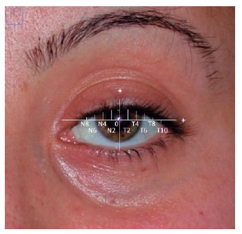
METHODS: We reviewed the charts of unilateral ptosis patients who underwent external levator advancement or Müller’s muscle conjunctival resection. Eyelid contour analysis was conducted on preoperative and 6-month postoperative digital images. This was performed with the multiple margin reflex distances technique, measuring the vertical distance from a line intersecting the center of the pupil to the eyelid margin at 10 positions at 2 mm intervals. The marginal peak point changes were analyzed digitally using the coordinates of the peak point according to the pupil center. Each position’s mean distance was compared preoperatively, postoperatively, and with the fellow eyelid.
RESULTS: Sixteen patients underwent external levator advancement and 16 patients had Müller’s muscle conjunctival resection. The mean margin reflex distance was improved by both techniques (1.46 vs. 2.43 mm and 1.12 vs. 2.25 mm, p=0.008 and p=0.0001 respectively) and approached that of the fellow eyelid (2.43 vs. 2.88 and 2.25 vs. 2.58 mm, p=0.23 and p=0.19, respectively). However, statistically significant lid margin elevation was limited to between the N6 and T6 points in the external levator advancement group. Whereas, significant elevation was achieved along the whole lid margin in the Müller’s muscle conjunctival resection group. The marginal peak point was shifted slightly laterally in the external levator advancement group (p=0.11).
CONCLUSIONS: Both techniques provide effective lid elevation, however, the external levator advancement’s effect lessens toward the canthi while Müller’s muscle conjunctival resection provides more uniform elevation across the lid margin. The margin reflex distance alone is not sufficient to reflect contour changes.
Keywords: Blepharoptosis; Eyelids; Conjunctiva; Oculomotor muscles; Image processing, computer-assisted; Treatment outcome


 ABO is licensed under a Creative Commons Attribution-NonComercial 4.0 Internacional.


09-fig01tb.jpg)
07-tab01tb.jpg)
06-tab01tb.jpg)

11-fig01.jpg)
12-fig01.jpg)
02-fig01.jpg)