Arq. Bras. Oftalmol. 2024;87 (3 )
:1-8
| DOI: 10.5935/0004-2749.2022-0076
Abstract
MÉTODOS: Córneas humanas de treinamento disponibilizadas foram randomizadas em quatro grupos: Pachy-100 (profundidade de incisão = espessura corneana central - margem de segurança de 100 µm), Pachy-50 (margem de segurança de 50 µm), Pachy-0 (sem margem de segurança) e Pachy+50 (profundidade de incisão = espessura corneana central + 50 µm). Todas as lamelas foram dissecadas através um método padronizado e já publicado (Pachy-DSEK). As espessuras das lamelas (centro, zona de 3,0mm e zona de 6,0mm) foram medidas com tomografia de coerência óptica. A razão de espessura centro-periferia foi calculada aos 3,0 e 6,0 mm de diâmetro.
RESULTADOS: Perfuração endotelial ocorreu apenas no grupo Pachy+50 (n=3, 30%). A espessura central da lamela nos grupos Pachy-100, Pachy-50, Pachy-0 e Pachy+50 foi de 185 ± 42 µm, 122 ± 29 µm, 114 ± 29 µm, e 58 ± 31 µm, respectivamente (p<0,001). As razões C/P aos 3,0 e 6,0 mm foram de 0,97 ± 0,06 e 0,92 ± 0,14, respectivamente. Os parâmetros de características do doador não se correlacionaram com os resultados de espessura de lamela. A profundidade planejada de incisão se correlacionou com a maioria dos parâmetros de espessura de lamela (p<0,001). A espessura de lamela se correlacionou negativamente com a profundidade planejada da incisão (p<0.001, r=-0,580). O melhor resultado foi observado no grupo Pachy-0, em que 75% das lamelas mediram abaixo de 130 µm e não houve perfuração endotelial.
CONCLUSÃO: Através de um método padronizado de dissecção, a maioria das lamelas endoteliais apresentou uma configuração planar. O planejamento de profundidade de incisão igual à espessura corneana central resultou em alta porcentagem de lamelas ultrafinas sem ocorrência de perfuração.
Keywords: Transplante de córnea; Ceratoplastia lamelar; Endotélio corneano; Dissecção; Tomografia de coerência óptica
Arq. Bras. Oftalmol. 2026;89 (4 )
:1-9
| DOI: 10.5935/0004-2749.2025-0034
Abstract
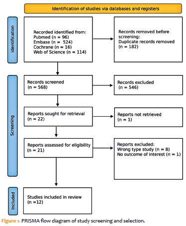
PURPOSE: To evaluate the impact of the COVID-19 pandemic and characterize the serological profile of discarded corneal donations in the coverage area of the Banco de Olhos de Londrina, through reverse transcription-polymerase chain reaction testing for COVID-19 and serological screening of cornea donors excluded because of positive test results.
METHODS: This observational retrospective study included 776 cornea donors who’s serological and reverse transcription-polymerase chain reaction test results were processed at the Hospital of Universidade Estadual de Londrina between May 2020 and 2022. The number of corneal donations and tissue utilization rates throughout the years of operation of the Banco de Olhos de Londrina were also analyzed.
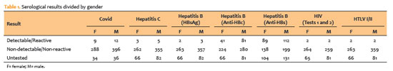
RESULTS: The mean donor age was 53.14 years; 332 donors (43%) were female, and 444 (57%) were male. Positive results were identified in 15.76% of donors for hepatitis B core antibody antibodies, 0.65% for hepatitis B surface antigen, 1.03% for hepatitis C antibodies, and 0.52% for human immunodeficiency virus and human T-lymphotropic vírus. Positive reverse transcription-polymerase chain reaction results for SARS-CoV-2 were observed in 2.7% of cases. Older adults were 2.6 times more likely to test positive for SARS-CoV-2 (95% CI, 1.06-6.34) and 3.0 times more likely to test positive for hepatitis B core antibody (95% CI, 1.95-4.41) than younger individuals. A 75.2% reduction in corneal donations was observed in 2020 compared with 2019, accompanied by a 5% increase in tissue utilization, possibly associated with the effectiveness of donor screening during the pandemic.
CONCLUSION: The COVID-19 pandemic had a profound impact on the number of corneal transplants worldwide, in Brazil, and at the Banco de Olhos de Londrina because of the substantial decline in donations during this period. Hepatitis B was the leading cause of corneal tissue discard due to positive serology in both this study and previous reports, highlighting the importance of prevention programs and improved vaccination coverage. Strict legislation, comprehensive serological screening, and appropriate processing of donated tissue remain essential to eliminate potential sources of infection and ensure transplantation safety.
Keywords: Cornea; Corneal transplantation; COVID-19; Eye banks; Serology
Arq. Bras. Oftalmol. 2025;88 (5 )
:1-9
| DOI: 10.5935/0004-2749.2024-0326
Abstract
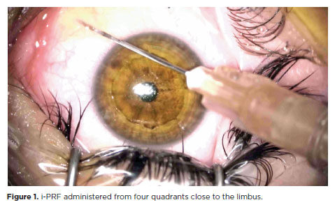
PURPOSE: This study was conducted to investigate the effect of injectable platelet-rich fibrin on the recovery of compromised epithelium due to crosslinking treatment.
METHODS: In this comparative study, the epithelial closure rates and in vivo confocal biomicroscopy results of 26 patients with keratoconus who underwent subconjunctival injection of injectable platelet-rich fibrin near the limbus after epithelium-off corneal crosslinking treatment were compared with those of 25 patients who did not receive the injection of injectable platelet-rich fibrin.
RESULTS: The average time to epithelial defect closure in the injectable platelet-rich fibrin group was 2.76 ± 0.90 days compared to 3.56 ± 0.86 days in the non-injectable platelet-rich fibrin group (p=0.003). At the end of the 1st month, the mean subbasal nerve plexus density was 1.26 ± 1.61 nerves/mm2 in the injectable platelet-rich fibrin group, whereas it was 0.72 ± 0.89 nerves/mm2 in the non-injectable platelet-rich fibrin group (p=0.016). By the 3rd month, the density increased to 3.42 ± 1.13 nerves/mm2 in the injectable platelet-rich fibrin group and 2.36 ± 1.15 nerves/mm2 in the non-injectable platelet-rich fibrin group (p=0.002). Similarly, the anterior stromal keratocyte density at the end of the 1st month was 93.6 ± 33.5 cells/mm2 in the injectable platelet-rich fibrin group compared to 67.3 ± 26.4 cells/mm2 in the non-injectable platelet-rich fibrin group (p=0.001). By the end of the 3rd month, the density increased to 255.2 ± 45.7 cells/mm2 in the injectable platelet-rich fibrin group and 222.1 ± 43.6 cells/mm2 in the non-injectable platelet-rich fibrin group (p=0.011). In the non-injectable platelet-rich fibrin group, one patient developed a sterile infiltrate at the end of the 1st week, whereas no complications were observed in the injectable platelet-rich fibrin group.
CONCLUSION: Subconjunctival injectable platelet-rich fibrin application is an effective and safe method for corneal epithelial healing after crosslinking treatment.
Keywords: Keratoconus; Platelet-rich fibrin; Epithelium; corneal; Corneal crosslinking; Wound healing
Arq. Bras. Oftalmol. 2024;87 (3 )
:1-7
| DOI: 10.5935/0004-2749.2023-0049
Abstract
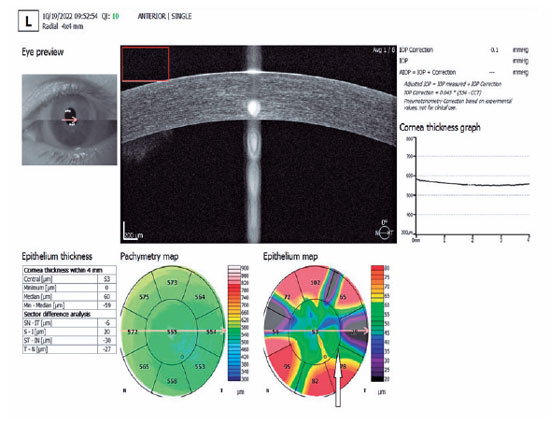
PURPOSE: To investigate the association of pre-photorefractive keratectomy Schirmer-1 test value with post-photorefractive keratectomy central corneal epithelial thickness, ocular surface disease index score, and uncorrected distance visual acuity.
METHODS: Patients were categorized according to preoperative Schirmer-1 value: the normal Schirmer Group (n=54; Schirmer-1 test value, >10 mm) and the low Schirmer Group (n=52; Schirmer-1 test value, between 6 and 10 mm). We analyzed ablation depth, visual acuity, result of Schirmer-1 test (with anesthesia), tear film break-up time, ocular surface disease index score, central corneal epithelial thickness, and spherical equivalent refraction.
RESULTS: We found significant differences between the groups in Schirmer-1 test value, tear film break-up time, and ocular surface disease index score, both preoperatively and postoperatively (p<0.001). The preoperative central corneal epithelial thicknesses of the two groups were similar (p>0.05). After photorefractive keratectomy, the Schirmer-1 test value and spherical equivalent refraction decreased in both groups (p<0.05), and ocular surface disease index scores and central corneal epithelial thickness values increased in the low Schirmer Group (p<0.001) but not in the normal Schirmer Group (p>0.05). The postoperative central corneal epithelial thicknesses of the low Schirmer Group were significantly higher than those of the normal Schirmer Group (p<0.001). Postoperative uncorrected distance visual acuity did not differ significantly between the two groups (p>0.05).
CONCLUSIONS: In patients with low Schirmer-1 test values before photorefractive keratectomy, the corneal epithelium thickened and ocular surface complaints increased during the postoperative period. However, changes in the corneal epithelium did not affect the postoperative uncorrected distance visual acuity. To reduce postoperative problems on the ocular surface in these patients, we recommend that dry eye be treated before photorefractive keratectomy.
Keywords: Epithelium, corneal; Cornea; Photorefractive keratectomy; Schirmer test; Visual acuity
Arq. Bras. Oftalmol. 2024;87 (3 )
:1-8
| DOI: 10.5935/0004-2749.2023-0109
Abstract
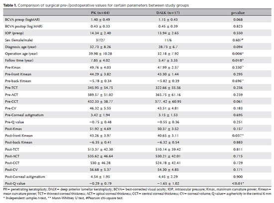
PURPOSES: This study aims to assess and compare the postoperative visual and topographic outcomes, complications, and graft survival rates following deep anterior lamellar keratoplasty and penetrating keratoplasty in patients with macular corneal dystrophy.
METHODS: In this study we enrolled 59 patients (23 male; and 36 female) with macular corneal dystrophy comprising 81 eyes. Out of these, 64 eyes underwent penetrating keratoplasty, while 17 eyes underwent deep anterior lamellar keratoplasty. The two groups were analyzed and compared based on best-corrected visual acuity, corneal tomography parameters, pachymetry, complication rates, and graft survival rates.
RESULTS: After 12 months, 70.6% of the patients who underwent deep anterior lamellar keratoplasty (DALK) and 75% of those who had penetrating keratoplasty (PK) achieved a best-corrected visual acuity of 20/40 or better (p=0.712). Following surgery, DALK group showed lower front Kmean (p=0.037), and Q values (p<0.01) compared to the PK group. Postoperative interface opacity was observed in seven eyes (41.2%) in the DALK group. Other topography values and other complications (graft rejection, graft failure, cataract, glaucoma, microbial keratitis, optic atrophy) did not show significant differences between the two groups. The need for regrafting was 9.4% and 11.8% in the PK and DALK groups, respectively (p=0.769). Graft survival rates were 87.5% and 88.2% for PK and DALK; respectively (p=0.88 by Log-rank test).
CONCLUSION: Both PK and DALK are equally effective in treating macular corneal dystrophy, showing similar visual, topographic, and survival outcomes. Although interface opacity occurs more frequently after DALK the visual results were comparable in both groups. Therefore, DALK emerges as a viable surgical choice for patients with macular corneal dystrophy without Descemet membrane involvement is absent.
Keywords: Macular corneal dystrophy; Corneal dystrophies; Hereditary; Keratoplasty; Penetrating; Corneal transplantation
Arq. Bras. Oftalmol. 2024;87 (4 )
:1-8
| DOI: 10.5935/0004-2749.2023-0144
Abstract
PURPOSE: To assess the outcomes of deep anterior lamellar keratoplasty or penetrating keratoplasty at the scar and the edema stages.
METHODS: Forty-five patients (45 eyes) with keratoconus scar stage (scar group, n=26; penetrating keratoplasty a subgroup, n=7; deep anterior lamellar keratoplasty b subgroup, n=19) and keratoconus edema stage (edema group, n=19; penetrating keratoplasty c subgroup, n=12; deep anterior lamellar keratoplasty d group, n=7) who received penetrating keratoplasty or deep anterior lamellar keratoplasty from 2000 to 2022 were retrospectively studied. At 1, 6, and 12 months after surgery, the best-corrected visual acuity, astigmatism, spherical equivalent, corneal endothelial cell density, and complications were analyzed.
RESULTS: The best-corrected visual acuity and average corneal endothelial cell loss rate were not significantly different between the scar and edema groups (p>0.05). At 6 and 12 months after surgery, the astigmatism and spherical equivalent in the scar group were significantly lower than those in the edema group (p<0.05). The spherical equivalent of the deep anterior lamellar keratoplasty b subgroup was lower than that of the penetrating keratoplasty a subgroup in the scar group 6 months after surgery (p<0.05). In the edema group, there was no significant difference in spherical equivalent between subgroups (p>0.05). There were no significant differences in best-corrected visual acuity and astigmatism between subgroups within the two groups (p>0.05). In comparison to the scar group, the edema group experienced more complications. According to a survival analysis, there was no statistically significant difference between the scar group and the edema group regarding the progression of vision.
CONCLUSIONS: In terms of the outcomes and prognosis for vision after keratoplasty with edema stage and scar stage, deep anterior lamellar keratoplasty may be as effective as penetrating keratoplasty.
Keywords: keratoconus; Edema; Cicatrix; keratoplasty, penetrating; Corneal transplantation; Astigmatism; Corneal endothelial cell loss; Endothelial cells
Arq. Bras. Oftalmol. 2024;87 (2 )
:1-7
| DOI: 10.5935/0004-2749.2022-0084
Abstract
OBJETIVOS: Foram estudadas cinco técnicas de cultivo primário de células epiteliais de córnea humana para se determinar o melhor protocolo para a obtenção do maior rendimento de meio de cultivo condicionado para ser utilizado na diferenciação de células tronco mesenquimais para células epiteliais de córnea.
MÉTODOS: As técnicas de cultivo estudadas foram: explantes em frascos de cultivo com e sem tratamento hidrofílico de superfície, sobre membrana amniótica, com digestão enzimática e por raspado de córnea. O meio de cultivo condicionado foi coletado e as células tronco mesenquimais induzidas a se diferenciarem em células epiteliais da córnea utilizando o meio de cultivo condicionado. As células foram caracterizadas por citometria de fluxo com pan-citoqueratina e com os marcadores específicos da córnea, citoqueratina 3 e citoqueratina 12.
RESULTADOS: A técnica utilizando frascos com o tratamento de superfície apresentou o maior rendimento de meio de cultivo condicionado. Os frascos sem tratamento de superfície levaram a uma taxa de sucesso muito baixa. A digestão enzimática e a raspagem da córnea mostraram contaminação das culturas com fibroblastos de córnea. A cultura sobre membranas amnióticas só permitiu a coleta do meio de cultivo condicionado durante a 1ª confluência celular. A análise de citometria de fluxo confirmou o sucesso da diferenciação celular utilizando o meio de cultivo condicionado coletado, demonstrada pela expressão de citoqueratina 3 (95,3%), citoqueratina 12 (93,4%) e pan-citoqueratina (95,3%).
CONCLUSÃO: O cultivo de explantes de células tronco mesenquimais em frascos com tratamento hidrofílico de superfície é a melhor técnica para a obtenção de um alto rendimento de meio de cultivo condicionado.
Keywords: Cultivo de células; Células tronco mesenquimais; Diferenciação celular; Células epiteliais; Córnea; Meio de cultivo condicionado; Técnicas de cultivo
Arq. Bras. Oftalmol. 2024;87 (2 )
:1-8
| DOI: 10.5935/0004-2749.2022-0328
Abstract
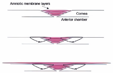
PURPOSE: Wet bio-amniotic membrane plugging combined with transplantation is a novel option that combined amniotic membrane plugging with amniotic membrane transplantation for the treatment of small corneal perforations. This study aimed to evaluate the efficacy of wet bio-amniotic membrane plugging in the treatment of small corneal perforations and compared it with that of the penetrating keratoplasty procedure.
METHODS: Forty patients (41 eyes) with small corneal perforations <3 mm in diameter treated at our hospital between July 2018 and January 2021 were retrospectively included. Among them, 21 eyes were treated with wet bio-amniotic membrane plugging (wet bio-amniotic membrane plugging group), and 20 eyes were treated with penetrating keratoplasty procedure (penetrating keratoplasty procedure group). The best-corrected visual acuity, anterior chamber formation, corneal thickness, primary disease control, postoperative complications, and graft survival rate were assessed.
RESULTS: No significant difference in baseline characteristics was found between the wet bio-amniotic membrane plugging and penetrating keratoplasty procedure groups (p>0.05). The postoperative control rates of primary diseases in the wet bio-amniotic membrane plugging and penetrating keratoplasty procedure groups were 95.2% and 90.0%, respectively (p=0.481). Visual acuity was improved 6 months after the operation in the wet bio-amniotic membrane plugging group and was improved at postoperative 1 month in the penetrating keratoplasty procedure group. The formation time of the anterior chamber in the wet bio-amniotic membrane plugging group was significantly shorter than that in the penetrating keratoplasty procedure group (p=0.023). The corneal thickness of the two groups significantly increased 12 months after the operation; however, the degree of thickening in the penetrating keratoplasty procedure group was higher than that in the wet bio-amniotic membrane plugging group (p<0.001). During the follow-up, postoperative complications were not different between the two groups (p>0.999).
CONCLUSION: The results suggest that wet bio-amniotic membrane plugging is effective and safe in the treatment of small corneal perforations. Thus, it can be used as an emergency treatment alternative to penetrating keratoplasty procedure for small corneal perforations.
Keywords: Amnion; Transplantation; Amniotic membrane; Keratoplasty, penetrating; Corneal perforation; Wet bio-amniotic membrane plugging; Wet bio-amniotic membrane transplantation
Arq. Bras. Oftalmol. 2024;87 (2 )
:1-6
| DOI: 10.5935/0004-2749.2023-2022-0341
Abstract
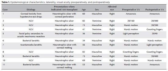
PURPOSE: To evaluate the clinical results of cryopreserved amniotic membrane transplantation as a treatment option for refractory neurotrophic corneal ulcers.
METHODS: This prospective study included 11 eyes of 11 patients who underwent amniotic membrane transplantation for the treatment of refractory neurotrophic corneal ulcers at Hospital de Clínicas da Universidade Federal do Paraná, in the city of Curitiba, from May 2015 to July 2021. Patients underwent different surgical techniques in which the amniotic membrane was applied with the epithelium facing upward to promote corneal re-epithelialization.
RESULTS: The median age of the patients was 60 years (range, 34-82 years), and 64% were men. The predominant etiology of corneal ulcers was herpes zoster (45% of cases). Approximately one-third of the patients (27%) were chronically using hypotensive eye drops, and more than half (54%) had previously undergone penetrating corneal transplantation. At the time of amniotic membrane transplantation, 18% of the eyes had corneal melting, 9% had corneal perforation, and the others had corneal ulceration without other associated complications (73%). The time between clinical diagnosis and surgical treatment ranged from 9 days to 2 years. The corrected visual acuity was worse than 20/400 in 90% of the patients preoperatively, with improvement in 36% after 3 months of the procedure, worsening in 18% and remaining stable in 36%. Of the patients, 81% complained of preoperative pain, and 66% of them reported total symptom relief after the surgical procedure. In one month, 54.6% of the patients presented a closure of epithelial defect, and half of the total group evolved with corneal thinning. The failure rate was 45.5% of the cases.
CONCLUSION: Cryopreserved amniotic membrane transplantation can be considered a good alternative for treating refractory neurotrophic corneal ulcers, as it resulted in significant improvement in pain (66%) and complete epithelial closure (60%) in many patients at 1 month postoperatively. Notably, the high failure rate highlights the need for further studies to identify patient- and ulcer-related factors that may influence the outcomes of this procedure.
Keywords: Amnion/transplantation; Corneal ulcer; Anterior eye segment; Keratitis
 ABO is licensed under a Creative Commons Attribution-NonComercial 4.0 Internacional.
02-fig01.jpg)
14-fig01.jpg)
06-tab01tb.jpg)


01-fig01.jpg)