Showing of 1 until 14 from 239 result(s)
Search for: Amnion; Stevens-Johnson syndrome; Cornea; Epithelium, corneal; Eye burns
Abstract
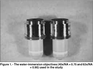
Objetivo: Este estudo prospectivo compara as imagens de microscopia confocal do epitélio corneano do coelho e do homem, obtidas através de 2 objetivas com aberturas numéricas (AN) diferentes. Métodos: Dez olhos de coelhos foram enucleados e fixados através de um suporte pneumático para garantir o melhor desempenho de cada objetiva. Cinco pacientes normais foram selecionados após consentimento. Os olhos de coelhos e dos pacientes foram previamente examinados na lâmpada de fenda. O exame de microscopia confocal (Tomey, Erlangen-Tennenlohe, Alemanha) foi realizado com as objetivas Achroplan 40x/AN = 0,75 e 63x/AN = 0,9 (Zeiss, Oberkochen, Alemanha). Imagens selecionadas do epitélio corneano foram avaliadas qualitativamente com relação ao tamanho, forma e refletividade das células. Resultados: As células no epitélio superficial dos coelhos e dos pacientes, previamente à descamação, tiveram uma refletividade maior que as células adjacentes. Este aspecto foi claramente observado somente com a objetiva 63x/AN = 0,9. As camadas basal e intermediária do epitélio em coelhos foram visualizadas somente através desta objetiva. Estas camadas nos pacientes tornaram-se mais nítidas com a objetiva de abertura numérica maior (63x/AN = 0,9). Conclusão: Uma objetiva de abertura numérica elevada produz melhor resolução dos cortes ópticos, facilitando a análise das camadas do epitélio no coelho e no homem.
Keywords: Corneal epithelium; Confocal microscopy; Epitélio corneano; Microscopia confocal
Abstract
PURPOSE: To report the ophthalmological signs, symptoms, and clinical management observed during an unprecedented outbreak of chemical ocular injuries related to cosmetic hair ointments in Brazil.
METHODS: This descriptive, cross-sectional study reviewed medical records of patients treated at the emergency center of Fundação Altino Ventura for chemical ocular trauma associated with cosmetic hair ointment use between February 2022 and February 2023. Records with incomplete medical information were excluded.
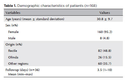
RESULTS: The study included 168 patients (95.2% [n=160] female), with a mean age of 30.8 ± 9.7 years. The most frequently reported symptoms at presentation were pain (167/168, 99.4%) and photophobia (92/168, 54.8%). Severe pain was reported by 137 patients (80%). Keratitis was present in 280 of 336 eyes (83.3%), conjunctival hyperemia in 256 eyes (76.4%), and corneal abrasions in 174 eyes (51.8%). A decrease in visual acuity (worse than 20/25) was documented in 18.5% (31/168) of cases. Lubricants, antibiotics, and re-epithelialization
ointments were prescribed to 64.8% (109/168) of the patients. Topical corticosteroids and oral vitamin C were administered to 34% (57/168) and 1.2% (2/168) of patients, respectively. Followup visits were required in 19% (33/168) of cases.
CONCLUSION: The outbreak of chemical ocular injuries linked to cosmetic ointments used for braiding and hair modeling in Brazil was marked by intense ocular pain, conjunctival hyperemia, keratitis, and corneal abrasions. Most patients were treated with lubricants, antibiotics, and re-epithelialization ointments, although approximately one-fifth required followup care, and one-third received additional treatment with either topical corticosteroids and/or oral vitamin C.
Keywords: Cosmetics; Hair preparations; Eye injuries; Burns, chemical; Eye burns; Keratitis; Cornea; Corneal diseases; Visual low.
12-fig01.jpg)
Abstract
Objetivo: O nosso objetivo neste estudo foi comparar as alterações na unidade funcional lacrimal em dois modelos de síndrome do olho seco neurogênica: desnervação sensorial da córnea versus desnervação autonômica da glândula lacrimal.
Métodos: A rede neural é um importante suporte para a unidade funcional lacrimal. Pode ser dividido em vias aferentes (sensoriais) e eferentes (autonômicas), sujeitas a doenças graves que comprometem a unidade funcional lacrimal. Ratos Wistar machos, com 8 semanas de idade, foram divididos em três grupos: 1) Controle naïve (n=16 animais); 2) Desnervação autonômica: onde os ratos foram submetidos à ablação do nervo da glândula lacrimal direita e avaliados após um e dois meses (1 M a 2 M) do procedimento (n=7 animais por subgrupo, desnervação autonômica 1M e desnervação autonômica 2M, respectivamente); 3) Desnervação sensorial induzida por colírio a 0,2% de cloreto de benzalcônio, duas vezes ao dia por 7 dias no olho direito (n=10 animais). A sensibilidade da córnea foi medida pelo teste de movimento pata-olho com capsaicina (10 μM). A PCR quantitativa em tempo real foi aplicada para comparar a expressão relativa de mRNA de citocinas pró-inflamatórias: Il1b, Il6, Tnf, Mmp9, na córnea, gânglio trigêmio e glândula lacrimal. O mRNA dos agentes pró-mitóticos Bmp7, Runx1, Runx3, Fgf10 e Smad1 foram comparados na glândula lacrimal.
Resultados: A desnervação sensorial induziu hiperalgesia da córnea (p=0,001). Desnervação sensorial e desnervação autonômica aumentaram o mRNA de citocinas pró-inflamatórias no córnea e glândula lacrimal (p<0,05), mas apenas desnervação sensorial aumentou o mRNA de Il1b e Tnf no gânglio trigêmio (p<0,05) quando comparado ao controle naïve.
Conclusões: Os modelos de desnervação autonômica e desnervação sensorial podem ter características comuns, como inflamação de diferentes partes da unidade funcional lacrimal. No entanto, a hiperestesia e os marcadores inflamatórios no gânglio trigêmio de desnervação sensorial e a expressão de mediadores regenerativos na glândula lacrimal na desnervação autonômica são características que distinguem essas doenças, podendo ser investigadas em estudos futuros que abordam o olho seco secundário ao dano neural da unidade funcional lacrimal.
Keywords: Córnea; Unidade functional lacrimal; Aparelho lacrimal; Hipersensibilidade; Ratos Wistar; Síndrome do olho seco
13-tab01tb.jpg)
Abstract
Objetivos: Explorar sistematicamente as mudanças dinâmicas e a sequência temporal no processo de apoptose de células epiteliais corneanas após excesso de irradiação com ultravioleta B.
Métodos: A radiação ultravioleta B (144 mJ/cm2) foi utilizada para irradiar células epiteliais da córnea de rato durante 2h. A morfologia celular foi observada por meio de microscópio de contraste de interferência diferencial, e os números de diferentes tipos de células apoptóticas foram contados e registrados pelo software ImageJ. A viabilidade celular foi medida pelo método brometo de 3- (4, 5-dimetil-2-tiazolil) -2, 5-difenil-2-H-tetrazólio. A taxa apoptótica celular e a perda do potencial da membrana mitocondrial foram detectadas por meio de análises citométricas de fluxo. Os níveis de expressão de três genes apoptóticos foram medidos por reação em cadeia da polimerase quantitativa em tempo real em diferentes momentos dentro de 0-24 h após a irradiação.
Resultados: Após 144 mJ/cm2 de irradiação com ultravioleta B por 2h, os níveis de expressão de caspase-8 e Bax foram maiores em 0h; o potencial da membrana mitocondrial diminuiu a 0h e permaneceu constante por 6h na cultura subsequente. Às 6h, a caspase-3 foi ativada. A diminuição da viabilidade celular e o aumento da taxa apoptótica atingiu o pico em 6h. A expressão de caspase-3 diminuiu dentro de 12 - 24 h, levando a um declínio na taxa apoptótica e alteração no estágio apoptótico.
Conclusões: As células epiteliais da córnea apresentaram uma apoptose rápida após excesso de irradiação com ultravioleta B, e esse processo foi associado tanto à via extrínseca como à via intrínseca.
Keywords: Irradiação com ultravioleta B; Radiação; Epitélio anterior; Célula epitelial; Sobrevivência celular; Apoptose; Ratos
Karolyna Andrade de Carvalho; Lucas Paolera; Bernardo Kaplan Moscovici; Luiz Antônio Brito; Luiz Felipe Ramos Bueno; Sergio Felberg
Abstract
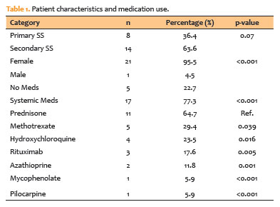
PURPOSE: The purpose of this study is to look into the relationship between tear film osmolarity, tear crystallization, and corneal esthesiometry findings in Sjögren's syndrome patients.
METHODS: This cross-sectional observational study included 43 eyes from patients with a confirmed diagnosis of Sjögren's syndrome. Tear osmolarity was measured with an iPen osmometer, tear crystallization was graded using Roland's classification, and corneal sensitivity was evaluated with a Cochet–Bonnet aesthesiometer. Ocular symptoms were assessed using the Ocular Surface Disease Index questionnaire. Patients who had undergone keratoplasty or worn contact lenses within 4 hours of testing were excluded.
RESULTS: The cohort's mean tear osmolarity was 292.5±15.0 mOsm/L (median: 293 mOsm/L, IQR: 17.5). There was no significant difference between patients with primary Sjögren's syndrome (mean: 289.4 mOsm/L) and those with secondary Sjögren's syndrome (mean: 294.5 mOsm/L; p=0.413). Tear crystallization patterns were more severe in patients with primary Sjögren's syndrome (mean: 3.25, median: 3.5, IQR: 1.25) than in those with secondary Sjögren's syndrome (mean: 3.19, median: 3.0, IQR: 1.0), though the difference was not statistically significant (p=0.87). Corneal sensitivity was reduced by 3.5±1.7 mm (median: 4.0 mm, IQR: 2.13). Tear crystallization has a significant negative correlation with corneal sensitivity (r=−0.313, p=0.041), suggesting that poorer tear quality leads to decreased corneal sensitivity.
CONCLUSION: Tear crystallization patterns and corneal sensitivity were found to be significantly correlated in Sjögren's syndrome patients. The findings also indicate that systemic medication use may affect tear film quality.
Keywords: Sjogren's syndrome, Tear crystallization, Dry eye disease, Cornea, Tears, Osmolarity
Abstract
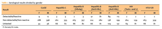
PURPOSE: To evaluate the impact of the COVID-19 pandemic and characterize the serological profile of discarded corneal donations in the coverage area of the Banco de Olhos de Londrina, through reverse transcription-polymerase chain reaction testing for COVID-19 and serological screening of cornea donors excluded because of positive test results.
METHODS: This observational retrospective study included 776 cornea donors who’s serological and reverse transcription-polymerase chain reaction test results were processed at the Hospital of Universidade Estadual de Londrina between May 2020 and 2022. The number of corneal donations and tissue utilization rates throughout the years of operation of the Banco de Olhos de Londrina were also analyzed.
RESULTS: The mean donor age was 53.14 years; 332 donors (43%) were female, and 444 (57%) were male. Positive results were identified in 15.76% of donors for hepatitis B core antibody antibodies, 0.65% for hepatitis B surface antigen, 1.03% for hepatitis C antibodies, and 0.52% for human immunodeficiency virus and human T-lymphotropic vírus. Positive reverse transcription-polymerase chain reaction results for SARS-CoV-2 were observed in 2.7% of cases. Older adults were 2.6 times more likely to test positive for SARS-CoV-2 (95% CI, 1.06-6.34) and 3.0 times more likely to test positive for hepatitis B core antibody (95% CI, 1.95-4.41) than younger individuals. A 75.2% reduction in corneal donations was observed in 2020 compared with 2019, accompanied by a 5% increase in tissue utilization, possibly associated with the effectiveness of donor screening during the pandemic.
CONCLUSION: The COVID-19 pandemic had a profound impact on the number of corneal transplants worldwide, in Brazil, and at the Banco de Olhos de Londrina because of the substantial decline in donations during this period. Hepatitis B was the leading cause of corneal tissue discard due to positive serology in both this study and previous reports, highlighting the importance of prevention programs and improved vaccination coverage. Strict legislation, comprehensive serological screening, and appropriate processing of donated tissue remain essential to eliminate potential sources of infection and ensure transplantation safety.
Keywords: Cornea; Corneal transplantation; COVID-19; Eye banks; Serology
03-tab01.jpg)
Abstract
Objetivos: Descrever as características demográficas e clínicas das vítimas de trauma ocular por fogos de artifício atendidas nas emergências oftalmológicas de dois centros de referência em Pernambuco e identificar fatores relacionados a mau prognóstico visual.
Métodos: Avaliação retrospectiva dos prontuários de pacientes admitidos na emergência oftalmológica com história de trauma por fogos de artifício entre janeiro de 2012 e dezembro de 2018. A coleta de dados incluiu idade, gênero, procedência, mês e ano do acidente, estruturas oculares acometidas e características das lesões, além do tipo de tratamento a que os pacientes foram submetidos. Naqueles pacientes acompanhados por mais de 30 dias, analisou-se a acuidade visual final e a associação com sua procedência.
Resultados: Foram incluídos 370 olhos de 314 pacientes. Destes, 248 (79,0%) vítimas eram do sexo masculino e 160 (51,0%) da região metropolitana do Recife, com uma média de idade de 25.6 ± 18.8 anos. Em 56 (17,8%) dos casos o trauma foi bilateral. No mês de junho ocorreu um total de 152 (48,4%) casos. Os sítios mais acometidos foram pálpebras em 91 (24,6%) olhos e superfície ocular em 252 (68,1%). O tratamento cirúrgico foi necessário em 87 (23,5%) olhos. Após manejo clínico-cirúrgico, 37 (10.0%) olhos desenvolveram visão pior do que 20/400. Destes, 34 (91,9%) olhos eram de pacientes do interior do estado de Pernambuco ou de outro estado. Os pacientes provenientes do interior do estado apresentaram maior chance de desenvolver cegueira quando comparados aos que eram provenientes da região metropolitana (Odds Ratio de 5,46).
Conclusões: As vítimas de trauma ocular por fogos de artificio foram em sua maioria do sexo masculino, procedentes da região metropolitana do estado e das faixas etárias pediátrica e economicamente ativa. Aqueles provenientes do interior ou de outros estados apresentaram maior chance de desenvolver cegueira.
Keywords: Emergências; Queimaduras oculares/epidemiologia; Incêndios;Traumatismos por explosões; Substâncias explosivas
09-tab01.jpg)
Abstract
OBJETIVO: Estudar os dados epidemiológicos, resultados laboratoriais e fatores de risco associados às ceratites infecciosas.
MÉTODOS: Estudo retrospectivo das amostras de cultura de córnea em pacientes com ceratites infecciosas entre Janeiro/2010 a Dezembro/2019. Os resultados foram analisados de acordo com o diagnóstico etiológico de infecção bacteriana, fúngica ou parasitária e correlacionado com os fatores de risco relacionados.
RESULTADOS: Quatro mil, oitocentas e dez amostras corneanas de 4047 pacientes (média de idade de 47,79 ± 20,68 anos; homens em sua maioria (53,7%) foram incluídas.
A prevalência de infecções por bactéria, fungo e Acanthamoeba foram de 69.80%, 7,31%, and 3,51%, respectivamente. A maioria das bactérias mais frequentemente isoladas foram Staphylococcus coagulase-negativo (CoNS) (45,14%), S. aureus (10,02%), Pseudomonas spp. (8,80%), e Corynebacterium spp. (6,21%). Dentre CoNS, o principal agente foi S. epidermidis (n=665). Nas ceratites fúngicas, Fusarium spp. (35,42%) e Candida parapsilosis (16,07%) foram os agentes mais comuns entre os filamentosos e leveduriformes, respectivamente. O uso de lentes de contato foi associado à cultura positiva para Acanthamoeba spp. (OR=19,04; p<0,001) e Pseudomonas spp (OR=3,20; p<0,001). Trauma ocular prévio foi associado a culturas positivas para fungo (OR=1,80; p=0,007), e idade avançada foi associada a culturas positivas para bactéria (OR=1,76; p=0,001).
CONCLUSÕES: Nossos achados demonstraram uma maior positividade para bactérias em amostras de cultura corneana. Dentre estas, CoNS foi mais frequentemente identificado, sendo S. epidermidis o principal agente. Nas ceratites fúngicas, Fusarium spp. Foi o mais comumente isolado. O risco de positividade para Acanthamoeba spp. e Pseudomonas spp. foi maior em usuários de lentes de contato. Trauma ocular aumentou o risco de cultura positiva para fungo, ao passo que idade mais avançada aumentou o risco de infecção bacteriana.
Keywords: Úlcera da córnea/epidemiologia; Úlcera da córnea/microbiologia; Ceratite; Infecções oculares
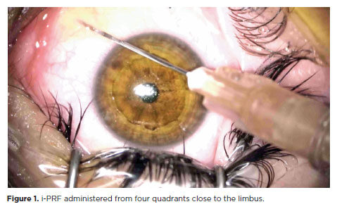
Abstract
PURPOSE: This study was conducted to investigate the effect of injectable platelet-rich fibrin on the recovery of compromised epithelium due to crosslinking treatment.
METHODS: In this comparative study, the epithelial closure rates and in vivo confocal biomicroscopy results of 26 patients with keratoconus who underwent subconjunctival injection of injectable platelet-rich fibrin near the limbus after epithelium-off corneal crosslinking treatment were compared with those of 25 patients who did not receive the injection of injectable platelet-rich fibrin.
RESULTS: The average time to epithelial defect closure in the injectable platelet-rich fibrin group was 2.76 ± 0.90 days compared to 3.56 ± 0.86 days in the non-injectable platelet-rich fibrin group (p=0.003). At the end of the 1st month, the mean subbasal nerve plexus density was 1.26 ± 1.61 nerves/mm2 in the injectable platelet-rich fibrin group, whereas it was 0.72 ± 0.89 nerves/mm2 in the non-injectable platelet-rich fibrin group (p=0.016). By the 3rd month, the density increased to 3.42 ± 1.13 nerves/mm2 in the injectable platelet-rich fibrin group and 2.36 ± 1.15 nerves/mm2 in the non-injectable platelet-rich fibrin group (p=0.002). Similarly, the anterior stromal keratocyte density at the end of the 1st month was 93.6 ± 33.5 cells/mm2 in the injectable platelet-rich fibrin group compared to 67.3 ± 26.4 cells/mm2 in the non-injectable platelet-rich fibrin group (p=0.001). By the end of the 3rd month, the density increased to 255.2 ± 45.7 cells/mm2 in the injectable platelet-rich fibrin group and 222.1 ± 43.6 cells/mm2 in the non-injectable platelet-rich fibrin group (p=0.011). In the non-injectable platelet-rich fibrin group, one patient developed a sterile infiltrate at the end of the 1st week, whereas no complications were observed in the injectable platelet-rich fibrin group.
CONCLUSION: Subconjunctival injectable platelet-rich fibrin application is an effective and safe method for corneal epithelial healing after crosslinking treatment.
Keywords: Keratoconus; Platelet-rich fibrin; Epithelium; corneal; Corneal crosslinking; Wound healing
Abstract
PURPOSE: To investigate the association of pre-photorefractive keratectomy Schirmer-1 test value with post-photorefractive keratectomy central corneal epithelial thickness, ocular surface disease index score, and uncorrected distance visual acuity.
METHODS: Patients were categorized according to preoperative Schirmer-1 value: the normal Schirmer Group (n=54; Schirmer-1 test value, >10 mm) and the low Schirmer Group (n=52; Schirmer-1 test value, between 6 and 10 mm). We analyzed ablation depth, visual acuity, result of Schirmer-1 test (with anesthesia), tear film break-up time, ocular surface disease index score, central corneal epithelial thickness, and spherical equivalent refraction.
RESULTS: We found significant differences between the groups in Schirmer-1 test value, tear film break-up time, and ocular surface disease index score, both preoperatively and postoperatively (p<0.001). The preoperative central corneal epithelial thicknesses of the two groups were similar (p>0.05). After photorefractive keratectomy, the Schirmer-1 test value and spherical equivalent refraction decreased in both groups (p<0.05), and ocular surface disease index scores and central corneal epithelial thickness values increased in the low Schirmer Group (p<0.001) but not in the normal Schirmer Group (p>0.05). The postoperative central corneal epithelial thicknesses of the low Schirmer Group were significantly higher than those of the normal Schirmer Group (p<0.001). Postoperative uncorrected distance visual acuity did not differ significantly between the two groups (p>0.05).
CONCLUSIONS: In patients with low Schirmer-1 test values before photorefractive keratectomy, the corneal epithelium thickened and ocular surface complaints increased during the postoperative period. However, changes in the corneal epithelium did not affect the postoperative uncorrected distance visual acuity. To reduce postoperative problems on the ocular surface in these patients, we recommend that dry eye be treated before photorefractive keratectomy.
Keywords: Epithelium, corneal; Cornea; Photorefractive keratectomy; Schirmer test; Visual acuity
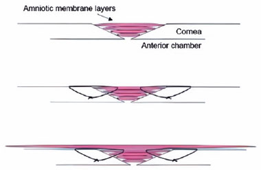
Abstract
PURPOSE: Wet bio-amniotic membrane plugging combined with transplantation is a novel option that combined amniotic membrane plugging with amniotic membrane transplantation for the treatment of small corneal perforations. This study aimed to evaluate the efficacy of wet bio-amniotic membrane plugging in the treatment of small corneal perforations and compared it with that of the penetrating keratoplasty procedure.
METHODS: Forty patients (41 eyes) with small corneal perforations <3 mm in diameter treated at our hospital between July 2018 and January 2021 were retrospectively included. Among them, 21 eyes were treated with wet bio-amniotic membrane plugging (wet bio-amniotic membrane plugging group), and 20 eyes were treated with penetrating keratoplasty procedure (penetrating keratoplasty procedure group). The best-corrected visual acuity, anterior chamber formation, corneal thickness, primary disease control, postoperative complications, and graft survival rate were assessed.
RESULTS: No significant difference in baseline characteristics was found between the wet bio-amniotic membrane plugging and penetrating keratoplasty procedure groups (p>0.05). The postoperative control rates of primary diseases in the wet bio-amniotic membrane plugging and penetrating keratoplasty procedure groups were 95.2% and 90.0%, respectively (p=0.481). Visual acuity was improved 6 months after the operation in the wet bio-amniotic membrane plugging group and was improved at postoperative 1 month in the penetrating keratoplasty procedure group. The formation time of the anterior chamber in the wet bio-amniotic membrane plugging group was significantly shorter than that in the penetrating keratoplasty procedure group (p=0.023). The corneal thickness of the two groups significantly increased 12 months after the operation; however, the degree of thickening in the penetrating keratoplasty procedure group was higher than that in the wet bio-amniotic membrane plugging group (p<0.001). During the follow-up, postoperative complications were not different between the two groups (p>0.999).
CONCLUSION: The results suggest that wet bio-amniotic membrane plugging is effective and safe in the treatment of small corneal perforations. Thus, it can be used as an emergency treatment alternative to penetrating keratoplasty procedure for small corneal perforations.
Keywords: Amnion; Transplantation; Amniotic membrane; Keratoplasty, penetrating; Corneal perforation; Wet bio-amniotic membrane plugging; Wet bio-amniotic membrane transplantation
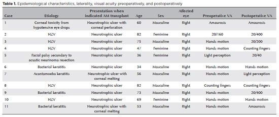
Abstract
PURPOSE: To evaluate the clinical results of cryopreserved amniotic membrane transplantation as a treatment option for refractory neurotrophic corneal ulcers.
METHODS: This prospective study included 11 eyes of 11 patients who underwent amniotic membrane transplantation for the treatment of refractory neurotrophic corneal ulcers at Hospital de Clínicas da Universidade Federal do Paraná, in the city of Curitiba, from May 2015 to July 2021. Patients underwent different surgical techniques in which the amniotic membrane was applied with the epithelium facing upward to promote corneal re-epithelialization.
RESULTS: The median age of the patients was 60 years (range, 34-82 years), and 64% were men. The predominant etiology of corneal ulcers was herpes zoster (45% of cases). Approximately one-third of the patients (27%) were chronically using hypotensive eye drops, and more than half (54%) had previously undergone penetrating corneal transplantation. At the time of amniotic membrane transplantation, 18% of the eyes had corneal melting, 9% had corneal perforation, and the others had corneal ulceration without other associated complications (73%). The time between clinical diagnosis and surgical treatment ranged from 9 days to 2 years. The corrected visual acuity was worse than 20/400 in 90% of the patients preoperatively, with improvement in 36% after 3 months of the procedure, worsening in 18% and remaining stable in 36%. Of the patients, 81% complained of preoperative pain, and 66% of them reported total symptom relief after the surgical procedure. In one month, 54.6% of the patients presented a closure of epithelial defect, and half of the total group evolved with corneal thinning. The failure rate was 45.5% of the cases.
CONCLUSION: Cryopreserved amniotic membrane transplantation can be considered a good alternative for treating refractory neurotrophic corneal ulcers, as it resulted in significant improvement in pain (66%) and complete epithelial closure (60%) in many patients at 1 month postoperatively. Notably, the high failure rate highlights the need for further studies to identify patient- and ulcer-related factors that may influence the outcomes of this procedure.
Keywords: Amnion/transplantation; Corneal ulcer; Anterior eye segment; Keratitis
17-equ01.jpg)
Abstract
Esta é uma revisão crítica do efeito da radiação ultravioleta no olho. Trata da classificação dessa radiação, nível no meio ambiente e os fatores que o determinam, penetração no olho humano, toxicidade às estruturas dos oculares, morbidades associadas, eventos passíveis de aumentar a vulnerabilidade do olho e filtros oculares artificiais. Discute, ainda, o risco real dessas radiações ao olho humano à luz do conhecimento atual.
Keywords: Radiação eletromagnética; Raios ultravioleta; Queimaduras oculares; Filtros ultravioletas; Transtornos da visão
Abstract
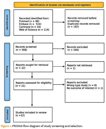
This study aimed to evaluate the efficacy and safety of topical losartan for treating corneal injuries and stromal fibrosis based on preclinical and clinical evidence. A systematic search was conducted in October 2024 following PRISMA guidelines across Embase, PubMed, Web of Science, and the Cochrane Library. Studies assessing topical losartan use in animal models or human patients with corneal injury were included. No randomized clinical trials were identified. Of 750 articles screened, 12 met the inclusion criteria – seven preclinical studies and five case reports. Preclinical evidence indicated that topical losartan at 0.2-0.8 mg/dL reduced stromal opacity and myofibroblast differentiation. Higher concentrations (8-80 mg/dL) offered no additional benefit and were associated with ocular surface irritation. The five case reports included 11 patients (12 eyes); eight eyes showed visual improvement, and no adverse effects were observed at a dose of 0.8 mg/dL. Topical losartan demonstrates potential as an antifibrotic agent for corneal injuries. However, variability in outcomes and dose-related toxicity at higher concentrations highlight the need for controlled clinical trials to confirm efficacy, establish optimal dosing, and ensure safety.
Keywords: Cornea; Epithelial cells; Myofibroblasts; Corneal opacity; Losartan
 ABO is licensed under a Creative Commons Attribution-NonComercial 4.0 Internacional.
About
Issues
Editorial Board
Submission
Arquivos Brasileiros de Oftalmologia
Official publication of Brazilian Council of Ophthalmology - Conselho Brasileiro de Oftalmologia (CBO)
Rua Casa do Ator, 1.117 - 2nd floor - Zip Code: 04546-004
São Paulo - SP, Brazil
TEL: +55 11 3266-4000
E-mail: [email protected]