Arq. Bras. Oftalmol. 2025;88 (4 )
:1-7
| DOI: 10.5935/0004-2749.2023-0356
Abstract
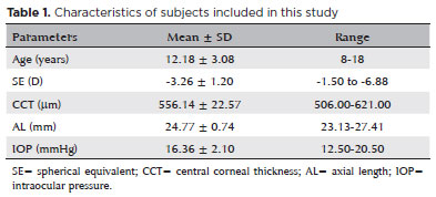
PURPOSE: Although the orthokeratology effects on corneal biomechanics have been proven with clinical trials, reports of stiffness parameter change are scarce. This study investigated the short-term orthokeratology effects in pediatric myopia and compared stiffness parameter changes to those published in recent clinical investigations. This prospective study aimed to investigate corneal biomechanics changes induced by short-term overnight orthokeratology treatment, focusing on stiffness parameter at A1 and stress-strain index
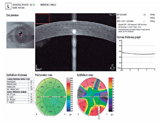
METHODS: Twenty-six children aged 8 to 18 were included in this study using orthokeratology lenses for two different durations: 1 day and 1 week. Corneal biomechanics were assessed using corneal visualization (Corvis) Scheimpflug technology. Measurements were taken at baseline and after each wearing session. Changes in corneal stiffness parameters and corneal curvature were analyzed.
RESULTS: All parameters changed significantly after 1 week of lens wear (p<0.05), except for velocity of corneal apex at the first and second applanation times highest concavity time, radius, stiffness parameter at A1 and stress-strain index. After 1 day, central corneal thickness, first applanation time, second applanation time, deformation amplitude ratio (2 mm), and Corvis biomechanical index (CBI) remained stable (p>0.05). After 1 week, central corneal thickness and first applanation time decreased, whereas second applanation time, deformation amplitude ratio, and Corvis Biomechanical Index significantly increased. With intraocular pressure and central corneal thickness as control variables, no significant correlation was found between stress-strain index and curvature changes (p>0.05). With age as the control variable, no significant correlation was found between stress-strain index and curvature changes (p>0.05).
CONCLUSIONS: Short-term orthokeratology treatment induced notable changes in several corneal biomechanical parameters. Stiffness parameter at A1 and stress-strain index are unaffected by increasing lens wear duration and do not influence the orthokeratology effect.
Keywords: Orthokeratologic procedures; Epithelium, corneal; Corneal topography; Myopia/therapy; Diagnostic techniques, ophthalmological; Biomechanical phenomena; Refraction, ocular; Visual acuity; Humans; Children; Adolescent
Arq. Bras. Oftalmol. 2022;85 (5 )
:465-471
| DOI: 10.5935/0004-2749.20220067
Abstract
Objetivos: Relatar a distribuição dos motivos de encaminhamento de crianças para ambulatório de glaucoma infantil em um serviço oftalmológico terciário.
Métodos: Dados médicos de pacientes menores que 18 anos encaminhados para ambulatório de glaucoma infantil na cidade de São Paulo, Brasil, de 2012 a 2018 foram retrospectivamente analisados. Os dados incluíram os motivos de encaminhamento, a idade, a origem e quem detectou a alteração ocular. Para definição diagnóstica, a classificação do Childhood Glaucoma Research Network foi usada.
Resultados: 563 olhos de 328 pacientes foram incluídos no estudo. O diagnóstico de glaucoma foi confirmado em 162 crianças (49%). 83 (25%) pacientes tiveram diagnóstico de glaucoma descartado, e 83 (25%) continuaram em acompanhamento como glaucoma suspeito. Os principais motivos de encaminhamento foram relação escavação-disco >0,5 ou assimetria ≥0,2 (24%), pressão intraocular >21 mmHg (21%) e opacidade corneana (15%). No período neonatal, os motivos de encaminhamento foram opacidade corneana, buftalmo, lacrimejamento e fotofobia. Após o período neonatal, além desses sinais externos, outros sinais foram também motivos de encaminhamento, como escavação-disco >0,5 ou assimetria ≥0,2, pressão intraocular >21 mmHg e miopização. Os encaminhamentos ocorreram por profissionais de saúde em 69% e preocupação dos pais em 30%. Os pais foram os primeiros a identificar as alterações e procurar cuidado médico em 97% dos casos de glaucoma congênito primário.
Conclusões: Os motivos de encaminhamento de crianças para um serviço de glaucoma de glaucoma terciário foram determinados e diferem em diferentes faixas etárias e grupos. Os autores reforçam a necessidade de alertar diferentes grupos para os sinais mais comuns, a fim de evitar, não só o adiamento do diagnóstico, o que impacta no prognóstico, mas também despender recursos importantes direcionados ao enfrentamento dessas doenças, com encaminhamentos imprecisos.
Keywords: Glaucoma/congênito; Glaucoma/fisiopatologia; Opacidade da córnea; Criança; Acuidade visual; Encaminhamento e consulta; Serviços de saúde ocular
Arq. Bras. Oftalmol. 2023;86 (4 )
:353-358
| DOI: 10.5935/0004-2749.20230055
Abstract
Objetivo: Examinar a eficácia da ceratectomia fototerapêutica para o tratamento de patologias variáveis que apresentarem opacidades anteriores da córnea, e avaliar a distribuição das indicações de ceratectomia fototerapêutica nos últimos 10 anos.
Métodos: Foram revisados retrospectivamente os prontuários de 276 pacientes, com 334 olhos tratados com ceratectomia fototerapêutica entre março de 2010 e o ano de 2020. As etiologias dos pacientes submetidos à ceratectomia fototerapêutica foram anotadas e suas alterações foram examinadas. Os resultados refrativos e de acuidade visual antes e após a operação foram registrados e analisados de acordo com a etiologia.
Resultados: A idade média dos pacientes foi de 40,7 ± 16,2 anos (faixa: 19-84). O tempo médio de acompanhamento foi de 25,5 ± 19,1 meses (faixa: 3-96). A ceratectomia fototerapêutica foi aplicada com mais frequência para distrofias estromais corneanas (44%, 151 olhos de 111 pacientes); entre as distrofias corneanas como um todo, a distrofia granular foi a indicação terapêutica mais comum desse procedimento. Ao contrário de outras indicações, nos últimos 10 anos houve um aumento na aplicação de ceratectomia fototerapêutica em casos de opacidade subepitelial persistente causada por conjuntivite adenoviral. Houve um aumento significativo na acuidade visual em todos os grupos, exceto para o grupo com defeito epitelial recorrente (p<0,05). A maior melhora na acuidade visual foi detectada em casos de distrofia estromal, no subgrupo das distrofias granulares.
Conclusão: Apesar das mudanças nas tendências de indicação, a ceratectomia fototerapêutica continua sendo uma abordagem terapêutica eficaz e confiável para tratar lesões da córnea anterior.
Keywords: Ceratectomia fotorrefrativa; Opacidade da córnea; Lesões da córnea; Distrofias da córnea; Fototerapia.
Arq. Bras. Oftalmol. 2025;88 (6 )
:1-7
| DOI: 10.5935/0004-2749.2025-0120
Abstract
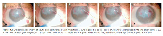
PURPOSE: To describe the technique and outcomes of intrastromal autologous blood injection in patients with severe corneal hydrops.
METHODS: Nineteen patients with corneal hydrops underwent intrastromal autologous blood injection. Postoperative assessments included best-corrected visual acuity and time to resolution of corneal edema
RESULTS: Corneal edema resolved within 1 week in 5 patients, within 1 month in 11, and within 3 months in 3. The mean duration of edema persistence was 37.94 ± 33.05 days (range, 6–124). Corneal thickness decreased from 2.06 ± 0.71-mm preoperatively to 1.34 ± 0.65-mm at day 7, 0.85 ± 0.56-mm at day 30, and 0.57 ± 0.13-mm at day 90 (p<0.001). Descemet’s membrane (DM) detachment decreased from 1.01 ± 0.75-mm to 0.44 ± 0.57-mm, 0.24 ± 0.36-mm, and 0.08 ± 0.11-mm on postoperative days 7, 30, and 90, respectively (p<0.001). DM break size decreased from 1.12 ± 1.19-mm to 0.62 ± 0.84-mm at 3 months (p<0.005). Three patients developed hematocornea; no other major complications were observed. At 3 months, mean best-corrected visual acuity improved from 2.37 ± 0.66 to 0.41 ± 0.17 logMAR with hard contact lenses (p<0.001).
CONCLUSIONS: Intrastromal autologous blood injection is an effective treatment for severe corneal hydrops, promoting faster edema resolution and visual improvement with minimal complications.
Keywords: Corneal edema; Corneal diseases; Edema; Visual acuity; keratoconus.
Arq. Bras. Oftalmol. 2025;88 (3 )
:1-7
| DOI: 10.5935/0004-2749.2023-0309
Abstract
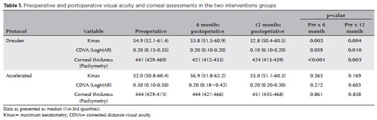
PURPOSE: Keratoconus presents certain peculiarities in pediatric patients when compared with adults. The greatest challenge in children is that the disease is more severe and faster in progression. In this retrospective study, we aimed to compare the accelerated and Dresden protocols for corneal crosslinking in patients aged <18 years who were followed-up for at least 12 months.
METHODS: A total of 36 eyes from 27 patients were included in the study. The best corrected and uncorrected visual acuity, maximal keratometry, corneal thickness, foveal thickness, and endothelial microscopy findings were evaluated at baseline and during the postoperative period at one, three, and six months. Thereafter, the patients were evaluated at one, three, six and twelve months postoperative. Corneal crosslinking was performed in all patients via the Dresden protocol (n=21 eyes) or the accelerated protocol (n=15 eyes). Data between the two groups were compared and XY statistical analysis was used.
RESULTS: Both protocols were effective in halting keratoconus progression. No patient had progression at the 12-month follow-up. A significant reduction in Kmax and improvement in the corrected distance visual acuity were observed only in the Dresden protocol group. Although the Dresden protocol was superior to the accelerated protocol in reducing Kmax (p=0.002), there was no significant difference in corrected distance visual acuity between the two groups.
CONCLUSION: The accelerated protocol is as efficient as the Dresden protocol in stabilizing keratoconus progression. Although the Dresden protocol was superior to the accelerated protocol in reducing the Kmax, it did not produce better clinical results. Thus, the accelerated protocol is an efficient option. Furthermore, considering the advantages of reduced surgical time, the accelerated protocol is effective in halting keratoconus progression in the pediatric age group.
Keywords: Keratoconus; Corneal diseases; Ultraviolet rays; Cross-linking reagents; Visual acuity
Arq. Bras. Oftalmol. 2024;87 (3 )
:1-7
| DOI: 10.5935/0004-2749.2023-0049
Abstract
PURPOSE: To investigate the association of pre-photorefractive keratectomy Schirmer-1 test value with post-photorefractive keratectomy central corneal epithelial thickness, ocular surface disease index score, and uncorrected distance visual acuity.
METHODS: Patients were categorized according to preoperative Schirmer-1 value: the normal Schirmer Group (n=54; Schirmer-1 test value, >10 mm) and the low Schirmer Group (n=52; Schirmer-1 test value, between 6 and 10 mm). We analyzed ablation depth, visual acuity, result of Schirmer-1 test (with anesthesia), tear film break-up time, ocular surface disease index score, central corneal epithelial thickness, and spherical equivalent refraction.
RESULTS: We found significant differences between the groups in Schirmer-1 test value, tear film break-up time, and ocular surface disease index score, both preoperatively and postoperatively (p<0.001). The preoperative central corneal epithelial thicknesses of the two groups were similar (p>0.05). After photorefractive keratectomy, the Schirmer-1 test value and spherical equivalent refraction decreased in both groups (p<0.05), and ocular surface disease index scores and central corneal epithelial thickness values increased in the low Schirmer Group (p<0.001) but not in the normal Schirmer Group (p>0.05). The postoperative central corneal epithelial thicknesses of the low Schirmer Group were significantly higher than those of the normal Schirmer Group (p<0.001). Postoperative uncorrected distance visual acuity did not differ significantly between the two groups (p>0.05).
CONCLUSIONS: In patients with low Schirmer-1 test values before photorefractive keratectomy, the corneal epithelium thickened and ocular surface complaints increased during the postoperative period. However, changes in the corneal epithelium did not affect the postoperative uncorrected distance visual acuity. To reduce postoperative problems on the ocular surface in these patients, we recommend that dry eye be treated before photorefractive keratectomy.
Keywords: Epithelium, corneal; Cornea; Photorefractive keratectomy; Schirmer test; Visual acuity
Arq. Bras. Oftalmol. 2024;87 (3 )
:1-8
| DOI: 10.5935/0004-2749.2023-0109
Abstract
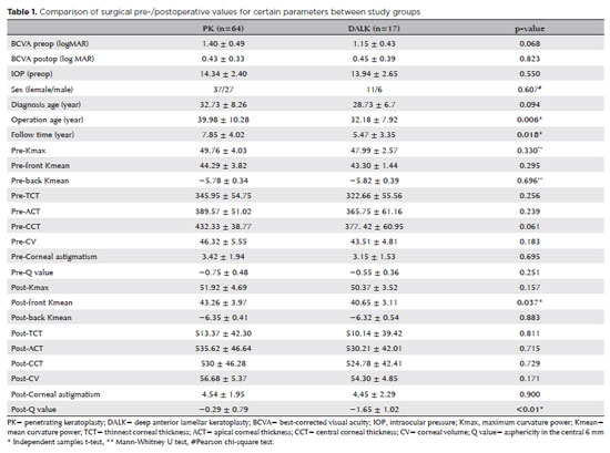
PURPOSES: This study aims to assess and compare the postoperative visual and topographic outcomes, complications, and graft survival rates following deep anterior lamellar keratoplasty and penetrating keratoplasty in patients with macular corneal dystrophy.
METHODS: In this study we enrolled 59 patients (23 male; and 36 female) with macular corneal dystrophy comprising 81 eyes. Out of these, 64 eyes underwent penetrating keratoplasty, while 17 eyes underwent deep anterior lamellar keratoplasty. The two groups were analyzed and compared based on best-corrected visual acuity, corneal tomography parameters, pachymetry, complication rates, and graft survival rates.
RESULTS: After 12 months, 70.6% of the patients who underwent deep anterior lamellar keratoplasty (DALK) and 75% of those who had penetrating keratoplasty (PK) achieved a best-corrected visual acuity of 20/40 or better (p=0.712). Following surgery, DALK group showed lower front Kmean (p=0.037), and Q values (p<0.01) compared to the PK group. Postoperative interface opacity was observed in seven eyes (41.2%) in the DALK group. Other topography values and other complications (graft rejection, graft failure, cataract, glaucoma, microbial keratitis, optic atrophy) did not show significant differences between the two groups. The need for regrafting was 9.4% and 11.8% in the PK and DALK groups, respectively (p=0.769). Graft survival rates were 87.5% and 88.2% for PK and DALK; respectively (p=0.88 by Log-rank test).
CONCLUSION: Both PK and DALK are equally effective in treating macular corneal dystrophy, showing similar visual, topographic, and survival outcomes. Although interface opacity occurs more frequently after DALK the visual results were comparable in both groups. Therefore, DALK emerges as a viable surgical choice for patients with macular corneal dystrophy without Descemet membrane involvement is absent.
Keywords: Macular corneal dystrophy; Corneal dystrophies; Hereditary; Keratoplasty; Penetrating; Corneal transplantation
 ABO is licensed under a Creative Commons Attribution-NonComercial 4.0 Internacional.
14-fig01tb.jpg)
06-tab01.jpg)
04-fig01.jpg)
09-fig01.jpg)