Arq. Bras. Oftalmol. 2020;83 (6 )
:463-472
| DOI: 10.5935/0004-2749.20200087
Abstract
Objetivo: O objetivo deste estudo foi caracterizar os estreptococos alfa-hemolíticos isolados de endoftalmite infecciosa e ceratite e determinar sua distribuição.
Métodos: A amostra incluiu 27 e 35 isolados não-duplicados de estreptococos alfa-hemolíticos recuperados de pacientes com endoftalmite infecciosa (2002-2013) e ceratite (2008-2013), respectivamente. Os isolados foram identificados pelos testes de suscetibilidade à optoquina e bile solubilidade, utilizando um sistema de identificação bioquímica. A concentração inibitória mínima foi determinada pelo método de microdiluição em caldo. A identificação molecular foi realizada pela análise de três genes constitutivos e análise complementar de sequências multilocus. A epidemiologia molecular do Streptococcus pneumoniae foi investigada por tipagem de sequência multilocus, e a presença do gene codificador do polissacarídeo capsular foi avaliada por reação em cadeia da polymerase convencional. Os resultados foram avaliados utilizando os prontuários médicos dos pacientes.
Resultados: Os testes fenotípicos diferenciaram S. pneumoniae dos outros estreptococos alpha-hemolíticos, consistentes com identificações moleculares posteriores. S. oralis foi significativamente prevalente entre os isolados de endoftalmite, assim como S. pneumoniae nos isolados de ceratite. Foram observados altos níveis de suscetibilidade a antibióticos, incluindo vancomicina, cefalosporinas e fluoroquinolonas. Alta variabilidade genética foi detectada entre as 19 cepas de S. pneumoniae, com 15 previstas para serem encapsuladas. Os prontuários médicos dos pacientes com endoftalmite infecciosa foram revisados (n=15/27; 56%), e a acuidade visual final foi avaliada em 12 casos (44%). Muitos pacientes evoluiram para um estado final de acuidade visual de “sem percepção luminosa” (6/12; 50%), “percepção luminosa” (3/12; 25%) ou “movimentos de mãos” (1/12; 8%). Também foram revisados os prontuários médicos dos pacientes com ceratite infecciosa (n=24/35; 69%), e a acuidade visual final foi avaliada em 18 casos (51%). Da mesma foram, a maioria dos pacientes evoluiu para um estado final de acuidade visual de “sem percepção luminosa” (6/18; 33%), “percepção luminosa” (1/18; 6%) ou “movimentos de mãos” (6/18; 33%). No geral, a maioria dos pacientes evoluiu para um estado final de acuidade visual de “sem percepção luminosa” (12/30), “percepção luminosa” (4/30) ou “movimentos de mãos” (7/30).
Conclusões: A distribuição de estreptococos alfa-hemolíticos nas infecções oculares sugeriu a presença de um tropismo de tecido específico da espécie. Os prognósticos dos pacientes com infeções oculares por estreptococos foram altamente desfavoráveis e a resistência a antibióticos contribuiu não para as progressões clínicas desfavoráveis e os maus resultados.
Keywords: Endoftalmite; Ceratite; Infecções oculares bacterianas; Infecções estreptocócicas; Estreptococos viridans/isolamento & purificação; Resistência antimicrobiana a medicamentos; Fluoroquinolonas
Arq. Bras. Oftalmol. 2025;88 (4 )
:1-7
| DOI: 10.5935/0004-2749.2024-0229
Abstract
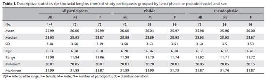
PURPOSE: The volume of the vitreous chamber varies with the size of the eye. The space created in the vitreous cavity by a vitrectomy is called the vitrectomized space. The volume of the vitrectomized space is strongly correlated with the axial length of the eye. This study aims to present guidelines for estimating the using participants stratified by axial length, sex, and history of cataract surgery.
METHODS: This retrospective, observational, cross-sectional study included 144 randomly selected participants who underwent vitrectomies between 2013 and 2023. Before surgery, the axial lengths of participants' eyes were measured using optical biometrics. The axial lengths of the eyes in our sample were between 20-32 mm. In all cases, a complete vitrectomy was performed, followed by complete fluid-air exchange and injection of a balanced saline solution. The volume infused was recorded.
RESULTS: The median (interquartile range; range) volume of the vitrectomized space was 6.1 (3.8; 3.1-11.3) mL in men and 6.1 (3.3; 3.2-11.2) mL in women (p=0.811). The median volume of the vitrectomized space was 5.9 (3.6; 3.1-11.2) mL in patients with phakic lenses and 6.25 (3.6; 3.3-11.3) mL in those with pseudophakic lenses (p=0.533). A positive correlation was found between the axial length and the volume of the vitrectomized space in this sample (r=0.968; p<0.001). In a cubic polynomial regression, the coefficient of determination was 0.948. Similar results were observed in both sexes and in both phakic and pseudophakic patients. The estimated cubic polynomial regression equation for this sample was VVS = 0.000589052857847605 × AL3 - 0.025114926401582700 × AL2 + 0.685961117595624000 × AL - 5.088226672620790000.
CONCLUSION: We developed this axial length estimation of the volume of vitrectomized space as a guideline for the determination of vitrectomized space volume using axial length.
Keywords: Cataract extraction; Retinal perforations/surgery; Epiretinal membrane/surgery; Vitreous body; Axial length, eye; Vitrectomy; Biometry/methods; Diagnostic techniques, ophthalmological; Guidelines as topic.
Arq. Bras. Oftalmol. 2025;88 (6 )
:1-5
| DOI: 10.5935/0004-2749.2025-0085
Abstract
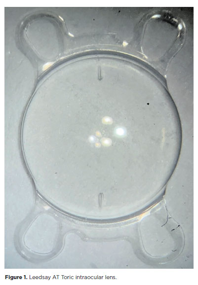
PURPOSE: The purpose of this study was to assess visual outcomes and patient satisfaction following cataract surgery involving the implantation of quad-loop intraocular lenses, including trifocal, bifocal, and toric variants.
METHODS: Information was obtained from both physical and electronic medical records of patients who underwent phacoemulsification cataract surgery with implantation of different intraocular lenses between January 1, 2022, and December 31, 2023. The study included individuals aged over 18 who received bilateral implantation of bifocal, trifocal, or monofocal toric intraocular lenses. Visual acuity was assessed at various postoperative time points using the logMAR scale. Quantitative variables were analyzed using mean and standard deviation.
RESULTS: A total of 92 eyes received premium intraocular lenses: 4 bifocal, 32 trifocal, 52 toric monofocal, and 4 trifocal toric lenses. The average preoperative corrected visual acuity was logMAR 0.478 ± 0.259. On the first postoperative day, the average uncorrected visual acuity was logMAR 0.301 ± 0.207. By day 30, 67.4% of eyes achieved uncorrected distance visual acuity of logMAR 0.2 or better. Patient satisfaction was high, with few reports of glare or halos.
CONCLUSION: Quad-loop intraocular lenses-including trifocal, bifocal, and toric models-demonstrated effective improvement in visual acuity and high levels of patient satisfaction. These lenses represent a suitable option for enhancing visual outcomes after cataract surgery. Additional studies with larger cohorts are recommended to confirm these results.
Keywords: Cataract extraction; Aberrometry/methods; Lenses, intraocular; Lens implantation, intraocular; Prosthesis design
Arq. Bras. Oftalmol. 2026;89 (2 )
:1-8
| DOI: 10.5935/0004-2749.2025-0175
Abstract
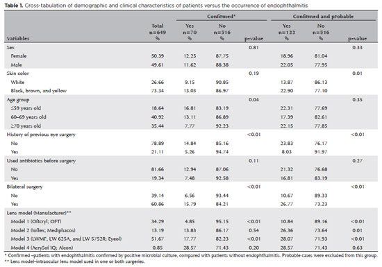
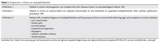
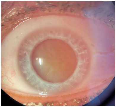
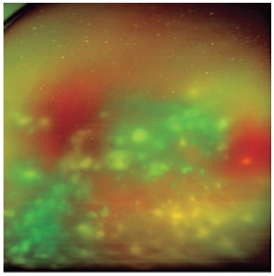
PURPOSE: Endophthalmitis is one of the most important adverse events after cataract surgery, as it can lead to total vision loss. This study aimed to describe the occurrence of endophthalmitis after phacoemulsification with intraocular lens implantation in patients treated in a community setting in Porto Velho, Rondônia, Brazil.
METHODS: This retrospective cohort study was conducted using a database of 649 medical records of patients who underwent surgery and were followed for three months. Poisson regression analysis was used to estimate relative risks and 95% confidence intervals (95% CIs).
RESULTS: The incidence of confirmed endophthalmitis was 11.94% (95% CI, 9.50-14.76), while the incidence of confirmed and probable cases was 20.50% (95% CI, 17.52-23.73). For confirmed cases, bilateral surgery and the use of lens model 3 were identified as risk factors for endophthalmitis, whereas age over 70 yr and preoperative antibiotic use were protective factors. For confirmed and probable cases, brown and yellow skin color, bilateral surgery, and the use of lens model 3 were also identified as risk factors. Gram-negative bacteria were the predominant etiological agents, and corneal edema was the main clinical manifestation. The mean duration of treatment was eight days, and 27.12% of patients used antibiotics.
CONCLUSION: The incidence observed was substantially higher than that reported in the literature, with a predominance of Gram-negative agents and an association with bilateral surgeries and the Eyeol intraocular lens model. These findings reinforce the need for continuous epidemiological surveillance and the implementation of specific biosafety and infection control protocols during cataract surgery campaigns.
Keywords: Endophthalmitis; Disease outbreaks; Phacoemulsification; Lens implantation, intraocular; Lenses, intraocular; Cataract; Risk factors; Anti-bacterial agents
Arq. Bras. Oftalmol. 2025;88 (6 )
:1-8
| DOI: 10.5935/0004-2749.2024-0394
Abstract
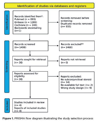
The advantages and disadvantages of using perioperative subconjunctival steroid injections in dropless cataract surgery continue to be debated. A systematic review of PubMed, EMBASE, and the Cochrane Central database identified five studies—two randomized controlled trials and three non-randomized studies—encompassing 70,751 eyes. Among these, 12,319 eyes (17.4%) received subconjunctival steroid injections, while 58,432 eyes (82.6%) were managed with topical steroids. The Cochrane Collaboration’s RoB 2 tool was applied for bias assessments in randomized controlled trials, and heterogeneity was assessed using the I² statistics. No statistically significant differences were found between the two groups regarding macular edema (p=0.249), visual acuity (p=0.73), or laser flare count (p=0.45). Both subconjunctival injections and topical steroids demonstrated comparable efficacy and safety in controlling postoperative inflammation after cataract surgery. Additional research is warranted to validate these conclusions.
Keywords: Cataract extraction; Phacoemulsification; Lens implantation, intraocular; Postoperative care; Intravitreal injections; Anti-inflammatory agents, non-steroidal/administration & dosage; Glucocorticoids; Triamcinolone acetonide; Research design; Randomiz
Arq. Bras. Oftalmol. 2025;88 (3 )
:1-6
| DOI: 10.5935/0004-2749.2023-0345
Abstract
PURPOSE: To determine the impact of prophylactic intracameral cefuroxime administration on the post-cataract surgery endophthalmitis rates and analyze its safety.
METHODS: The incidence of post-phacoemulsification endophthalmitis before and after the introduction of antibiotic prophylaxis with cefuroxime was compared. Data were extracted from the electronic medical records of patients who underwent cataract surgery between July 2019 and July 2022 at a tertiary-care hospital. Data were also collected from the Hospital Infection Control Service database. Statistical analysis was performed to assess the efficacy of cefuroxime prophylaxis in reducing endophthalmitis rates.
RESULTS: Of the 4459 cataract surgeries included in the study, 2247 were included in the control group (pre-cefuroxime), and 2212 were included in the post-cefuroxime (ATB-P) Group. In the control group, 6 (0.13%) cases of endophthalmitis were reported. In the ATB-P Group, there were no cases of acute endophthalmitis. The frequency of endophthalmitis was significantly higher in the control group than in the ATB-P Group (p=0.016). Furthermore, Staphylococcus sp. was the most identified causative agent (75%). No adverse effects were reported after cefuroxime administration.
CONCLUSION: The introduction of intracameral prophylaxis with cefuroxime significantly reduced the incidence of post-cataract surgery endophthalmitis. Additionally, its administration is safe.
Keywords: Cataract extraction; Endophthalmitis; Antibiotic prophylaxis; Injections; Cefuroxime
Arq. Bras. Oftalmol. 2025;88 (5 )
:1-7
| DOI: 10.5935/0004-2749.2024-0368
Abstract
PURPOSE: To compare endothelial corneal cell changes following cataract surgery performed by phacoemulsification with intraocular lens implantation, conducted by surgeons with varying levels of experience.
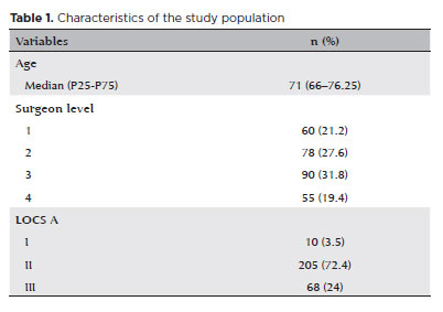
METHODS: Two hundred and eighty-three eyes diagnosed with cataract were included. Lens opacity was classified into three categories (I, II, and III). Surgeons were categorized into four experience levels (1, 2, 3, and 4), based on years of practice and lifetime surgeries performed. Corneal endothelial characteristics were assessed using non-contact specular microscopy, with measurements taken before surgery and 30-60 days post-surgery.
RESULTS: Pre- and postoperative endothelial analysis showed no significant differences between surgeon levels regarding visual acuity achieved, corneal thickness, and endothelial hexagonality. However, the central endothelial cell density index showed a significantly greater reduction among level 1 surgeons (p=0.026). Grade II cataracts exhibited significant variations in the central endothelial cell density (p=0.011) and average cell size, with level 1 surgeons showing the largest increases (p=0.024).
CONCLUSIONS: The analysis revealed significant differences in visual acuity and endothelial indices between surgeon experience levels, with less experienced surgeons showing greater variations and poorer performance. Clinical protocols should consider these data to establish safer training protocols.
Keywords: Cataract extraction; Phacoemulsification; Endothelium; corneal; Lens implantation, intraocular; Visual acuity; Internship and residency; Surgeons
 ABO is licensed under a Creative Commons Attribution-NonComercial 4.0 Internacional.
01-fig01tb.jpg)
02-fig01.jpg)
05-fig01.jpg)
04-fig01.jpg)
13-fig01tb.jpg)