Arq. Bras. Oftalmol. 2021;84 (6 )
:538-542
| DOI: 10.5935/0004-2749.20210090
Abstract
Objetivo: Investigar se as aberrações da córnea e as amplitudes de acomodação alteram mais em pacientes com esclerose múltipla do que em populações normais.
Métodos: Vinte pacientes previamente diagnosticados com esclerose múltipla com envolvimento do nervo óptico (grupo com eslerose múltipla) e 20 indivíduos saudáveis pareados por sexo e idade (grupo controle) foram incluídos no estudo. Pacientes com menos de 40 anos de idade foram incluídos em ambos os grupos devido à deterioração significativa de acomodação em pacientes com mais de 40 anos de idade. Para cada participante, a amplitude de acomodação foi medida em dioptrias pelo teste de lentes negativas e as aberrações de alta ordem foram avaliadas com o aberrômetro iDesign. Em seguida, a amplitude de acomodação e a média da raiz quadrada de aberrações de alta ordem foram comparadas entre os grupos.
Resultados: As médias da idade dos grupos com esclerose múltipla e controle foram 35,25 ± 4,52 anos e 32,28 ± 6,83 anos, respectivamente (p=0,170). A amplitude de acomodação foi de 4,05 ± 1,25 D no grupo com esclerose múltipla e 6,00 ± 1,03 D no grupo controle. A diferença entre os com esclerose múltipla e o grupo controle foi estatisticamente significativa (p<0, 001). A média da raiz quadrada das aberrações de alta ordem não foi significativamente diferente entre os grupos (com esclerose múltipla, 0,44 ± 0,22; controle, 0,43 ± 0,10, p<0,824). Não houve diferenças estatisticamente significativas entre os grupos em termos de alterações de aberrações entre a linha de base e o estímulo 5 D.
Conclusões: Este estudo mostra que a amplitude de acomodação diminuiu em pacientes com esclerose múltipla. Portanto, esses resultados podem causar possíveis razões de deficiências visuais transitórias em pacientes com esclerose múltipla. No entanto, esta diferença de amplitude de acomodação não fez uma diferença significativa entre os grupos quanto à alteração das aberrações de alta ordem durante a acomodação.
Keywords: Acomodação ocular; Esclerose múltipla; Nervo óptico
Arq. Bras. Oftalmol. 2023;86 (2 )
:131-136
| DOI: 10.5935/0004-2749.20230036
Abstract
Objetivo: Avaliar o impacto dos tumores córneo-conjuntivais na superfície ocular e na qualidade de vida dos pacientes antes e após o tratamento cirúrgico.
Métodos: Este estudo prospectivo conduziu uma avaliação pré-operatória e com 30 e 90 dias de pós-operatório de pacientes com diagnóstico de tumores de córnea e conjuntiva. Os dados demográficos foram coletados no pré-operatório. Os questionários Health Survey Short-Form (SF-12) e Ocular Surface Disease Index (OSDI) foram aplicados para avaliar a qualidade de vida dos pacientes e a percepção de suas funções relacionadas à visão. Os testes tear break-up time (TBUT) e Schirmer foram realizados para avaliação da superfície ocular. A extensão do tumor foi medida usando o programa ImageJ.
Resultados: Vinte e três pacientes foram incluídos. A média de idade foi de 52,8 ± 17,3 anos (27-79 anos). O tipo mais comum de tumor foi o carcinoma de células escamosas (61,5%). A acuidade visual dos pacientes melhorou significativamente em 1 mês e 3 meses (p=0,018 e p=0,036, respectivamente). Não houve diferenças significativas entre os testes tear break-up time e Schirmer no pré-operatório e com 3 meses de pós-operatório (p=0,150 e p=0,490, respectivamente). Os escores do SF-12 demonstraram que o componente mental apresentou diferença estatisticamente significante entre o pré-operatório e no 30 e 90 dias de pós-operatório (p=0,008 e p=0,026, respectivamente). A extensão do tumor foi de 868,7 ± 344,9 pixels (intervalo, 224,6-1481,6 pixels) e foram significativamente correlacionados com o componente mental de SF-12 no pré-operatório (p=0,011), 30 (p=0,017) e 90 dias de pós-operatório (p=0,012), e o componente emocional no 30º dia de pós-operatório (p=0,016).
Conclusão: Pacientes com tumores córneo-conjuntivais melhoraram os sintomas oculares, a acuidade visual e o componente emocional da qualidade de vida após a excisão cirúrgica do tumor.
Keywords: Neoplasias oculares; Neoplasias da túnica conjuntiva; Doenças da córnea; Acuidade visual; Qualidade de vida.
Arq. Bras. Oftalmol. 2021;84 (2 )
:140-148
| DOI: 10.5935/0004-2749.20210022
Abstract
OBJETIVO: Determinar o grau de deficiência visual em crianças com tumores da via óptica incapazes de informar a acuidade visual de reconhecimento.
MÉTODO: A acuidade visual de grades, em logMAR, foi estimada por potenciais visuais evocados de varredura em crianças com tumores das vias ópticas. O déficit da acuidade visual de grades binocular foi calculado em relação ao valor mediano normativo esperado para a idade e a deficiência visual, classificada como leve (0,10 a 0,39 logMAR), moderada (0,40 a 0,79 logMAR) ou grave (≥0,80 logMAR). Diferenças inter-oculares foram calculadas por subtração e consideradas aumentadas se >0,10 logMAR.
RESULTADOS: Foram avaliadas 25 crianças (13 meninos; média de idade ± DP=35,1± 25,9 meses; mediana=32,0 meses) com tumores da via óptica (24 gliomas e 1 tumor embrionário) localizados particularmente na transição hipotalâmico-quiasmática (n=21; 84,0%) e com anormalidades visuais detectadas pelos pais (n=17; 68,0%). A média do déficit da acuidade de grades foi 0,60 ± 0,36 logMAR (mediana=0,56 logMAR). Observou-se deficiência visual leve em 10 (40,0%), moderada em 8 (32,0%) e grave em 7 (28,0%), além de aumento da diferença interocular da acuidade visual (n=16; 64,0%). As principais alterações oftalmológicas encontradas foram: nistagmo (n=17; 68,0%), aumento da escavação do disco óptico e/ou palidez (n=13; 52,0%), estrabismo (n=12; 48,0%) e comportamento visual pobre (n=9; 36,0%).
CONCLUSÃO: Em crianças com tumor da via óptica e incapazes de responder aos testes de acuidade visual de reconhecimento, foi possível quantificar deficiência visual por meio dos potenciais visuais evocados de varredura e avaliar a diferença interocular da acuidade visual de grades. A gravidade do déficit da acuidade visual de grades relacionado à idade e a diferença interocular da acuidade visual de grades foram congruentes com alterações oftalmológicas e neuroimagem. O déficit da acuidade visual de grades foi útil à caracterização do estado visual em crianças com tumores da via óptica e ao embasamento da assistência neuro-oncológica.
Keywords: Transtornos da visão; Potenciais evocados visuais; Acuidade visual; Vias visuais; Glioma do nervo óptico; Criança
Arq. Bras. Oftalmol. 2025;88 (6 )
:1-7
| DOI: 10.5935/0004-2749.2025-0120
Abstract
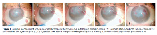
PURPOSE: To describe the technique and outcomes of intrastromal autologous blood injection in patients with severe corneal hydrops.
METHODS: Nineteen patients with corneal hydrops underwent intrastromal autologous blood injection. Postoperative assessments included best-corrected visual acuity and time to resolution of corneal edema
RESULTS: Corneal edema resolved within 1 week in 5 patients, within 1 month in 11, and within 3 months in 3. The mean duration of edema persistence was 37.94 ± 33.05 days (range, 6–124). Corneal thickness decreased from 2.06 ± 0.71-mm preoperatively to 1.34 ± 0.65-mm at day 7, 0.85 ± 0.56-mm at day 30, and 0.57 ± 0.13-mm at day 90 (p<0.001). Descemet’s membrane (DM) detachment decreased from 1.01 ± 0.75-mm to 0.44 ± 0.57-mm, 0.24 ± 0.36-mm, and 0.08 ± 0.11-mm on postoperative days 7, 30, and 90, respectively (p<0.001). DM break size decreased from 1.12 ± 1.19-mm to 0.62 ± 0.84-mm at 3 months (p<0.005). Three patients developed hematocornea; no other major complications were observed. At 3 months, mean best-corrected visual acuity improved from 2.37 ± 0.66 to 0.41 ± 0.17 logMAR with hard contact lenses (p<0.001).
CONCLUSIONS: Intrastromal autologous blood injection is an effective treatment for severe corneal hydrops, promoting faster edema resolution and visual improvement with minimal complications.
Keywords: Corneal edema; Corneal diseases; Edema; Visual acuity; keratoconus.
Arq. Bras. Oftalmol. 2025;88 (3 )
:1-7
| DOI: 10.5935/0004-2749.2023-0309
Abstract
PURPOSE: Keratoconus presents certain peculiarities in pediatric patients when compared with adults. The greatest challenge in children is that the disease is more severe and faster in progression. In this retrospective study, we aimed to compare the accelerated and Dresden protocols for corneal crosslinking in patients aged <18 years who were followed-up for at least 12 months.
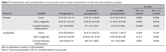
METHODS: A total of 36 eyes from 27 patients were included in the study. The best corrected and uncorrected visual acuity, maximal keratometry, corneal thickness, foveal thickness, and endothelial microscopy findings were evaluated at baseline and during the postoperative period at one, three, and six months. Thereafter, the patients were evaluated at one, three, six and twelve months postoperative. Corneal crosslinking was performed in all patients via the Dresden protocol (n=21 eyes) or the accelerated protocol (n=15 eyes). Data between the two groups were compared and XY statistical analysis was used.
RESULTS: Both protocols were effective in halting keratoconus progression. No patient had progression at the 12-month follow-up. A significant reduction in Kmax and improvement in the corrected distance visual acuity were observed only in the Dresden protocol group. Although the Dresden protocol was superior to the accelerated protocol in reducing Kmax (p=0.002), there was no significant difference in corrected distance visual acuity between the two groups.
CONCLUSION: The accelerated protocol is as efficient as the Dresden protocol in stabilizing keratoconus progression. Although the Dresden protocol was superior to the accelerated protocol in reducing the Kmax, it did not produce better clinical results. Thus, the accelerated protocol is an efficient option. Furthermore, considering the advantages of reduced surgical time, the accelerated protocol is effective in halting keratoconus progression in the pediatric age group.
Keywords: Keratoconus; Corneal diseases; Ultraviolet rays; Cross-linking reagents; Visual acuity
Arq. Bras. Oftalmol. 2025;88 (3 )
:1-11
| DOI: 10.5935/0004-2749.2024-0049
Abstract
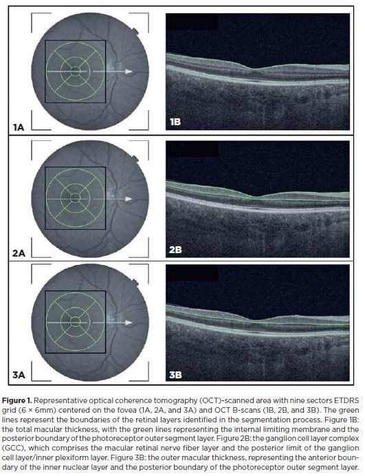
PURPOSE: This study aimed to evaluate the total macular thickness as well as the thickness of the inner and outer retinal layers in patients with Parkinson's disease. It also aimed to verify the correlation of these parameters with motor symptoms and cognitive function.
METHODS: A total of 46 eyes of 23 patients with Parkinson's disease and 40 eyes of 20 healthy controls were included in the study. The patients' cognitive, functional, and nonmotor symptoms were evaluated using the Katz Index of Independence and Pfeffer's Activities of Daily Living, Mini-Mental State Examination, Frontal Assessment Battery, Schwab and England Staging Scales, and Movement Disorders Society Nonmotor Symptoms Scale. The macular thickness measurements obtained via total, inner, and outer optical coherence tomography were recorded. Furthermore, the correlation of the parameters of optical coherence tomography with cognitive, functional, and nonmotor symptoms was assessed.
RESULTS: The scores of the Katz Index of Independence and Pfeffer's Activities of Daily Living as well as the Movement Disorders Society Nonmotor Symptoms Scale were significantly lower in patients with Parkinson's disease than in healthy controls. Moreover, the former had greater total macular thickness. The temporal and inferior outer sectors were significantly greater for the ganglion cell complex thickness in patients. A significant correlation was observed between the total macular thickness and the Movement Disorder Society-Unified Parkinson's Disease Rating Scale, Parte III (MDS-UPDRS-III) values. Contrarily, there was a negative correlation between the outer macular thickness and the MDS-UPDRS-III values. Meanwhile, the total macular thickness and ganglion cell complex thickness were significantly correlated with the scores of the Mini-Mental State Examination, Schwab and England Staging Scale, Frontal Assessment Battery, and Katz Index of Independence and Pfeffer's Activities of Daily Living. In addition, the Schwab and England scale was correlated with the outer macular thickness.
CONCLUSION: The total and inner macular thicknesses at the temporal and inferior outer sectors were greater in patients with Parkinson's disease than in the control group. These findings indicate that macular thickness may be greater in those with Parkinson's disease, particularly when associated with mild motor symptoms. In addition, the parameters of the total, inner, and outer optical coherence tomography were significantly associated with motor and nonmotor symptoms as well as cognitive function impairment.
Keywords: Parkinson's disease; Tomography, optical coherence; Neurodegenerative diseases; Cognitive dysfunction; Cognition; Motor perception; Visual acuity; Retina
 ABO is licensed under a Creative Commons Attribution-NonComercial 4.0 Internacional.
02-tab01.jpg)
13-tab01.jpg)
09-fig01.jpg)
08-fig01.jpg)
10-fig01.jpg)
14-fig01.jpg)
01-fig01tb.jpg)
03-fig01.jpg)
13-fig01tb.jpg)