Arq. Bras. Oftalmol. 2022;85 (1 )
:13-18
| DOI: 10.5935/0004-2749.20220003
Abstract
Objetivo: Fornecer informações sobre a ocorrência e a eficácia do aconselhamento sobre o uso de tabaco por oftalmologistas a pacientes com doenças oculares associadas à tireoide.
Métodos: Analisamos os prontuários médicos eletrônicos de uma coorte digital de pacientes atendidos por oftalmologistas no Sistema de Saúde da Universidade da Pensilvânia entre o início de 2012 e o final de 2017 com os códigos da Classificação Internacional de Doenças (CID) para a doença de Graves, exoftalmia tireotóxica ou doença ocular associada à tireoide. Os históricos de uso de tabaco foram registrados na primeira e na última visita ao consultório de Oftalmologia, ou na visita mais próxima no tempo. A quantidade de maços/dia (mpd) e todas as anotações feitas nas visitas ao consultório de Oftalmologia foram analisadas para aconselhamento sobre o uso de tabaco.
Resultados: Um total de 435 indivíduos preencheram os critérios de inclusão, dos quais 72 (16,6%) estavam fumando ativamente no momento do primeiro encontro. Apenas 57 (79,2%) desses indivíduos que fumam ativamente registraram queixas relacionadas ao tabagismo, sendo que 34 (59,6%) deles receberam alguma forma de aconselhamento sobre o uso de tabaco. Ao todo, 9 (26,5%) indivíduos dentre os que receberam aconselhamento sobre tabaco e 1 (4,3%) que não teve aconselhamento registrado pararam de fumar (diferença de risco de 22,1%; IC 95%, [1,7%, 39,1%]; p=0,04). Dentre aqueles que receberam aconselhamento, 17 (50,0%) reduziram seus mpd, além de 7 (30,4%) daqueles que não tiveram aconselhamento (diferença de risco de 19,6%; IC 95% [-6,3%, 41,3%]; p=0,18). No geral, 14 (25,5%) dos 55 oftalmologistas que tiveram um paciente fumante ativo registraram evidências de aconselhamento sobre o uso de tabaco.
Conclusões: Os resultados deste estudo revelam tanto as oportunidades perdidas de aconselhamento sobre o uso do tabaco quanto a eficácia do aconselhamento no contexto de doenças oculares associadas à tireoide.
Keywords: Uso de tabaco; Aconselhamento; Doenças da glândula tireóide; Doença de Graves; Oftalmopatias
Arq. Bras. Oftalmol. 2023;86 (2 )
:131-136
| DOI: 10.5935/0004-2749.20230036
Abstract
Objetivo: Avaliar o impacto dos tumores córneo-conjuntivais na superfície ocular e na qualidade de vida dos pacientes antes e após o tratamento cirúrgico.
Métodos: Este estudo prospectivo conduziu uma avaliação pré-operatória e com 30 e 90 dias de pós-operatório de pacientes com diagnóstico de tumores de córnea e conjuntiva. Os dados demográficos foram coletados no pré-operatório. Os questionários Health Survey Short-Form (SF-12) e Ocular Surface Disease Index (OSDI) foram aplicados para avaliar a qualidade de vida dos pacientes e a percepção de suas funções relacionadas à visão. Os testes tear break-up time (TBUT) e Schirmer foram realizados para avaliação da superfície ocular. A extensão do tumor foi medida usando o programa ImageJ.
Resultados: Vinte e três pacientes foram incluídos. A média de idade foi de 52,8 ± 17,3 anos (27-79 anos). O tipo mais comum de tumor foi o carcinoma de células escamosas (61,5%). A acuidade visual dos pacientes melhorou significativamente em 1 mês e 3 meses (p=0,018 e p=0,036, respectivamente). Não houve diferenças significativas entre os testes tear break-up time e Schirmer no pré-operatório e com 3 meses de pós-operatório (p=0,150 e p=0,490, respectivamente). Os escores do SF-12 demonstraram que o componente mental apresentou diferença estatisticamente significante entre o pré-operatório e no 30 e 90 dias de pós-operatório (p=0,008 e p=0,026, respectivamente). A extensão do tumor foi de 868,7 ± 344,9 pixels (intervalo, 224,6-1481,6 pixels) e foram significativamente correlacionados com o componente mental de SF-12 no pré-operatório (p=0,011), 30 (p=0,017) e 90 dias de pós-operatório (p=0,012), e o componente emocional no 30º dia de pós-operatório (p=0,016).
Conclusão: Pacientes com tumores córneo-conjuntivais melhoraram os sintomas oculares, a acuidade visual e o componente emocional da qualidade de vida após a excisão cirúrgica do tumor.
Keywords: Neoplasias oculares; Neoplasias da túnica conjuntiva; Doenças da córnea; Acuidade visual; Qualidade de vida.
Arq. Bras. Oftalmol. 2025;88 (6 )
:1-5
| DOI: 10.5935/0004-2749.2024-0321
Abstract
PURPOSE: To report the ophthalmological signs, symptoms, and clinical management observed during an unprecedented outbreak of chemical ocular injuries related to cosmetic hair ointments in Brazil.
METHODS: This descriptive, cross-sectional study reviewed medical records of patients treated at the emergency center of Fundação Altino Ventura for chemical ocular trauma associated with cosmetic hair ointment use between February 2022 and February 2023. Records with incomplete medical information were excluded.
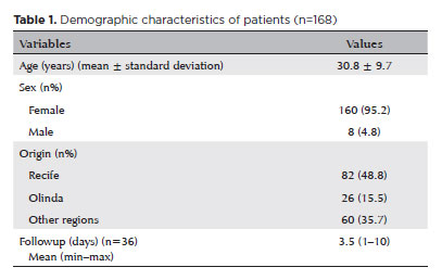
RESULTS: The study included 168 patients (95.2% [n=160] female), with a mean age of 30.8 ± 9.7 years. The most frequently reported symptoms at presentation were pain (167/168, 99.4%) and photophobia (92/168, 54.8%). Severe pain was reported by 137 patients (80%). Keratitis was present in 280 of 336 eyes (83.3%), conjunctival hyperemia in 256 eyes (76.4%), and corneal abrasions in 174 eyes (51.8%). A decrease in visual acuity (worse than 20/25) was documented in 18.5% (31/168) of cases. Lubricants, antibiotics, and re-epithelialization
ointments were prescribed to 64.8% (109/168) of the patients. Topical corticosteroids and oral vitamin C were administered to 34% (57/168) and 1.2% (2/168) of patients, respectively. Followup visits were required in 19% (33/168) of cases.
CONCLUSION: The outbreak of chemical ocular injuries linked to cosmetic ointments used for braiding and hair modeling in Brazil was marked by intense ocular pain, conjunctival hyperemia, keratitis, and corneal abrasions. Most patients were treated with lubricants, antibiotics, and re-epithelialization ointments, although approximately one-fifth required followup care, and one-third received additional treatment with either topical corticosteroids and/or oral vitamin C.
Keywords: Cosmetics; Hair preparations; Eye injuries; Burns, chemical; Eye burns; Keratitis; Cornea; Corneal diseases; Visual low.
Arq. Bras. Oftalmol. 2023;86 (5 )
:1-6
| DOI: 10.5935/0004-2749.20230067
Abstract
Objetivos: O objetivo do estudo é avaliar o perfil das visitas ao Pronto-Socorro de Oftalmologia (PS) do Hospital São Paulo, serviço público de atendimento terciário aberto 24 horas em São Paulo - Brasil, pertencente à Universidade Federal de São Paulo, nos últimos 11 anos.
Métodos: Foi realizado um estudo transversal retrospectivo, com base em todos os pacientes (n=634.726) admitidos no pronto-socorro de oftalmologia do Hospital São Paulo entre janeiro de 2009 e dezembro de 2019.
Resultados: De 2009 a 2019, houve um aumento no influxo de 39,2% com importante variação nos atendimentos ao longo dos anos, a mediana de idade foi de 38 ± 20,4 anos, o sexo masculino representou 53,3% e os pacientes únicos representaram 53,1%. Verificou-se que 79,5% das visitas ocorreram das 7h às 17h e 80,8% nos dias da semana. Os diagnósticos mais frequentes foram conjuntivite aguda seguida de blefarite, ceratite, hordéolo / calázio e corpo estranho corneano.
Conclusão: Ao longo do período de análise do estudo, houve importante aumento nas apresentações, com predominância de atendimentos não urgentes e baixa proporção de pacientes com uma única visita. Nossos resultados evidenciam o perfil das consultas oftalmológicas, podendo gerar mudanças no sistema público de saúde visando a melhoria da qualidade do atendimento e acesso às emergências oftalmológicas na cidade de São Paulo.
Keywords: Serviço hospitalar de emergência; Epidemiologia; Traumatismos oculares; Oftalmopatias.
Arq. Bras. Oftalmol. 2025;88 (1 )
:1-5
| DOI: 10.5935/0004-2749.2023-0083
Abstract
PURPOSE: This study aimed to determine whether early-stage intraocular pressure can be modulated using a thermal face mask.
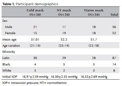
METHODS:In this prospective clinical study, healthy participants were randomized on a 1:1:1 allocation ratio to three mask groups: hypothermic (G1), normothermic (G2), and hyperthermic (G3). After randomization, 108 eyes from 108 participants were submitted to clinical evaluations, including measurement of initial intraocular pressure (T1). The thermal mask was then applied for 10 minutes, followed by a second evaluation of intraocular pressure (T2) and assessment of any side effects.
RESULTS:The hypothermic group (G1) showed a significant reduction in mean intraocular pressure between T1 (16.97 ± 2.59 mmHg) and T2 (14.97 ± 2.44 mmHg) (p<0.001). G2 showed no significant pressure difference between T1 (16.50 ± 2.55 mmHg) and T2 (17.00 ± 2.29 mmHg) (p=0.054). G3 showed a significant increase in pressure from T1 (16.53 ± 2.69 mmHg) to T2 (18.58 ± 2.95 mmHg) (p<0.001). At T1, there was no difference between the three study groups (p=0.823), but at T2, the mean values of G3 were significantly higher than those of G1 and G2 (p<0.00).
CONCLUSION:Temperature was shown to significantly modify intraocular pressure. Thermal masks allow the application of temperature in a controlled, reproducible manner. Further studies are needed to assess the duration of these effects and whether they are reproducible in patients with pathologies that affect intraocular pressure.
Keywords: Intraocular pressure; Temperature; Masks; Glaucoma; Eye diseases
Arq. Bras. Oftalmol. 2024;87 (3 )
:1-5
| DOI: 10.5935/0004-2749.2021-0490
Abstract
OBJETIVO: Analisar a teleconsulta em um hospital público de ensino oftalmológico, durante o período da pandemia do COVID-19.
MÉTODOS: Foram revisados os registros médicos dos pacientes que solicitaram teleconsulta oftalmológica, no período de Junho de 2020 a Março de 2021. Os resultados incluem dados demográficos, sintomas de queixas oculares e hipóteses diagnósticas. Além disso, foram analisados dados da pesquisa de satisfação aplicada após cada teleconsulta.
RESULTADOS: Um total de 161 prontuários foram revisados. A idade média dos pacientes foi de 45.98 ± 17.57 (8 a 90) anos, a maioria mulheres, 113 (70,20%). Apenas 57 (35,60%) eram pacientes acompanhados no hospital previamente. A principal razão pela busca pela teleconsulta foi o erro refracional, 73 (45.43%), seguido de olho seco, 16 (9.93%), pterígio, 13 (8.07%). Outros motivos foram o acompanhamento de doenças prévias como glaucoma, retinopatias, miopia, estrabismo e ceratocone. Quanto a pesquisa de satisfação, 151(93,87%) pacientes responderam a pesquisa on-line. A maioria deles mostrou-se satisfeito com a teleconsulta (94.03%) e fariam uma nova teleconsulta (90.06%).
CONCLUSÃO: A teleconsulta pode auxiliar a saúde pública em oftalmologia podendo ser utilizada em hospitais universitários. Embora o erro refracional tenha sido o motivo mais frequente nas consultas, os pacientes mostraram-se satisfeitos com essa modalidade de atendimento que serve como um serviço de orientação.
Keywords: COVID-19; Telemedicina; Pandemias; Consulta remota; Oftalmopatias/diagnóstico; Hospitais públicos
Arq. Bras. Oftalmol. 2025;88 (3 )
:1-5
| DOI: 10.5935/0004-2749.2023-0174
Abstract
PURPOSE: To compare objective and subjective intraocular pressure measurements immediately after cataract surgery and intraocular pressure measurements between less experienced surgeons (Group 1) and experienced surgeons (Group 2).
METHODS: Surgeons were asked to estimate the IOP after corneal sealing after surgery based on their tactile perception of eye tension (subjective intraocular pressure) Objective intraocular pressure was measured using a Perkins tonometer while patients were still in the surgical field. Objective intraocular pressure was compared to subjective intraocular pressure. Results from less experienced surgeons were compared to more experienced surgeons.
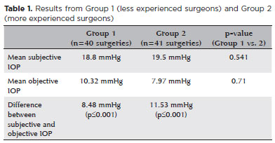
RESULTS: The study comprised 81 surgeries (81 eyes) performed by 27 surgeons. The mean objective intraocular pressure (9.14 mmHg; SD=5.86) was statistically significantly lower (p<0.001) than the mean subjective intraocular pressure (19.21 mmHg; SD=4.82). Hypotony (intraocular pressure <6mmHg) was observed in 25 eyes (30.86%). The mean subjective intraocular pressure was 18.8 mmHg (SD=5.19) for less experienced surgeons and 19.5 mmHg (SD=4.46) for more experienced, without statistically significant difference (p=0.541). No statistically significant difference (p=0.71) was observed when comparing objective intraocular pressure in Group 1 (10.32 mmHg; SD=6.65) and Group 2 (7.97 mmHg; SD=4.7).
CONCLUSION: Objective intraocular pressure was significantly lower than subjective intraocular pressure, regardless of surgeons' experience. This study showed that the subjective method is unreliable compared to the gold standard (Perkins tonometer) and does not improve with surgeons' experience. Establishing standard training methods is paramount to developing surgeons' skills.
Keywords: Cataract; Intraocular pressure; Hypotony, Tonometry; Eye diseases; Training
Arq. Bras. Oftalmol. 2025;88 (2 )
:1-7
| DOI: 10.5935/0004-2749.2024-0029
Abstract
PURPOSE: To evaluate the effect of upper eyelid ptosis repair with Muller muscle-conjunctival resection on meibomian gland function and ocular surface parameters.
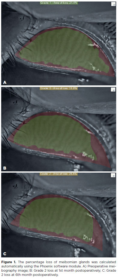
METHODS: Thirty-eight patients who underwent ptosis repair with Muller muscle-conjunctival resection were retrospectively reviewed. Meibomian gland loss, Ocular Surface Disease Index OXFORD score, meiboscore, and noninvasive keratograph break-up time were measured preoperatively and at 1st, 3rd, and 6th months postoperatively.
RESULTS: Noninvasive keratograph break-up time values decreased significantly at 1st and 3rd months postoperatively compared to the preoperative level, but were similar to the preoperative level at 6th months postoperatively (p<0.001 and p=0.628, respectively). Ocular surface disease index, OXFORD score, meibomian gland loss, and meiboscore values increased significantly in the 1st and 3rd postoperative months compared to the preoperative period, but these values decreased to preoperative levels in the 6th postoperative month (p<0.001 and p>0.05, respectively).
CONCLUSION: There is a transient deterioration in meibography findings and OSDI score in the early postoperative period after Muller muscle-conjunctival resection. Patients undergoing Muller muscle-conjunctival resection may require topical lubricants, especially in the first 3 postoperative months.
Keywords: Meibomian glands; Blepharoptosis; Preoperative period; Conjunctiva; Muscles; Eyelid diseases; Diagnostic techniques, ophthalmological
Arq. Bras. Oftalmol. 2025;88 (3 )
:1-6
| DOI: 10.5935/0004-2749.2024-0207
Abstract
PURPOSE: This study aimed to report the use, efficacy, and safety of intracameral voriconazole as an adjuvant treatment for deep fungal keratitis.
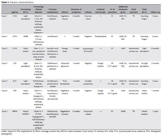
METHODS: This was a prospective case series of seven eyes with fungal keratitis with anterior chamber involvement or a corneal ulcer refractory to conventional topical treatment. In addition to topical treatment with 0.15% amphotericin B eye drops, voriconazole 50 μg/ 0.1 mL
was administered to the anterior chamber of each affected eye up to four times within 72 h. The primary outcome measures were healing (fungal eradication) and the need for therapeutic keratoplasty. Best-corrected visual acuity was a secondary outcome measure.
RESULTS: Three cases were confirmed by confocal microscopy, and four were diagnosed from positive culture tests. At presentation, one patient had a best-corrected visual acuity of 20/80, while all others had hand motion or worse. Four cases received one intracameral injection, two cases received three, and one case received four injections. There were no complications after any of the intracameral voriconazole injections. Four patients had imminent corneal perforations and were treated with cyanoacrylate adhesive and bandage contact lenses. Four patients recovered from the infection, and three underwent therapeutic keratoplasty. The final best-corrected visual acuity was improved in two cases but all patients had a final visual acuity of counting fingers or worse.
CONCLUSION: As an adjuvant treatment for deep fungal keratitis, intracameral voriconazole injection is a feasible option. Although fungal eradication was achieved in all patients, three required therapeutic keratoplasty and all patients had unsatisfactory visual acuity outcomes.
Keywords: Antifungal agents; Fungi; Corneal transplantation; Keratitis; Eye infections, fungal; Voriconazole


 ABO is licensed under a Creative Commons Attribution-NonComercial 4.0 Internacional.


06-tab01tb.jpg)
13-tab01.jpg)
09-tab01tb.jpg)
10-tab01.jpg)
02-fig01.jpg)