Arq. Bras. Oftalmol. 2025;88 (4 )
:1-12
| DOI: 10.5935/0004-2749.2023-0263
Abstract
PURPOSE: Amblyopia is a cortical neurological disorder caused by abnormal visual experiences during the critical period for visual development. Recent works have shown that, in addition to the well-known visual alterations, such as changes in visual acuity, several perceptual aspects of vision are affected. This study aims to analyze and compare the effects of different types of amblyopia on visual color processing and determine whether these effects are correlated with visual acuity.
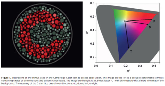
METHODS: Our study sample comprised 42 amblyopic individuals, aged 7-40 years, (strabismus, n=16; anisometropia, n=18; and mixed-cause, n=8) and 33 age-matched controls. Color vision was tested by measuring the chromaticity threshold of each patient on the protan, deutan, and tritan axes using version 02 of the Cambridge Color Test. Spatial stimulation cues were eliminated using spatial noise and luminance.
RESULTS: The color discrimination thresholds on the protan, deutan, and tritan axes were similar between control participants and amblyopic patients (p>0.05). There was no correlation between VA values and color thresholds (p>0.05).
CONCLUSION: Patients with amblyopia have normal color vision in contexts that include luminance and spatial noise. Our results may be indicative of independent neural pathways for spatial and chromatic visual processing.
Keywords: Amblyopia; Anisometropia; Color vision; Strabismus; Vision disorders; Visual acuity
Arq. Bras. Oftalmol. 2021;84 (4 )
:374-379
| DOI: 10.5935/0004-2749.20210065
Abstract
Objetivo: Sincinesias são resultado de inervações anômalas e ocasionam movimentos aberrantes dos músculos envolvidos. Apresentamos uma série com casos raros de sincinesias oculares congênitas dos músculos extraoculares e do levantador da pálpebra superior e especulamos a possibilidade de classificá-las dentro do espectro das desordens congênitas da desnervação cranianana.
Métodos: Prontuários de pacientes com diagnóstico de sincinesia ocular congênita foram estudados retrospectivamente. Analisamos sexo, lateralidade e as características completas do exame de motilidade de cada paciente.
Resultados: Nove pacientes com sincinesias oculares congênitas foram incluídos. Houve discreta predominância no sexo feminino. Em termos de lateralidade, o olho direito foi o único envolvido em 4 casos, o olho esquerdo também em 4 casos e 1 caso apresentou acometimento bilateral. 55,5% dos pacientes eram ortotrópicos na posição primária. Os III, VI e IV nervos participaram da sincinesia em 100%, 44,4% e 11,1% dos casos, respectivamente.
Conclusões: Sincinesias oculares congênitas podem se apresentar de modo bastante eclético e incomum. A inervação aberrante presente em cada um desses casos os coloca na lista de candidatos a integrar o grupo das desordens congênitas da desenervação craniana.
Keywords: Sincinesia; Nervo troclear; Nervos cranianos/ anormalidades; Músculos oculomotores; Transtornos da motilidade ocular/congênito
Arq. Bras. Oftalmol. 2025;88 (4 )
:1-6
| DOI: 10.5935/0004-2749.2024-0236
Abstract
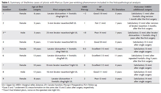
PURPOSE: This study was conducted to report the histopathological and clinical features of the Marcus Gunn phenomenon and other similar conditions of upper eyelid misfiring.
METHODS: This was a retrospective study of patients with congenital ptosis with Marcus Gunn phenomenon who have undergone surgical repair over a period of 12 years and another two patients with upper eyelid misfiring in association with extraocular movements to identify their histopathological findings as subtypes representing ocular congenital cranial dysinnervation disorder.
RESULTS: Among 136 patients with congenital ptosis, 11 (8%) patients with Marcus Gunn phenomenon or misfiring were identified, of whom 9 (6.6%) had typical known Marcus Gunn phenomenon and 2 (1.4%) had eyelid misfiring similar to Marcus Gunn phenomenon. In all patients, the histopathological changes of the excised levator muscle included overall loss and/or atrophy of muscle fibers and irregular-modified Gomori trichrome staining.
CONCLUSION: The Marcus Gunn phenomenon and similar misfiring conditions with synkinetic extraocular muscle movements share findings that are consistent with the neurogenic type of muscle atrophy. This result suggests a common underlying etiology with variable clinical findings, representing the ocular counterpart of congenital cranial dysinnervation disorder, which has been reported as ocular congenital cranial dysinnervation disorder.
Keywords: Eyelid diseases; Ocular motility disorders/surgery; Ophthalmologic surgical procedures
Arq. Bras. Oftalmol. 2026;89 (1 )
:1-6
| DOI: 10.5935/0004-2749.2025-0071
Abstract
PURPOSE: This study aimed to evaluate the outcomes of strabismus surgical correction in patients with Down syndrome.
METHODS: We conducted a retrospective chart review of patients with Down syndrome who underwent strabismus surgery between January 1997 and May 2024 at an Ophthalmology Outpatient Clinic in São Paulo, Brazil. The data collected included age, sex, medical and ocular history, surgical details, and follow-up outcomes. The patients were categorized by strabismus type into esotropia, fourth nerve palsy, and mixed groups. Surgical success was defined as final alignment within 10Δ of orthotropia and, where applicable, whether there was resolution of abnormal head posture of ocular origin. Patients with postoperative follow-up <6 months were excluded.
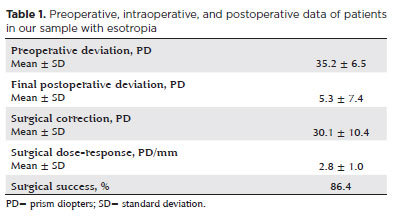
RESULTS: A total of 37 patients (21 females) were included. Of these, 22 (59.5%) were in the esotropia group, 10 (27.0%) in the fourth nerve palsy group, and 5 (13.5%) in the mixed group. The surgical success rate in the esotropia group was 86.4%, with a mean preoperative deviation of 35.2 (± 6.5)Δ, and mean surgical correction of 30.1 (± 10.4)Δ. The success rate in the fourth nerve palsy group was 40.0%, with a mean preoperative deviation of 10.4 (± 4.3)Δ. Overall, success was achieved with a single surgical procedure in 73.0% of the sample. No significant associations were found between surgical success and the clinical and demographic variables, including sex, age at surgery, oblique muscle overaction, pattern strabismus, visual acuity, amblyopia, preoperative deviation, or postoperative follow-up duration (p>0.05).
CONCLUSIONS: When standard surgical tables are applied, strabismus surgery in patients with Down syndrome appears to be safe and effective. We found high success rates, particularly among patients with esotropia. We observed no tendencies toward over- or under-correction. These findings support the use of conventional surgical protocols with this patient population.
Keywords: Down Syndrome/complications; Strabismus/surgery; Esotropia/surgery; Oculomotor nerve diseases/physiopathology; Vision disorders; Humans; Brazil.
 ABO is licensed under a Creative Commons Attribution-NonComercial 4.0 Internacional.
15-tab01tb.jpg)
12-fig01.jpg)
08-fig01.jpg)
08-fig01.jpg)
09-fig01.jpg)
13-fig01.jpg)
11-fig01tb.jpg)
10-fig01.jpg)

01-fig01.jpg)