Arq. Bras. Oftalmol. 1999;62 (6 )
:697-700
| DOI: 10.1590/S0004-27491999000600008
Abstract
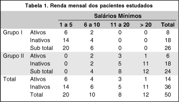
Objetivo: Estudar os custos de correção dos vícios de refração em grupos de pessoas de distinto poder aquisitivo. Métodos: Os autores estudaram cinqüenta pacientes portadores de vícios de refração. Estes foram separados em dois grupos: grupo I com pacientes escolhidos de forma aleatória na primeira consulta ao ambulatório de Oftalmologia do Hospital Evangélico de Curitiba (HUEC), e grupo II com voluntários médicos do HUEC e acadêmicos de medicina da Faculdade Evangélica de Medicina do Paraná (FEMPAR). Foram analisados dados referentes a sexo, faixa etária, profissão, renda, grau de instrução, uso de correção (óculos ou lentes) e seu custo, consultas oftalmológicas. Os pacientes foram submetidos ao exame oftalmológico de rotina. Resultados: Encontramos no grupo I predominância de pacientes de meia idade (48,5 anos), com renda entre 1 a 5 salários mínimos (SM) e hipermétropes; e no grupo II, pacientes jovens (24,4 anos), com renda acima de 20 SM e míopes foram mais freqüentes. Conclusão: O gasto médio anual com óculos fica no mínimo em R$ 46,50 (0,3 SM); com lentes de contato, no mínimo R$ 196,66 (1,4 SM); e com cirurgia refrativa em R$ 800,00 (5,9 SM). O estudo sugere a cirurgia refrativa como boa indicação para ambos os grupos.
Keywords: Vícios de refração; Aspectos socioeconômicos
Arq. Bras. Oftalmol. 2026;89 (2 )
:1-9
| DOI: 10.5935/0004-2749.2025-0113
Abstract
PURPOSE: This study aimed to identify the strategies adopted by Brazilian ophthalmologists to control myopia in clinical practice.
METHODS: This was a prospective cross-sectional study. Data were collected using an online questionnaire.
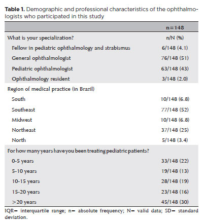
RESULTS: Responses from 148 participants were collected between March and May 2024. The majority of respondents were general ophthalmologists (51%) and pediatric ophthalmologists (43%). They came from all regions of Brazil, but more than half (52%) were from the Southeast region. Most participants (30%) had over 20 years of clinical practice experience. A significant proportion (89.2%) treated progressive myopia. The most requested complementary exams were optical biometry (83.78%) and corneal topography or tomography (69.59%). Behavioral measures were considered the most effective myopia treatment strategies by 41.2% of the respondents, followed by optical (33.8%) and pharmacological interventions (25%). Most recommended spending more time outdoors (94.59%) and reducing screen time (93.92%). Spectacle lenses for myopia (83.11%) and 0.025% atropine eye drops (54.73%) were the most prescribed treatments after the recommendation of environmental and behavioral changes.
CONCLUSION: This study presents a novel analysis of the clinical strategies for myopia control among Brazilian ophthalmologists. Understanding current clinical practices and identifying possible improvements are essential steps toward developing evidence-based guidelines and professional education aimed at improving patient care.
Keywords: Myopia/epidemiology; Refractive errors; Contact lenses; Myopia/drug therapy; Atropine/therapeutic use; Ophthalmologists; Practice patterns, physicians’; Surveys and questionnaires; Brazil/epidemiology
Arq. Bras. Oftalmol. 2020;83 (6 )
:490-496
| DOI: 10.5935/0004-2749.20200090
Abstract
Objetivo: Comparar a espessura central foveal, a da camada de fibras nervosas da retina e a da coróide subfoveal através da tomografia de coerência óptica swept-source em crianças de 5 anos de idade com história de retinopatia da prematuridade (RP) tratada com bevacizumabe intravítreo, ou com fotocoagulação a laser, com crianças em regressão espontânea da retinopatia da prematuridade, e com crianças saudáveis da mesma idade.
Métodos: Um total de 79 crianças foi dividido em quatro grupos. Grupo 1: crianças que receberam tratamento com bevacizumabe intravítreo. Grupo 2: crianças que foram tratadas com fotocoagulação a laser. Grupo 3: crianças que tiveram regressão espontânea da retinopatia da prematuridade . Grupo 4: crianças da mesma idade saudáveis e nascidas a termo. As funções visuais e o status refrativo foram avaliados aos 5 anos de idade. A análise de tomografia de coerência óptica foi feita por um dispositivo do tipo swept-source (DRI-OCT Triton; Topcon, EUA).
Resultados: Haviam 12 crianças (15,2%) no grupo 1, 23 crianças (29,1%) no grupo 2, 30 crianças (38%) no grupo 3 e 14 crianças (17,7%) no grupo 4. A distribuição por sexo foi semelhante em todos os grupos (p=0,420). A acuidade visual com a melhor correção mostrou-se significativamente maior no grupo 4 em comparação com os grupos 1, 2 e 3 (respectivamente, p=0,035, p=0,001 e p=0,001). Os resultados dos erros de refração foram semelhantes em todos os grupos (p=0,119). A espessura foveal central mostrou-se significativamente maior no grupo 2 do que no grupo 1 (p=0,023). Não foram observadas diferenças significativas entre os grupos quanto à espessura da camada de fibras nervosas da retina e à espessura da coroide subfoveal (p>0,05).
Conclusões: Os desfechos visuais funcionais foram melhores nas crianças saudáveis nascidas a termo, em comparação com aqueles observados nas crianças com história de retinopatia da prematuridade tratada ou com regressão espontânea. O tratamento com laser teve um efeito significativo na espessura foveal central em crianças de 5 anos de idade, nascidas prematuras, como revelado pela tomografia de coerência óptica swept-source.
Keywords: Retinopatia da prematuridade/tratamento farmacológico; Tomografia de coerência óptica; Bevacizumab/uso terapêutico; Fotocoagulação; Recém-nascido
Arq. Bras. Oftalmol. 2025;88 (1 )
:1-7
| DOI: 10.5935/0004-2749.2022-0367
Abstract
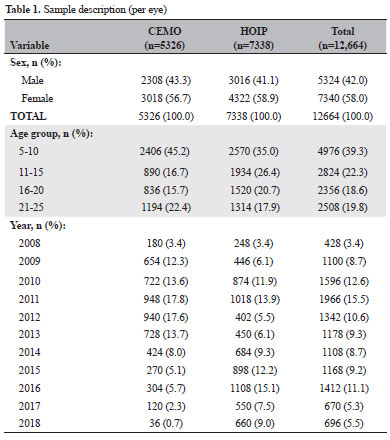
PURPOSE: This study aimed to examine the prevalence of myopic eyes over 11 years (2008-2018) in a private clinic and a public assistance service.
METHODS: We retrospectively evaluated 6332 individuals (12,664 eyes)
between 5 and 25 years old, seen at a private clinic-CEMO (2,663 individuals) and a public service-HOIP (3,669 individuals) from 2008 to 2018. We evaluated the prevalence of myopic eyes (EE ≤-0.50) and high myopic eyes (EE ≤-6.00).
RESULTS: Sex and services did not show statistical differences. The variation in the prevalence of myopic and high myopic eyes showed a random pattern during the study period (this prevalence could not be increased). Prevalences ranged from 20.7% (in 2017) to 32.4% (in 2015) for myopic eyes and from 1.6% (in 2009 and 2016) to 3.3% (in 2015) for eyes with high myopia. The prevalence of myopia showed a statistically significant increase based on the age group.
CONCLUSION: The prevalence of myopic eyes did not increase in our study. The mean prevalence of myopic eyes was similar in the private clinic and public service.
Keywords: Myopia; Refractive errors; Epidemiology; Prevalence
Arq. Bras. Oftalmol. 2023;86 (3 )
:1-8
| DOI: 10.5935/0004-2749.20230030
Abstract
Objetivo: Avaliar o desempenho clínico do Spot Vision Screener e estabelecer correlações clínicas entre a triagem automatizada e a retinoscopia após indução de cicloplegia em crianças pré-verbais.
Métodos: Neste estudo transversal prospectivo, crianças de 6 a 36 meses foram avaliadas usando o Spot Vision Screener. O exame oftalmológico completo, incluindo refração cicloplégica, foi então realizado, seguido de repetição da triagem automatizada e retinoscopia em todos os casos, a fim de estabelecer correlações quanto à hipermetropia, miopia e astigmatismo após a indução de cicloplegia.
Resultados: O estudo incluiu 185 crianças. A sensibilidade do dispositivo de triagem automática após cicloplegia foi de 100% (IC 95%: 85,18-100%) e a especificidade foi de 87,04% (IC 95%: 80,87-91,79%). Os valores preditivos positivos e negativos foram de 52,27% (42,36 - 62,01%) e 100%, respectivamente. Em comparação com a retinoscopia, o Spot Vision Screener superestimou os valores esféricos em 0,62 D (IC 95%: 0,56 - 0,69) no olho direito e em 0,60 (IC 95%: 0,54 - 0,66) no olho esquerdo e os valores cilíndricos em -0,38 D (IC 95%: -0,42 a -0,33) no olho direito e por -0,39 D (IC 95%: -0,43 a -0,34) no olho esquerdo. A diferença para os valores esféricos e cilíndricos de forma geral foi de 0,61 D (IC 95%: 0,57 - 0,65) e -0,38 D (IC 95%: -0,41 a -0,35), respectivamente.
Conclusão: Foi encontrada correlação substancial entre a retinoscopia e os dados objetivos captados pelo dispositivo. Isso mostra que a tecnologia pode ser usada em conjunto, contribuindo para um diagnóstico mais preciso e identificando os fatores de risco de ambliopia o mais precocemente possível. A técnica automatizada pode fazer a diferença em nível populacional para triagem e intervenção precoce.
Keywords: Erros de refração; Ambliopia; Estrabismo; Refratometria; Retinoscopia
Arq. Bras. Oftalmol. 2022;85 (1 )
:37-45
| DOI: 10.5935/0004-2749.20220006
Abstract
Objetivos: Avaliar a sensibilidade ao contraste em pacientes virgens de tratamento com retinopatia diabética proliferativa de não alto risco, submetidos a panfotocoagulação retiniana com injeções intravítreas de ranibizumabe versus panfotocoagulação isolada.
Métodos: Sessenta olhos de 30 pacientes foram randomizados em dois grupos: um submetido a panfotocoagulação com injeções de ranibizumabe (grupo estudo), e o outro submetimedo a panfotocoagulação isolada (grupo controle). Todos olhos foram tratados em 3 sessões de laser, seguindo recomendação do Early Treatment Diabetic Retinopathy Study (ETDRS). Avaliação da sensibilidade ao contraste foi realizada sob condições fotópicas (85 cd/m2) com tabela Visual Contrast Test Sensitivity 6500, permitindo avaliação de cinco frequências espaciais medidas com redes senoidais: 1.5, 3.0, 6.0, 12.0 e 18.0 ciclos por grau de ângulo visual (cpd). Foram realizadas medidas dos limiares de sensibilidade ao contraste intra e entre grupos na visita inicial, no 1º, 3º, e 6º mês de seguimento.
Resultados: Cinquenta e oito olhos, 28 do grupo estudo e 30 do grupo controle, atingiram o término do estudo. Análise comparativa da SC entre os grupos mostrou diferença estatisticamente significante, nas baixas frequências espaciais, no 1º mês em 1.5 cpd (p=0,001) e 3.0 cpd (p=0,04), no 3º mês em 1.5 cpd (p=0,016) e no 6º mês em 3.0 cpd (p=0,026) a favor do grupo estudo.
Conclusão: O tratamento com panfotocoagulação associada a injeção de ranibizumabe parece causar menos danos a sensibilidade ao contraste quando comparada com panfotocoagulação isolada em olhos com retinopatia diabética proliferativa de não alto risco. Dessa forma, os resultados apresentados podem justificar a associação do ranibizumabe à panfotocoagulação nestes pacientes.
Keywords: Retinopatia diabética; Fotocoagulação; Ranibizumab; Bevacizumab; Sensibilidade de contraste; Fator A de crescimento do endotélio vascular; Injeção intravítrea.
Arq. Bras. Oftalmol. 2025;88 (4 )
:1-8
| DOI: 10.5935/0004-2749.2024-0277
Abstract
PURPOSE: This retrospective study evaluated the safety and efficacy of real-world antiangiogenic therapy for ocular conditions in the private healthcare sector in southern Brazil.
METHODS: Medical records from patients who underwent intravitreal anti-vascular endothelial growth factor injections over the past 12 years were reviewed retrospectively. Data collection included the primary diagnoses, drugs administered, injection techniques, adverse effects, and treatment efficacy. Efficacy was assessed by comparing pre- and posttreatment visual acuity and central subfield thickness in eyes with followup exceeding 2 years.
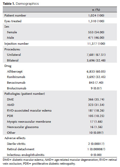
RESULTS: A total of 1,024 patients, 1,310 treated eyes, and 11,377 injections were analyzed. The injections included aflibercept (6,833), ranibizumab (3,692), bevacizumab (843), and brolucizumab (9), administered either bilaterally (3,696) or unilaterally (7,681). The most common diagnoses were diabetic macular edema, exudative age-related macular degeneration, retinal vein occlusion related macular edema, and proliferative diabetic retinopathy. No endophthalmitis cases were reported. Vitritis with transient visual acuity loss occurred in two cases following aflibercept injections. One retinal detachment case was successfully treated with vitrectomy. The median number of injections per patient was 6 (IQR [interquartile range], 3–13). Among 445 eyes from 328 patients with followup over 2 years (median, 4.05 years; IQR, 2.89–6.29), there was a significant improvement in best-corrected visual acuity from 0.3 to 0.4 (Snellen) (p<0.001) and a reduction in central subfield thickness from 361 to 267 microns (p<0.001). CST comparisons included patients with age-related macular degeneration, diabetic macular edema, and retinal vein occlusion related macular edema.
CONCLUSION: This real-world study, the largest of its kind in Brazil, confirms the safety and efficacy of antiangiogenic therapies in the southern Brazilian private healthcare system. The findings highlight a low incidence of severe adverse events and outcomes consistent with global studies, supporting the ongoing use of antiangiogenic agents as effective and well-tolerated treatments for various ocular conditions in developing countries.
Keywords: Antiangiogenic drugs; Macular edema; Age-related macular degeneration; Retinal vein occlusion; Patient safety
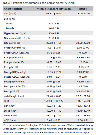
Arq. Bras. Oftalmol. 2025;88 (2 )
:1-7
| DOI: 10.5935/0004-2749.2023-0215
Abstract
PURPOSE: To compare the refractive prediction error of Hill-radial basis function 3.0 with those of 3 conventional formulas and 11 combination methods in eyes with short axial lengths.
METHODS: The refractive prediction error was calculated using 4 formulas (Hoffer Q, SRK-T, Haigis, and Hill-RBF) and 11 combination methods (average of two or more methods). The absolute error was determined, and the proportion of eyes within 0.25-diopter (D) increments of absolute error was analyzed. Furthermore, the intraclass correlation coefficients of each method were computed to evaluate the agreement between target refractive error and postoperative spherical equivalent.
RESULTS: This study included 87 eyes. Based on the refractive prediction error findings, Hoffer Q formula exhibited the highest myopic errors, followed by SRK-T, Hill-RBF, and Haigis. Among all the methods, the Haigis and Hill-RBF combination yielded a mean refractive prediction error closest to zero. The SRK-T and Hill-RBF combination showed the lowest mean absolute error, whereas the Hoffer Q, SRK-T, and Haigis combination had the lowest median absolute error. Hill-radial basis function exhibited the highest intraclass correlation coefficient, whereas SRK-T showed the lowest. Haigis and Hill-RBF, as well as the combination of both, demonstrated the lowest proportion of refractive surprises (absolute error >1.00 D). Among the individual formulas, Hill-RBF had the highest success rate (absolute error ≤0.50 D). Moreover, among all the methods, the SRK-T and Hill-RBF combination exhibited the highest success rate.
CONCLUSIONS: Hill-radial basis function showed accuracy comparable to or surpassing that of conventional formulas in eyes with short axial lengths. The use and integration of various formulas in cataract surgery for eyes with short axial lengths may help reduce the incidence of refractive surprises.
Keywords: Cataract; Lenses, intraocular; Axial length, eye; Refractive errors; Artificial intelligence
 ABO is licensed under a Creative Commons Attribution-NonComercial 4.0 Internacional.
05-fig01tb.jpg)
06-tab01.jpg)
10-tab01tb.jpg)
12-fig01.jpg)
10-fig01.jpg)

02-fig01.jpg)