Arq. Bras. Oftalmol. 2006; 69 (4): 10.1590/S0004-27492006000400003
Total: 2388
Sebastião Cronemberger1; Dollores Martins Ferreira2; Alberto Diniz Filho3; Rafael Vidal Mérula4; Nassim Calixto5
DOI: 10.1590/S0004-27492006000400003
ABSTRACT
PURPOSE: To present the prevalence of iridociliary cysts in eyes of patients submitted to UBM examination. METHODS: We analyzed retrospectively the UBM images of 1157 patients in the period from September 1995 to June 2004. The inclusion criterion was the UBM examination had been made on the four quadrants (superior, inferior, nasal and temporal) of the eyes. The cysts were assessed and classified in relation to their number in each quadrant; localization and morphology. Using the UBM Pro 2000 software we took the measurements of the largest diameter, wall thickness and area of the cysts besides the angle recession area (ARA). RESULTS: The prevalence of the cysts was 4.9%. No statistical difference was found between the prevalence of cysts regarding gender or between the right and left eyes. They were more prevalent in the inferior (39.8%) and temporal (29.1%) quadrants. The cysts with the largest diameter and area were located in the temporal and superior quadrants. No difference was found in relation to cysts' wall thickness in all quadrants. CONCLUSIONS: No statistical difference was found between the prevalence of cysts as concerns gender or between the right and left eyes. However, the cysts were more prevalent in the inferior and temporal quadrants. The locations of the largest cysts are preferentially the temporal and superior quadrants. The angle recession area was smaller in the temporal and superior quadrants. The cysts' wall thickness is practically the same in all quadrants. The diagnosis of iridociliary cysts was an occasional finding in 64.3% of the patients.
Keywords: Prevalence; Cysts; Iris neoplasms; Microscopy; Ultrasonography
RESUMO
OBJETIVO: Relatar a prevalência de cistos iridociliares em olhos de pacientes submetidos à biomicroscopia ultra-sônica (UBM). MÉTODOS: Analisaram-se retrospectivamente as imagens de UBM de 1.557 pacientes examinados de setembro de 1995 a junho de 2004. O critério de inclusão foi a UBM ter sido realizada nos quatro quadrantes (superior, inferior, nasal e temporal) do globo ocular. Avaliaram-se e classificaram-se os cistos quanto: a) ao número em cada quadrante; b) ao quadrante de localização; c) à morfologia, medindo o maior diâmetro (vertical ou horizontal), a maior espessura da parede e a área da lesão cística; d) à área de recesso angular (ARA) para cada um dos quadrantes em que havia cisto; e) à hipótese diagnóstica e/ou indicação da UBM. Utilizou-se o "software" UBM Pro 2000 para medir o diâmetro, a espessura da parede, a área do cisto e a ARA. RESULTADOS: Foram encontrados 103 cistos em 56 pacientes correspondendo à prevalência de 4,9% numa amostra de 1.132 pacientes selecionados. Dos 1.132 pacientes, 650 (57,4%) eram do sexo feminino e 482 (42,6%) eram do sexo masculino. Dos 56 pacientes com cisto, 37 (66,1%) eram do sexo feminino e 19 (33,9%) eram do sexo masculino. Dos 1.480 olhos examinados, 774 (52,3%) eram olhos direitos e 706 (47,7%) eram olhos esquerdos. Foram encontrados cistos em 38 (64,4%) olhos direitos e 21 (35,6%) olhos esquerdos. Não houve diferença estatisticamente significativa entre os sexos nem entre os olhos direito e esquerdo. Os cistos com maior média de diâmetro e área estavam localizados nos quadrantes temporal e superior, onde foram encontrados os menores valores de grau de abertura do seio camerular. CONCLUSÕES: Os cistos iridociliares são mais prevalentes nos quadrantes inferior e temporal. Os cistos de maior diâmetro e área situam-se nos quadrantes temporal e superior onde encontram-se menores valores médios de grau de abertura do seio camerular. A espessura da parede dos cistos é praticamente a mesma nos quatro quadrantes. Em 64,3% dos pacientes, a presença de cistos foi um achado ocasional do exame de UBM.
Descritores: Prevalência; Cistos; Neoplasias da íris; Microscopia; Ultra-sonografia
INTRODUÇÃO
A biomicroscopia ultra-sônica (BUS), também chamada de ultra-sonografia biomicroscópica (UBM), é um importante recurso propedêutico que permite o minucioso exame das estruturas das câmaras anterior e posterior do olho utilizando um ultra-som de alta freqüência (50-100 MHz)(1). É método não invasivo e indispensável na avaliação de cistos e tumores da íris e do corpo ciliar que geralmente não são detectados no exame de rotina com o biomicroscópio e o ultra-som convencional(1-3).
O equipamento possui uma sonda de freqüência variável entre 50 e 100 MHz que é incorporada a um modo B-scan(1). A UBM obtém imagens com resolução de 20 a 60 µm e com penetração máxima de 5 mm, sendo bastante valiosa no diagnóstico de lesões oculares localizadas dentro destes limites de penetração do feixe de ultra-som(1-3).
A UBM tem, entre as suas várias indicações, a de definir a relação entre as estruturas do segmento anterior, possibilitando o melhor entendimento dos mecanismos de oclusão do seio camerular e de diferentes tipos de glaucoma, a de fornecer informações importantes de estruturas intra-oculares anteriores antes de cirurgias em pacientes que apresentam opacidade da córnea, e a de avaliar cistos e tumores de íris e do corpo ciliar não visibilizados em exame de rotina(1-5).
Os cistos iridociliares primários são na sua maioria de origem neuroepitelial e de caráter benigno, a maioria deles não necessitando de tratamento(6-7). Mesmo assim, a sua avaliação é de extrema importância pois outras neoplasias de íris e de corpo ciliar podem ser confundidas com cistos devido à sua morfologia(8). Além disso, o acompanhamento dos cistos quanto ao seu crescimento pode detectar a existência de outro processo patológico adicional(1-3). Há relatos de glaucoma agudo de ângulo fechado secundário a cistos irianos(9-10).
OBJETIVO
Este trabalho tem como objetivo avaliar a prevalência de cistos iridociliares em exames de UBM e analisá-los quanto à sua localização, número, morfologia, grau de abertura do seio camerular e associação com doenças oculares.
MÉTODOS
Foram analisadas retrospectivamente as imagens de UBM de 1.557 pacientes que por diferentes indicações de estudo (suspeita de cistos irianos, uveíte intermediária, glaucoma de ângulo estreito, nevo iriano, melanose límbica, melanoma de íris, íris em platô, iridociclite crônica, sarcoidose ocular, avaliação pós-cirurgia de catarata, nanoftalmo e atalamia, etc.) foram submetidos a esse exame no período de setembro de 1995 a junho de 2004.
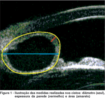
O critério de inclusão foi a UBM ter sido realizada nos quatro quadrantes (superior, inferior, nasal e temporal) do globo ocular. Os cistos foram avaliados e classificados quanto: a) ao número em cada quadrante; b) ao quadrante em que estava localizado; c) à morfologia (Figura 1), medindo-se o maior diâmetro (vertical ou horizontal) em mm, a maior espessura da parede em mm e a área da lesão cística em mm²; d) à área de recesso angular (ARA) em mm² para cada um dos quadrantes; e) à hipótese diagnóstica e/ou indicação da UBM. Para a realização dessas utilizou-se o "software" UBM Pro 2000 (Paradigm, Saint Louis, USA). Esse programa faz automaticamente a medida da ARA após a localização do esporão escleral, conforme técnica já descrita anteriormente(4). A espessura da parede do cisto foi incluída no cálculo da sua área.
As informações colhidas foram armazenadas em banco de dados apropriado(11) e analisadas descritivamente e por meio de testes estatísticos (teste para comparação de proporções(12) e ANOVA(11)), considerando-se o nível de significância de cinco por cento (p<0,05).
Todas as imagens foram analisadas e medidas pelo mesmo examinador (DMF) depois de adequado treinamento com um dos autores (SC).
RESULTADOS
Foram incluídos no estudo 1.132 pacientes, totalizando 1.480 olhos. Foram excluídos 425 pacientes porque não possuíam imagens de UBM dos quatro quadrantes. Em 56 (4,9%) pacientes e 59 (4,0%) olhos foi encontrado pelo menos um cisto em um dos quadrantes. Em 53 (94,6%) dos 56 pacientes, os cistos eram unilaterais e em apenas 3 (5,4%) eram bilaterais. Dos 1.132 pacientes, 650 (57,4%) eram do sexo feminino e 482 (42,6%) eram do sexo masculino. Dos 56 pacientes com cisto, 37 (66,1%) eram do sexo feminino e 19 (33,9%) eram do sexo masculino. Dos 1.480 olhos examinados, 774 (52,3%) eram olhos direitos e 706 (47,7%) eram olhos esquerdos. Foram encontrados cistos em 38 (64,4%) olhos direitos e 21 (35,6%) olhos esquerdos. Calculando-se a diferença de proporções(12) entre os sexos feminino e masculino verificou-se que ela não foi estatisticamente significativa (média= 0,118; erro-padrão= 0,01302 e p=0,1667). Também, a diferença de proporções entre os olhos direito e esquerdo não se mostrou estatisticamente significativa (média igual= 0,019; erro-padrão= 0,01019 e p=0,0623).
Nos 56 pacientes, foram encontrados 103 cistos dos quais 41 (39,8%) estavam situados no quadrante inferior, 30 (29,1%) no quadrante temporal, 21 (20,4%) no quadrante superior e 11 (10,7%) no quadrante nasal.
De maneira global, os valores das médias e dos desvios-padrão do diâmetro, da espessura da parede e da área dos cistos foram, respectivamente iguais a 0,853 ± 0,623 mm; 0,128 ± 0,049 mm e 0,796 ± 1,220 mm². Esses dados foram estratificados por quadrante (Tabelas 1, 2 e 3). Os quadrantes temporal e superior foram os que apresentaram cistos com maior média de diâmetro, respectivamente 1,316 e 1,039 mm (Tabela 1), assim como a maior média de área, respectivamente 1,310 e 0,888 mm (Tabela 2). Em relação às médias e desvios-padrão da espessura da parede dos cistos, observou-se que elas se mostraram muito semelhantes nos quatros quadrantes (Tabela 3).
Foram também calculados os valores médios e desvios-padrão da ARA nos quadrantes que apresentavam cistos. Esses valores foram comparados com os da ARA de indivíduos normais(4) (Tabela 4).
Analisando os valores médios e desvios-padrão encontrados para a área nos quatro quadrantes (Tabela 2), observa-se que em todos, os desvios-padrão foram maiores que as médias, o que significa que houve uma enorme dispersão das medidas das áreas dos cistos. De qualquer forma, os cistos apresentaram-se com diâmetros bem maiores nos quadrantes temporal e superior e bem menores (menos da metade da área encontrada nos quadrantes temporal e superior) no quadrante inferior.
Na tabela 4, observa-se que as médias e desvio-padrão da ARA foram diferentes em todos os quadrantes com significância estatística (p=0,0415 - ANOVA) e muito inferiores (cerca de 1/3 a 1/5) às médias e desvios-padrão da ARA encontrados em indivíduos normais(4), sendo as diferenças muito mais significativas. Chama a atenção que os desvios-padrão da ARA em pacientes com cistos são altos, às vezes até maiores que as médias (quadrantes superior e temporal) o mesmo ocorrendo em indivíduos normais, o que significa grande dispersão das medidas.
Com relação aos motivos do encaminhamento dos pacientes para exame de UBM verificou-se que foram muito variados. Dos 56 pacientes que apresentaram cisto, 20 (35,7%) foram encaminhados com suspeita de cistos irianos, 12 (21,4%), eram considerados normais, 5 (8,9%) apresentavam quadro de uveíte intermediária, 5 (8,9%) apresentavam glaucoma, 3 (5,4%) tinham hipótese diagnóstica de seio camerular estreito, em 2 (3,6%) havia suspeita diagnóstica de nevo iriano, em 1 (1,8%) o diagnóstico de suspeição era melanose límbica, em 1 (1,8%), de melanoma de íris, em 1 (1,8%), de íris em platô, 1 (1,8%) apresentava iridociclite crônica, 1 (1,8%) foi indicado para estudo de lesão pigmentada, 1 (1,8%), era portador de sarcoidose ocular, 1 (1,8%) foi encaminhado para avaliação após cirurgia de catarata, 1 (1,8%) tinha o diagnóstico de nanoftalmo e 1 (1,8%) de atalamia. Nenhum dos cistos encontrados nos pacientes deste estudo necessitou de tratamento.
DISCUSSÃO
Foram encontrados cistos em diferentes quadrantes do segmento anterior estando a maioria deles na junção iridociliar. São cistos neuroepiteliais mais ou menos redondos ou ovais, de conteúdo anecóico e parede ecogênica. Em 53 pacientes (94,6%) as lesões císticas eram unilaterais e em apenas 3 pacientes (5,4%) eram bilaterais, o que contrasta com os achados de outro estudo(7) em que os autores relataram ter encontrado em 19 (30,2%) pacientes cistos unilaterais e em 52 (82,5%) pacientes a presença de cistos em ambos os olhos.
É importante realçar que a maioria dos cistos, neste estudo, estava presente em pacientes do sexo feminino (66,1%) e no olho direito (64,4%). Pelo cálculo da diferença de proporções(12) entre os sexos, verificou-se que embora tenha havido maior prevalência de cistos no sexo feminino ela não se mostrou estatisticamente significativa (p=0,1667). Também, não se verificou diferença estatisticamente significativa (p=0,0623) na maior prevalência de cistos no olho direito em relação ao olho esquerdo.
A prevalência de cistos encontrada em nossa amostra foi de 4,9% enquanto que na casuística de outros autores(7-8) a prevalência foi respectivamente de 53,4% e 1,9%. Essas diferenças podem ser explicadas pelos diferentes critérios de inclusão de pacientes em cada trabalho. No presente trabalho, adotou-se como critério de inclusão a realização de UBM nos quatro quadrantes (superior, inferior, nasal e temporal) do globo ocular. Com esse critério, buscou-se estabelecer de maneira real a prevalência de cistos iridociliares. Noutro estudo(8), a maioria dos pacientes foi escaneada pela UBM somente nos meridianos verticais. Por outro lado, a alta prevalência de cistos encontrada em olhos normais por outros autores(7) ocorreu porque eles dividiram e escanearam pela UBM a circunferência do corpo ciliar em oito setores, o que a nosso ver, pode ser causa de confusão da presença de cistos. Nossos achados foram concordantes com os desses mesmos autores(7) no que diz respeito à maior prevalência de cistos nos quadrantes inferior (39,8% contra 66,1% daqueles autores) e temporal (29,1% contra 62,9% daqueles autores(7)).
Um achado importante do presente trabalho foi o de que os cistos apresentam áreas bem maiores nos quadrantes temporal e superior e bem menores no quadrante inferior (menos da metade da área encontrada nos quadrantes temporal e superior). Isto chama a atenção para a importância de realizar-se o escaneamento dos quatro quadrantes do globo ocular com a UBM. À exceção de um único estudo(7) no qual foram medidos os diâmetros dos cistos, não encontramos na literatura compulsada relatos das medidas do diâmetro, da área e da espessura dos cistos acrescidos da medida da ARA como foram feitas neste trabalho. Com essas medidas, observou-se também que qualquer que seja o quadrante de localização do cisto, a espessura da sua parede tem aproximadamente o mesmo valor (em média, 0,128 mm), sendo independente do seu diâmetro e área. Isto evidencia que os cistos diferem entre si pelo volume do seu conteúdo porém, sugere que, na eventualidade de indicação de abertura da sua parede com Yag Laser (iridocistotomia com laser), os parâmetros a serem utilizados deverão, teoricamente, ser os mesmos independentemente das dimensões dos mesmos.
Com relação à medida da ARA nos pacientes com cisto, observa-se (Tabela 4), que as médias e desvios-padrão desse parâmetro foram diferentes em todos os quadrantes com significância estatística (p=0,0415 – ANOVA), apresentando, como era de se esperar, menores valores respectivamente nos quadrantes temporal e superior nos quais os cistos apresentaram diâmetros e áreas bem maiores (Tabelas 1 e 2). As médias da ARA nos quadrantes com cistos foram muito inferiores (variando cerca de 1/3 a 1/5) às encontradas em indivíduos normais(4), sendo as diferenças ainda mais significativas. Isto significa que a presença de um cisto iridociliar em qualquer quadrante reduz significativamente ou elimina a área de recessão angular correspondente à localização do cisto. Outrossim, pode-se depreender que a presença de múltiplos cistos em diferentes quadrantes pode ser responsável pela elevação da pressão intra-ocular que pode se instalar de maneira súbita ou insidiosa em alguns pacientes. Alguns trabalhos relatam a ocorrência de glaucoma agudo de ângulo fechado secundário a cistos irianos(9-10).
Neste trabalho, foram estudados cistos iridociliares encontrados nos exames de UBM de pacientes que foram encaminhados para avaliação por diferentes causas. Nossa casuística difere portanto da de outros autores(7-8). Em 20 pacientes já havia a suspeita de cisto, 24 pacientes foram encaminhados devido a outras doenças oculares e 12 pacientes eram considerados normais antes da realização do exame. Chama a atenção o fato de que apenas 20 (35,7%) dos 56 pacientes com cistos foram realmente encaminhados com suspeita de cistos irianos. Nos outros pacientes (64,3%), a presença de cistos foi um achado ocasional do exame de UBM. Esse percentual é um pouco mais elevado que o encontrado por outros autores(7) cujos pacientes eram normais. Isto demonstra que a presença de cistos pode ser assintomática ou não, assim como pode ou não estar ligada a outras doenças oculares.
CONCLUSÕES
A prevalência de cistos neste estudo foi de 4,9%, não tendo havido diferença estatisticamente significativa entre os sexos nem entre os olhos direito e esquerdo. Os quadrantes inferior e temporal apresentaram maior ocorrência de cistos, respectivamente 39,8% e 29,1%. Os quadrantes temporal e superior foram os que apresentaram cistos com maiores valores médios de diâmetro e área. Nesses dois quadrantes, foram encontrados os menores valores de grau de abertura do seio camerular. A espessura da parede dos cistos não diferiu entre os quatro quadrantes. Em 64,3% dos pacientes, a presença de cistos foi um achado ocasional do exame de UBM.
REFERÊNCIAS
1. Pavlin CJ, Harasiewicz K, Sherar MD, Foster FS. Clinical use of ultrasound biomicroscopy. Ophthalmology. 1991;98(3):287-95.
2. Pavlin CJ. Practical application of ultrasound biomicroscopy. Can J Ophthalmol. 1995;30(4):225-9. Review.
3. Cronemberger S, Marigo FA, Calixto N. Biomicroscopia Ultra-sônica. In: Yamane R. (ed.). Semiologia Ocular. 2a ed. Rio de Janeiro: Cultura Médica; 2003. p.367-85.
4. Costa LT, Marigo FA, Souza Filho EC, Cronemberger S, Calixto N, Marigo PVB. Medida da área de recesso angular em indivíduos normais pela biomicroscopia ultra-sônica. Rev Bras Oftalmol. 2004;63(7/8):378-83.
5. Ishikawa H, Esaki K, Liebmann JM, Uji Y, Ritch R. Ultrasound biomicroscopy dark room provocative testing: a quantitative method for estimating anterior chamber angle width. Jpn J Ophthalmol. 1999;43(6):526-34.
6. Marigo FA, Esaki K, Finger PT, Ishikawa H, Greenfield DS, Liebmann JM, Ritch R. Differential diagnosis of anterior segment cysts by ultrasound biomicroscopy. Ophthalmology. 1999;106(11):2131-5.
7. Kunimatsu S, Araie M, Ohara K, Hamada C. Ultrasound biomicroscopy of ciliary body cysts. Am J Ophthalmol. 1999;127(1):48-55.
8. Chialant D, Damji KF, Browstein S, Coupal D. Demonstration of retroiridal extension of an epithelial membrane by ultrasound biomicroscopy: a clinicopathologic correlation. Am J Ophthalmol. 2001;131(5):654-6.
9. Kuchenbecker J, Motschmann M, Schmitz K, Behrens-Baumann W. Laser iridocystotomy for bilateral acute angle-closure glaucoma secondary to iris cysts. Am J Ophthalmol. 2000;129(3):391-3.
10. Badlani VK, Quinones R, Wilensky JT, Hawkins A, Edward DP. Angle-closure glaucoma in teenagers. J Glaucoma. 2003;12(3):198-203.
11. Dean AG, Dean JA, Coulombier D, Brendel KA, Smith DC, Burton AH, et al. Epi Info, Version 6: a word processing, database, and statistics program for epidemiology on microcomputers. Atlanta: Centers for Disease Control and Prevention; 1994.
12. Soares JF, Siqueira AL. Introdução à estatística médica. 2a ed. Belo Horizonte: COOPMED; 2002.
Endereço para correspondência:
Rua Martim de Carvalho, 410/501
Belo Horizonte (MG) CEP 30190-090
E-mail: [email protected]
Recebido para publicação em 01.04.2005
Versão revisada recebida em 20.11.2005
Aprovação em 17.02.2006
Trabalho realizado no Serviço de Glaucoma do Hospital São Gerado, Hospital das Clínicas - UFMG.
Os autores não têm interesse comercial nos produtos e equipamentos utilizados neste estudo.

How to cite this article:
 ABO is licensed under a Creative Commons Attribution-NonComercial 4.0 Internacional.