
Aline do Lago1; Rafael Andrade1; Cristina Muccioli1; Rubens Belfort Jr1
DOI: 10.1590/S0004-27492006000300022
ABSTRACT
Our aim was to study optical coherence tomographic findings in a case of Toxocara granuloma. A patient with a cicatricial macular lesion, diagnosed as ocular toxocariasis, was examined with optical coherence tomography. In optical coherence tomography images, the macular granuloma appeared as a highly reflective round mass protruding above the retinal pigment epithelium with two other surrounding masses. Optical coherence tomography may increase understanding of the pathophysiology of the retinal Toxocara granuloma and help in the clinical diagnosis and management of its macular complications.
Keywords: Granuloma; Retinal diseases; Eye infections, parasitic; Tomography, optical coherence; Toxocara; Case reports [publication type]
RESUMO
Estudar os achados em tomografia de coerência óptica num caso de granuloma por Toxocara. Paciente com uma lesão macular cicatricial no olho esquerdo foi submetido a retinografia e tomografia de coerência óptica. Nas imagens de tomografia de coerência óptica, o granuloma aparece como uma imagem de uma massa arredondada hiper-reflectiva acima do epitélio pigmentar e abaixo da retina neurossensorial acompanhado de duas lesões satélites menores. A tomografia de coerência óptica pode nos ajudar a melhorar o entendimento da fisiopatologia do granuloma retiniano do Toxocara, seu diagnóstico e conduta.
Descritores: Granuloma; Doenças retinianas; Infecções oculares parasitárias; Tomografia de coerência óptica; Toxocara; Relatos de casos [tipo de publicação]
RELATOS DE CASOS
Optical coherence tomography in presumed subretinal Toxocara granuloma: case report
Tomografia de coerência óptica em granuloma sub-retiniano por Toxocara: relato de caso
Aline do LagoI; Rafael AndradeI; Cristina MuccioliI; Rubens Belfort JrII
IMedical Doctor (MD) at Universidade Federal de São Paulo - UNIFESP, São Paulo (SP) - Brasil
IIMedical Doctor (MD), PhD, at UNIFESP, São Paulo (SP) - Brasil
ABSTRACT
Our aim was to study optical coherence tomographic findings in a case of Toxocara granuloma. A patient with a cicatricial macular lesion, diagnosed as ocular toxocariasis, was examined with optical coherence tomography. In optical coherence tomography images, the macular granuloma appeared as a highly reflective round mass protruding above the retinal pigment epithelium with two other surrounding masses. Optical coherence tomography may increase understanding of the pathophysiology of the retinal Toxocara granuloma and help in the clinical diagnosis and management of its macular complications.
Keywords: Granuloma/diagnosis; Retinal diseases; Eye infections, parasitic; Tomography, optical coherence/methods; Toxocara; Case reports [publication type]
RESUMO
Estudar os achados em tomografia de coerência óptica num caso de granuloma por Toxocara. Paciente com uma lesão macular cicatricial no olho esquerdo foi submetido a retinografia e tomografia de coerência óptica. Nas imagens de tomografia de coerência óptica, o granuloma aparece como uma imagem de uma massa arredondada hiper-reflectiva acima do epitélio pigmentar e abaixo da retina neurossensorial acompanhado de duas lesões satélites menores. A tomografia de coerência óptica pode nos ajudar a melhorar o entendimento da fisiopatologia do granuloma retiniano do Toxocara, seu diagnóstico e conduta.
Descritores: Granuloma/diagnóstico; Doenças retinianas; Infecções oculares parasitárias; Tomografia de coerência óptica/métodos; Toxocara; Relatos de casos [tipo de publicação]
INTRODUCTION
Visceral larva migrans was first described by Beaver et al., to define a clinical syndrome of man characterized by hepatomegaly, fever, chronic eosinophilia and hypergammaglobolulinemia(1). The common roundworms, Toxocara canis and Toxocara cati are still the most frequently incriminated agents.
Transmission to human beings occurs by ingestion of contaminated soil or eggs on hands and fomites. Direct contact with infected dogs and cats plays a secondary role in transmission because eggs need an extrinsic period to become infective. Neither worms nor eggs are eliminated in human feces and because larvae are difficult to detect in tissues, diagnosis is mostly based on serology(2).
There are two forms of ocular Toxocara: visceral and ocular. Ocular toxocariasis is rare and therefore the spectrum of clinical disease is difficult to establish(3). Toxocara canis is a nematode infection with potentially serious consequences for vision(4), and is typically found in children below the age of sixteen(5).Wilkinson and Welch classificated ocular Toxocara into: difuse endophtalmitis, posterior pole granuloma and peripheral(6).
Probably the most common presentation is the granuloma found in the posterior pole or at the periphery(7). Standard diagnostic methods for ocular Toxocara are fundoscopy, ultrasound and serologic tests. We studied the features of a presumed subretinal Toxocara granuloma using optical coherence tomography - OCT.
CASE REPORT
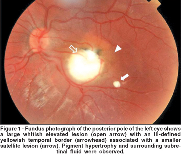
A 5-year-old boy with eye deviation for nine months was referred for examination. Both physical examination and a complete blood cell count with differential count were normal. Visual acuity was 20/20 in the right eye and counter fingers at 1m in the left eye and a medium angle endotropia was noticed. The anterior segment was quiet and the intraocular pressure was normal. An ill-defined whitish elevated lesion associated to smaller satellite lesions with a serous retinal detachment were observed in the macula of the left eye (Figure 1). No abnormalities were found in the right eye.

The patient's pupils were dilated with 0.5% cyclopentolate and 2.5% phenylephrine eye drops instilled at least 30 minutes before examination. Topical anesthetic eye drops (0.1% proximetain cloridate) were instilled immediately before examination. After the examination, the patient was submitted to OCT evaluation. The fovea was measured four times for the 6 radial scans. Fundoscopy, classification and OCT were performed on the same day.
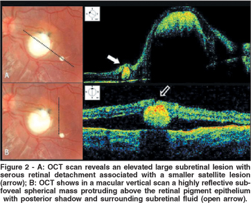
Upon OCT examination, a central highly reflective subretinal round mass and another one smaller satellite mass structure, surrounded by subretinal fluid, were seen protruding above the retinal pigment epithelium in the macular area of the left eye (Figure 1).
A clinical diagnosis of ocular toxocariasis was made and confirmed by the presence of circulating IgG antibodies by the enzyme-linked immunosorbent assay (ELISA).
DISCUSSION
Recently it was demonstrated that the OCT is useful for the differential diagnosis between subretinal Toxocara granuloma and idiopathic choroidal neovascularization(8).
The Toxocara larva is known to migrate across the retina, but the layer in which it migrates and its effect on the retina was unknown(9). OCT images of our patient demonstrated that the presumed Toxocara granuloma had a subretinal extension with elevation and an ill-defined yellowish border associated with atypical smaller satellite lesions as well as serous retinal detachment. The foveal involvement was clearly demonstrated by optical coherence tomography (Figure 2A). Cross-sectional optical coherence tomography images may increase understanding of the pathophysiology of presumed subretinal Toxocara granulomas and help in the clinical management of the retinal complications related to this disease.

REFERENCES
1. Beaver PC, Snyder CH, Carrera GM, Dent JH, Lafferty JW. Chronic eosinophilia due to visceral larva migrans; report of three cases. Pediatrics. 1952;9(1): 7-19.
2. Nunes CM, Tundisi RN, Heinemann MB, Ogassawara S, Richtzenhain LJ. Toxocariasis: serological diagnosis by indirect antibody competition ELISA. Rev Inst Med Trop Sao Paulo. 1999;41(2):95-100.
3. Gillespie SH, Dinning WJ, Voller A, Crowcroft NS. The spectrum of ocular toxocariasis. Eye. 1993;7( Pt 3):415-8. Comment in: Eye. 1993;7(Pt 6):810.
4. Molk R. Ocular toxocariasis: a review of the literature. Ann Ophthalmol. 1983; 15(3):216-9,222-7,230-1.
5. Ashton N. Larval granulomatosis of the retina due to Toxocara. Br J Ophthalmol. 1960;44:129-48.
6. Wilkinson CP, Welch RB. Intraocular toxocara. Am J Ophthalmol. 1971; 71(4):921-30.
7. Rubin ML, Kaufman HE, Tierney JP, Lucas HC. An intraretinal nematode (a case report). Trans Am Acad Ophthalmol Otolaryngol. 1968;72(6):855-66.
8. Higashide T, Akao N, Shirao E, Shirao Y. Optical coherence tomographic and angiographic findings of a case with subretinal Toxocara granuloma. Am J Ophthalmol. 2003;136(1):188-90.
9. Suzuki T, Joko, T, Akao N, Ohashi Y. Following the migration of a Toxocara larva in the retina by optical coherence tomography and fluorescein angiography. Jpn J Ophthalmol. 2005;49(2):159-61.
 Addres to correspondence:
Aline do Lago
Rua Sena Madureira, 1265, apt. 51
São Paulo (SP) - Brazil CEP 04021-051
E-mail: [email protected]
Recebido para publicação em 31.03.2005
Versão revisada recebida em 02.11.2005
Aprovação em 13.12.2005
Vision Institute, Department of Ophthalmology, Federal University of São Paulo – UNIFESP - Brazil.
None of the authors has a proprietary interest in any of the products mentioned in this article.
Nota Editorial: Depois de concluída a análise do artigo sob sigilo editorial e com a anuência do Dr. Walter Yukihiko Takahashi sobre a divulgação de seu nome como revisor, agradecemos sua participação neste processo.