
Arq. Bras. Oftalmol. 2026; 89 (6): 10.5935/0004-2749.2026-0195
Total: 16
Ana Paula Couto1; Igor Coelho2; Luciana Finamor Peixoto3; Raul N. G. Vianna1
DOI: 10.5935/0004-2749.2026-0195
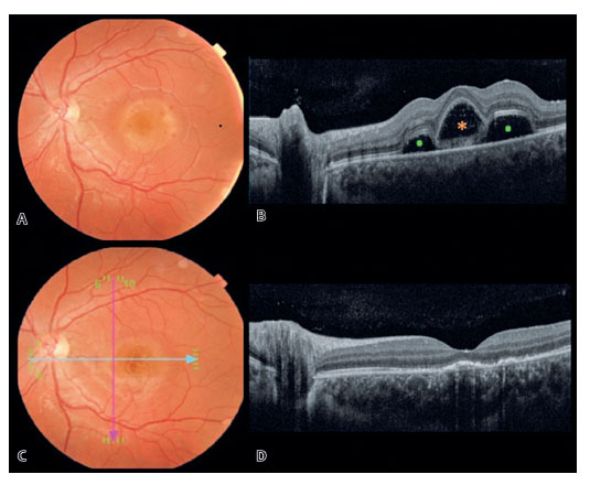
A 25-year-old man presented with acute visual loss (20/400) following a Coxsackievirus prodrome approximately 1 month earlier. Multimodal imaging revealed a round, yellowish, placoid macular lesion with pinpoint hemorrhages, subretinal fluid (dots), and bacillary layer detachment (asterisk; Figure A,B). The patient was managed with clinical observation alone. After 21 days, visual acuity improved to 20/25, with resolution of the macular abnormalities (Figure C, D), consistent with the self-limiting nature of unilateral acute idiopathic maculopathy (UAIM)(1). Coxsackievirus infection has been implicated as a potential cause of this condition(2,3).
REFERENCES
1. Yannuzzi LA, Jampol LM, Rabb MF, Sorenson JA, Beyrer C, Wilcox LM Jr. Unilateral acute idiopathic maculopathy. Arch Ophthalmol. 1991 Oct;109(10):1411-6.
2. Beck AP, Jampol LM, Glaser DA, Pollack JS. Is coxsackievirus the cause of unilateral acute idiopathic maculopathy? Arch Ophthalmol. 2004 Jan;122(1):121-3.
3. Milad D, Antaki F, Hammamji K. Unilateral acute idiopathic maculopathy in hand, foot, and mouth disease. Can J Ophthalmol. 2024 Apr;59(2):e176.
Submitted for publication:
June 29, 2026.
Accepted for publication:
August 12, 2026.
Data Availability Statement: The datasets generated and/or analyzed during the current study are already available.
Edited by
Editor-in-Chief: Newton Kara-Júnior
Associate Editor: Carlos Augusto Moreira Neto
Funding: This study received no specific financial support.
Disclosure of potential conflicts of interest: The authors declare no potential conflicts of interest.

How to cite this article:
 ABO is licensed under a Creative Commons Attribution-NonComercial 4.0 Internacional.