
Arq. Bras. Oftalmol. 2026; 89 (6): 10.5935/0004-2749.2026-0029
Total: 14
João Victor Borges Gomes; Ivan Maynart Tavares
DOI: 10.5935/0004-2749.2026-0029
ABSTRACT
This narrative review summarizes the pathophysiology, diagnosis, and management of neovascular glaucoma and provides a comprehensive guide for clinical ophthalmologists. An iterative snowballing approach, together with targeted searches of PubMed/MEDLINE, Embase, and the Cochrane Library, identified 81 studies addressing key aspects of neovascular glaucoma, including molecular mechanisms, advanced diagnostic modalities, and surgical strategies. Neovascular glaucoma is an aggressive form of secondary glaucoma driven by ischemia-induced upregulation of vascular endothelial growth factor and angiopoietin-2 through the hypoxia-inducible factor-1α pathway. Diagnosis is primarily based on clinical findings and is increasingly supported by advanced imaging techniques. Management requires a multimodal approach combining panretinal photocoagulation and anti-vascular endothelial growth factor therapy with intraocular pressure-lowering interventions, including glaucoma drainage devices, micropulse transscleral cyclophotocoagulation, and slow-coagulation transscleral cyclophotocoagulation. Glaucoma drainage devices achieve the greatest intraocular pressure reduction, whereas micropulse transscleral cyclophotocoagulation offers the most favorable safety profile. Slow-coagulation transscleral cyclophotocoagulation represents an intermediate option, with a reported success rate of 71.7% and favorable safety outcomes. Emerging therapies aim to provide sustained angiogenic control while targeting fibrosis and neuroprotection.
Keywords: Glaucoma, neovascular/diagnosis; Glaucoma, neovascular/physiopathology; Glaucoma neovascular/drug therapy; Glaucoma drainage implants; Angiogenesis inhibitors/therapeutic use; Vascular endothelial growth factor A; Light coagulation; Neuroprotection; Revie
INTRODUCTION
Glaucoma is a chronic, progressive optic neuropathy and a leading cause of irreversible blindness worldwide, with projections estimating that more than 111 million individuals will be affected by 2040(1). Among its subtypes, neovascular glaucoma (NVG) is one of the most severe and vision-threatening forms.
The term NVG was first introduced by Weiss et al.(2) in 1963. NVG is characterized by iris neovascularization (rubeosis iridis), formation of a fibrovascular membrane within the anterior chamber angle, and a subsequent increase in intraocular pressure (IOP)(3). This form of secondary glaucoma typically results from posterior segment ischemia caused by an imbalance between angiogenic and antiangiogenic factors and is most commonly associated with proliferative diabetic retinopathy, central retinal vein occlusion (RVO), and ocular ischemic syndrome (OIS).
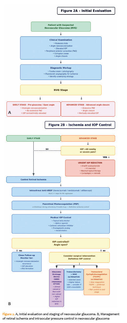
Clinically, NVG progresses through three stages(4). During the preglaucoma stage, fine neovascular tufts appear at the pupillary margin and occasionally within the angle, extending across the scleral spur onto the trabecular meshwork. In the open angle stage, a fibrovascular membrane covers the anterior iris surface and angle structures, mechanically impairing aqueous humor outflow despite the apparently open angle. In the angle closure stage, contraction of this membrane pulls the iris anteriorly, resulting in peripheral anterior synechiae (PAS), ectropion uveae, and eventual angle closure.
The prevalence of NVG varies among populations. In a tertiary hospital-based study conducted in China, NVG accounted for 5.8% of all glaucoma cases(5), although its prevalence in the general population remains relatively low, ranging from 0.01% to 0.12%. In high-risk populations, such as patients with proliferative diabetic retinopathy or severe posterior segment ischemia, the incidence is substantially higher, and NVG generally develops within months after the ischemic insult. Hospital-based studies have reported that NVG accounts for 9%–17.4% of secondary glaucoma cases. Among eyes with proliferative diabetic retinopathy, 33%–64% develop iris neovascularization. In patients with proliferative diabetic retinopathy and unilateral NVG, the fellow eye has a 33% risk of developing NVG(6).
Despite advances in treatment, NVG remains a major clinical challenge because of its aggressive course and poor visual prognosis, often demonstrating limited responsiveness to conventional therapies. Early detection of iris and angle neovascularization, together with prompt treatment of the underlying ischemic disorder, is essential for preserving vision and improving clinical outcomes(7).
METHODS
This narrative review employed a structured literature search strategy combining targeted searches of PubMed/MEDLINE, Embase, and the Cochrane Library with iterative backward and forward snowballing of retrieved articles. Studies published within the last 25 yr were considered eligible for inclusion, although several seminal studies published earlier were also included because they established key concepts related to NVG. Initial searches used Medical Subject Headings terms and keywords, including “neovascular glaucoma,” “NVG,” “iris neovascularization,” “rubeosis iridis,” “VEGF,” “anti-VEGF,” “glaucoma drainage devices,” “panretinal photocoagulation,” and “cyclophotocoagulation.”
Pathophysiology
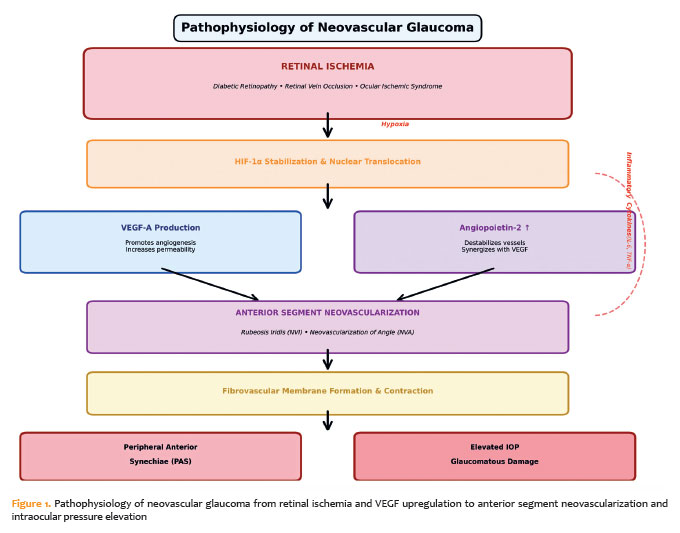
Retinal hypoxia is the central event in the pathogenesis of NVG, triggering a cascade of molecular events and the overexpression of proangiogenic factors. Under hypoxic conditions, hypoxia-inducible factor-1α (HIF-1α) becomes stabilized and translocates to the nucleus, where it activates the transcription of several genes, including vascular endothelial growth factor (VEGF)(8).
Vascular endothelial growth factor, particularly the VEGF-A isoform, is the principal mediator of this response. VEGF produced in the posterior segment diffuses into the aqueous humor and binds to VEGF receptors 1 and 2 (VEGFR-1 and VEGFR-2) on endothelial cells within the iris and angle vasculature, promoting endothelial proliferation, migration, and increased vascular permeability, ultimately leading to neovascularization(9).
Additional molecular pathways also contribute to disease progression. Angiopoietin-2 (Ang-2) acts synergistically with VEGF to destabilize existing blood vessels and promote pathological angiogenesis, whereas angiopoietin-1 (Ang-1) normally maintains vascular stability through activation of the Tie-2 receptor. Under ischemic and inflammatory conditions, Ang-2 levels increase and antagonize Ang-1 activity at the Tie-2 receptor, rendering blood vessels more responsive to VEGF(10). This dual mechanism has become a therapeutic target for bispecific antibodies such as faricimab.
Inflammation further amplifies this process. Proinflammatory cytokines, including interleukin-6 and tumor necrosis factor-α, are upregulated in the aqueous humor of eyes with NVG and potentiate angiogenesis. The interaction among ischemia, angiogenesis, and inflammation establishes a vicious cycle that contributes to the aggressive clinical course of NVG(11).
As VEGF and other angiogenic mediators accumulate, neovascularization extends into the anterior segment, involving the iris and trabecular meshwork. These fragile and permeable vessels are prone to bleeding and promote fibrovascular proliferation, which obstructs the trabecular meshwork, reduces aqueous humor outflow, and elevates IOP.
Over time, persistent angiogenesis and inflammation promote fibrovascular membrane formation across the angle. Contraction of these membranes results in PAS and angle closure, leading to sustained IOP elevation that, if left untreated, causes progressive optic nerve damage and irreversible vision loss (Figure 1).
Etiology of NVG
The development of NVG is primarily driven by retinal ischemia, which induces angiogenic responses and abnormal vessel proliferation in the anterior segment. Although the incidence and etiologic factors vary among populations, retinal ischemia remains the principal pathogenic mechanism. Proliferative diabetic retinopathy is one of the most common causes, particularly in patients with long-standing or poorly controlled diabetes, among whom NVG may develop in up to one-third of cases(12). Persistent hyperglycemia induces microvascular damage and retinal hypoxia, stimulating the release of angiogenic mediators such as VEGF, which promote abnormal vessel growth in the iris and angle, impair aqueous humor outflow, and increase IOP. Pars plana vitrectomy and cataract extraction, which are commonly performed in diabetic eyes, may further increase the risk of NVG by facilitating the anterior diffusion of vasoproliferative factors, thereby increasing rates of postoperative iris neovascularization and NVG.
RVO is another major cause of NVG, with approximately half of affected eyes developing some form of neovascularization following the occlusive event(13). Venous outflow obstruction compromises retinal perfusion, leading to ischemia, release of angiogenic mediators, and progressive neovascularization.
Carotid artery disease, particularly in the setting of OIS, is also strongly associated with NVG. Carotid stenosis or occlusion reduces ocular perfusion, resulting in diffuse retinal hypoxia and anterior segment neovascularization. NVG secondary to OIS may initially present with relatively low IOP but is frequently associated with serious systemic comorbidities, including cerebrovascular disease, thereby warranting prompt systemic evaluation.
Several less common conditions may also lead to NVG through ischemic or inflammatory mechanisms, including retinal detachment, intraocular or orbital tumors, radiation retinopathy, chronic uveitis, and severe ocular trauma. Although less frequent, these conditions highlight the diverse and multifactorial nature of NVG pathogenesis, unified by the central role of retinal ischemia and pathological neovascularization.
Treatment
Clinical Control of IOP
The management of NVG depends on early diagnosis and timely intervention because sustained elevation of IOP can result in glaucomatous optic neuropathy and severe ocular pain. The European Glaucoma Society recommends a treatment strategy based on two major principles: suppression of retinal ischemia through panretinal photocoagulation (PRP) or intravitreal anti-VEGF therapy and IOP control using medical or surgical approaches to prevent optic nerve damage. In the early stages of the disease, before the development of PAS, medical therapy combined with antiangiogenic treatment may stabilize disease progression. Once PAS obstructs the iridocorneal angle, pharmacologic treatment alone is generally insufficient, and surgical intervention becomes necessary. A multidisciplinary approach involving retina specialists is essential for adequate control of angiogenic stimuli and optimization of clinical outcomes.
Although definitive IOP control often requires surgical treatment, medical therapy remains crucial for slowing optic nerve damage, improving media clarity, relieving discomfort, and serving as a bridge until surgery can be performed. The primary pharmacologic strategy involves suppression of aqueous humor production. β-blockers, α2-adrenergic agonists, and carbonic anhydrase inhibitors (CAIs) effectively reduce IOP, usually within 1 hour after instillation(14). β-blockers such as timolol, available in 0.25% and 0.50% formulations, achieve approximately 20%–25% IOP reduction with twice-daily administration(15). Betaxolol, a cardioselective β1-blocker, demonstrates lower hypotensive efficacy because of its limited β2-receptor activity in ocular tissues(16) but may be preferable in patients with pulmonary disease(17). Systemic β-blocker therapy may attenuate the ocular hypotensive effect of topical agents such as timolol(18).
Selective α2-adrenergic agonists, including brimonidine, reduce IOP by approximately 20%–25% from baseline values(19) but may cause conjunctival hyperemia, follicular conjunctivitis, pruritus, and systemic adverse effects, such as dry mouth, fatigue, and dizziness(20). Topical CAIs, such as dorzolamide and brinzolamide, reduce IOP by about 15%–20% with twicedaily administration, without additional benefit from threetimesdaily dosing(21,22). Common adverse effects include blurred vision, tearing, burning sensation, and ocular discomfort(23). Although concerns regarding sulfonamide hypersensitivity persist, current evidence suggests that topical CAIs are generally safe in patients with self-reported sulfa allergy when careful monitoring is provided(24,25). Nevertheless, caution is advised in patients with chronic kidney disease because of the potential for systemic adverse effects.
Prostaglandin analogs, such as latanoprost, reduce IOP by enhancing uveoscleral outflow through relaxation of the ciliary muscle and remodeling of the extracellular matrix(26). These agents achieve approximately 25%–35% IOP reduction with once-dailyadministration, with onset of action generally occurring within 6 hours(27,28). Reported adverse effects include conjunctival hyperemia, increased eyelash growth, iris pigmentation changes, periorbital skin darkening, and potential exacerbation of intraocular inflammation. Importantly, prostaglandin analogs remain effective in eyes with PAS or angle closure and may therefore be used regardless of angle status(29).
Rho-kinase inhibitors, a newer class of topical medications, reduce IOP primarily by enhancing trabecular outflow and secondarily by decreasing episcleral venous pressure and aqueous humor production(30). Although these agents may theoretically be less effective in NVG because of compromised outflow structures, clinical evidence regarding their role in this setting remains limited.
Cholinergic agents such as pilocarpine, which increase trabecular outflow while reducing uveoscleral outflow, are generally avoided in NVG. Their use is contraindicated because they may exacerbate inflammation, induce anterior displacement of the iris–lens diaphragm, further narrow the anterior chamber angle, and precipitate angle closure(4). In addition, the miosis induced by these agents may impair visualization of the posterior segment, which is essential for NVG management.
Adjunctive therapies include topical corticosteroids, which reduce inflammation, vascular permeability, and neovascular proliferation, as well as cycloplegic agents such as atropine, which relieve pain associated with ciliary spasm and reduce the risk of hyphema(3,4).
Oral CAIs such as acetazolamide are frequently used for short-term IOP reduction and may lower IOP by approximately 19%–35% within hours of administration(31,32). However, systemic adverse effects, particularly metabolic acidosis, necessitate careful monitoring, especially in patients with renal impairment(11).
Hyperosmotic agents, including intravenous mannitol and oral glycerol, are reserved for emergency reduction of IOP. These agents act by increasing plasma osmolarity and reducing aqueous humor production through central nervous system-mediated effects(33,34). Consequently, close systemic monitoring is required because of the risk of fluid and electrolyte disturbances.
Trabeculectomy
Trabeculectomy, particularly when combined with antimetabolites such as mitomycin C (MMC) or 5-fluorouracil (5-FU), is a commonly performed filtering procedure for glaucoma. However, its efficacy is substantially reduced in NVG because of the intense inflammatory and angiogenic environment, which promotes subconjunctival fibrosis and intraoperative or postoperative bleeding, both of which are strongly associated with surgical failure.
Case series evaluating trabeculectomy without antifibrotic agents are limited, and reported failure rates may reach 80%(35). Identified risk factors for surgical failure include postoperative hyphema and previous vitrectomy(36). The introduction of off-label antifibrotic agents such as MMC and 5-FU was intended to reduce postoperative scarring and improve surgical outcomes. More recently, anti-VEGF agents have been investigated as adjunctive therapies because of their ability to modulate both neovascularization and wound healing(37).
Trabeculectomy with Mitomycin C
Despite the addition of MMC, trabeculectomy in eyes with NVG continues to demonstrate only modest long-term outcomes. In a Korean study, only 29% of eyes with NVG achieved adequate IOP control after 1 yr(38), although most patients had not undergone prior PRP. When trabeculectomy with MMC is preceded by PRP, surgical success rates improve considerably, reaching 67% at 1 yr(39). However, long-term success declines over time, decreasing to approximately 51% at 5 yr(36).
Trabeculectomy with 5-Fluorouracil (5-FU)
The use of 5-FU offers flexibility because it may be administered intraoperatively or postoperatively; however, available evidence remains limited. Early studies conducted during the 1980s reported success rates of 60%–70% at 1 yr(40,41). Nevertheless, surgical efficacy decreases substantially over time, with reported success rates of approximately 50% at 3 yr(42) and only 28% at 5 yr(43). Consequently, trabeculectomy with 5-FU is no longer recommended for the management of NVG(44).
Trabeculectomy with anti-VEGF Agents
Anti-VEGF agents such as bevacizumab and ranibizumab may improve surgical outcomes by reducing neovascularization and modulating fibroblast activity within Tenon’s capsule. In animal models, VEGF inhibition reduced conjunctival vascularization and collagen deposition(45), whereas histopathologic studies demonstrated decreased inflammation and vascular congestion within the trabecular meshwork of treated eyes(46).
Several routes of administration have been evaluated:
• Subconjunctival anti-VEGF: A randomized clinical trial demonstrated no significant overall benefit compared with placebo at 1 yr. However, a subgroup analysis of eyes with NVG showed a surgical success rate of 72%, compared with 66% in the control group(47).
• Intracameral anti-VEGF: Two case series reported encouraging outcomes when intracameral bevacizumab or ranibizumab was administered before trabeculectomy in eyes that had previously undergone PRP. In the study by De Moraes et al.(48), four eyes maintained an IOP between 10 and 14mmHg without medication over a mean follow-up period of 12.8 months. Elmekawey and Khafagy(49) reported complete IOP control in 53% of eyes and partial control in 40% at 6 months. Studies comparing different preoperative doses of bevacizumab (1.25 vs. 2.5mg) demonstrated comparable outcomes(50).
• Intravitreal anti-VEGF: Additional studies showed that preoperative intravitreal bevacizumab improved short-term surgical success rates (95% vs. 75% at 6 months)(51), although this benefit diminished after 1 yr(52).
The timing of anti-VEGF administration and adequate regression of iris neovascularization are critical for reducing hyphema, which is a major predictor of surgical failure(53).
Glaucoma Drainage Devices
Glaucoma drainage devices (GDDs) have become the preferred surgical option for refractory glaucoma, including NVG. These devices create a permanent fistula between the anterior chamber and an equatorial subconjunctival reservoir, thereby bypassing the compromised trabecular meshwork.
The principal advantages of GDDs over trabeculectomy include maintenance of a continuously patent fistula through the drainage tube, posterior aqueous drainage toward the equatorial region of the globe, which is generally less reactive, and formation of a well-defined encapsulated bleb over the endplate that facilitates controlled aqueous outflow.
Because NVG is frequently associated with postoperative hyphema and severe intraocular inflammation, both of which are linked to high trabeculectomy failure rates(36,53,54), implantation of a GDD is often preferred as the primary surgical treatment. This approach is particularly advantageous in eyes with marked intraocular inflammation(55).
In a retrospective study published in 2011, Shen et al.(56) compared MMC-augmented trabeculectomy with Ahmed glaucoma valve (AGV) implantation in 40 eyes with NVG. Surgical success rates were higher in the AGV group, reaching 70% at 1 yr and 60% at 2 yr, compared with 65% and 55%, respectively, in the trabeculectomy group.
Because eyes with NVG often present with markedly elevated preoperative IOP, valved GDDs are frequently preferred initially because they provide more immediate pressure reduction to levels of approximately 8–12mmHg, thereby reducing the risk of hypotony-related complications. However, compared with nonvalved implants such as the Baerveldt glaucoma implant (BGI), the AGV has demonstrated lower long-term survival rates. This difference may be attributable to early exposure of surrounding tissues to aqueous humor containing proinflammatory cytokines, which promotes the formation of a denser, more congested fibrous capsule and increases the incidence of the hypertensive phase(57).
The Ahmed Baerveldt Comparison Study compared AGV and BGI implantation in eyes with refractory glaucoma, including NVG. After 5 yr of follow-up, the study demonstrated higher rates of successful IOP control in the BGI group than in the AGV group. Eyes receiving nonvalved implants also required fewer adjunctive hypotensive medications. However, the BGI group showed a higher incidence of postoperative hypotony-related complications(58).
A more recent retrospective study published in 2020, which evaluated 152 eyes with NVG, demonstrated similar failure rates between AGV and BGI devices at 6 months. However, eyes implanted with a BGI required fewer topical hypotensive medications(59).
An additional therapeutic strategy frequently combined with surgery is the use of intravitreal anti-VEGF agents, which have been shown to improve surgical outcomes. Zhou et al.(60) reported that Ahmed valve implantation combined with preoperative intravitreal bevacizumab achieved comparable IOP reduction and reduced dependence on hypotensive medications compared with surgery alone. Importantly, anti-VEGF therapy significantly decreased the incidence of hyphema, a known risk factor for postoperative complications, thereby contributing to a safer postoperative course.
Accordingly, in our clinical practice, the preferred surgical approach for NVG consists of implantation of a valved GDD following a single intravitreal anti-VEGF injection and PRP.
Transscleral Cyclophotocoagulation
Each transscleral cyclophotocoagulation (TSCPC) technique offers distinct advantages with respect to efficacy and safety. Potential complications include pain, hyphema, persistent inflammation, corneal edema, hypotony, and even phthisis bulbi(61). Recent advances in TSCPC technology have broadened its indications in refractory glaucoma.
Conventional Continuous-Wave TSCPC (Pop-Titrated Technique)
The conventional approach delivers 1,750–2,000mW of laser energy for 2 s per application, with power titrated until an audible “pop” is heard, indicating tissue disruption. A retrospective study published in 2009 compared diode laser TSCPC with AGV implantation in 66 eyes with NVG and demonstrated comparable success rates between the two procedures after 2 yr of follow-up(62). Similar efficacy was reported in a more recent comparative study published in 2018 by Choy et al.(63), which demonstrated a success rate of approximately 86% in 12 eyes with NVG and no previous history of glaucoma surgery, with a minimum follow-up period of 6 months.
Slow-Coagulation TSCPC (Slow-Burn Technique)
Slow-coagulation TSCPC (SC-TSCPC) represents a refinement of the conventional technique in which a fixed lower laser power (1,250 mW) is delivered over a longer duration (4 s) without titration to an audible pop. This approach aims to achieve ciliary body ablation while minimizing collateral thermal damage to adjacent tissues.
In a retrospective study involving 53 patients with medically refractory NVG, Khodeiry et al.(64) reported that SC-TSCPC used as a primary surgical intervention achieved a significant reduction in IOP, from 40.7±8.6 to 18.4±12.2 mmHg. The cumulative success rates were 71% at 12 months and 64% at 24 months. Importantly, visual acuity remained relatively stable, and complication rates were lower than those historically reported with conventional continuous-wave TSCPC (CW-TSCPC). Decreased visual acuity occurred in 13% of cases, whereas anterior chamber inflammation occurred in 9%.
Comparative studies have demonstrated that SC-TSCPC provides superior IOP control compared with micropulse TSCPC (MP-TSCPC ). Gustianty et al.(65) found that SC-TSCPC achieved greater IOP reduction than MP-TSCPC without significant differences in complication rates. Similarly, Hwang et al.(66) reported that SC-TSCPC provided superior IOP control and lower surgical failure rates compared with micropulse laser treatment, although patients with NVG represented only approximately 10% of the study population. A recent randomized clinical trial by Beniz et al.(67) further supported these findings, identifying SC-TSCPC as a more effective option for refractory glaucoma, although eyes with NVG were not included in that study.
Micropulse TSCPC
Micropulse TSCPC delivers laser energy in short repetitive pulses (31.3-kHz frequency, 0.5-ms on time, 1.1-ms off time, 31.3% duty cycle) rather than by continuous application, thereby allowing tissue cooling between pulses and minimizing collateral thermal damage, including vision loss, hypotony, and phthisis bulbi(68).
Despite these advantages, MP-TSCPC is generally associated with higher costs and often produces less robust and shorter-lasting IOP reduction in clinical practice. This technique has shown promise in several glaucoma subtypes, including NVG, and demonstrates the most favorable safety profile among cyclodestructive procedures. However, MP-TSCPC appears to be less effective than SC-TSCPC for IOP reduction. In eyes with NVG, the 1-yr success rate was reported to be 54% for SC-TSCPC compared with 33% for MP-TSCPC(68). In addition, some studies have reported the need for rescue SC-TSCPC following failure of MP-TSCPC(69).
Comparative Summary of TSCPC Techniques
The three TSCPC modalities can be broadly ranked according to efficacy and safety. With respect to IOP reduction, conventional CW-TSCPC appears to be the most effective, followed by SC-TSCPC and then MP-TSCPC. In contrast, the safety profile follows the reverse order, with MP-TSCPC demonstrating the greatest safety.
SC-TSCPC therefore represents an intermediate option, combining a reproducible fixed-parameter technique with greater efficacy than MP-TSCPC and improved safety compared with conventional continuous-wave delivery. In the context of NVG, SC-TSCPC appears particularly promising as a primary surgical intervention, with reported success rates of approximately 71% at 1 yr and 64% at 2 yr(64).
In selected cases, TSCPC may offer specific advantages over GDDs. It may be particularly useful in eyes with intumescent cataracts and extensive PAS, in which tube implantation can be technically challenging. Favorable outcomes have also been reported in eyes filled with silicone oil(70). Moreover, TSCPC may be advantageous in patients with severe systemic comorbidities because it is a relatively rapid procedure associated with minimal postoperative restrictions and faster recovery than conventional incisional surgery.
Comparative Analysis of Surgical Modalities: Efficacy–Safety Trade-off
Comparative analysis of surgical approaches for NVG reveals a fundamental trade-off between efficacy and safety that should guide clinical decision-making. The available evidence is derived primarily from heterogeneous sources, including retrospective studies, case series, pilot trials, and cohorts of mixed or non-NVG refractory glaucoma, rather than from large randomized head-to-head clinical trials specifically designed for NVG.
GDDs remain the gold standard for surgical management, demonstrating the greatest IOP reduction, approximately 34 mmHg at 12 months. These findings are consistent with the landmark study by Shen et al., which reported superior outcomes for GDD implantation compared with trabeculectomy in this challenging population(56). However, GDD implantation requires incisional surgery and is associated with procedure-related risks, including tube exposure, corneal decompensation, and hypotony, thereby requiring careful patient selection and surgical expertise.
SC-TSCPC has emerged as a promising intermediate option, offering balanced efficacy with an improved safety profile compared with conventional CW-TSCPC. In eyes with NVG, SC-TSCPC achieved a mean IOP reduction of 22.3 mmHg. The fixed-parameter protocol used in SC-TSCPC (1,250 mW for 4 s) eliminates the need for pop titration and may reduce collateral thermal injury while maintaining substantial IOP reduction. Recent randomized controlled trial data demonstrated success rates of 70% with SC-TSCPC compared with 30% with MP-TSCPC in refractory glaucoma.
Although MP-TSCPC demonstrates the most favorable safety profile with relatively few complications, it achieves lower success rates and frequently requires retreatment.
The relatively poor long-term performance of MMC-augmented trabeculectomy in NVG reflects the aggressive neovascularization and fibrosis characteristic of this disease and reinforces current recommendations favoring GDD implantation over filtration surgery in this population(56).
This efficacy–safety spectrum enables individualized surgical planning. GDD implantation may be preferable for eyes with good visual potential and acceptable surgical risk; SC-TSCPC may represent an intermediate option balancing efficacy and invasiveness; MP-TSCPC may be more suitable for high-risk patients willing to accept the possibility of retreatment; and conventional CW-TSCPC may be reserved for palliative management of blind, painful eyes. The emergence of SC-TSCPC, with reported success rates of 71.7% in NVG, represents an important therapeutic advance that may bridge the gap between the high efficacy of GDD implantation and the superior safety of MP-TSCPC, warranting further evaluation in prospective comparative studies.
Minimally Invasive Glaucoma Surgeries
Studies evaluating minimally invasive glaucoma surgery (MIGS) in NVG remain scarce. This likely reflects the severe nature of NVG, which is typically associated with markedly elevated IOP and a predominantly pretrabecular mechanism resulting in secondary angle closure.
Nevertheless, a 2022 case report by Kanter et al.(71) described the potential role of gonioscopy-assisted transluminal trabeculotomy in a carefully selected case of NVG. The procedure was performed in a young patient with preserved visual potential and an open angle after complete regression of angle neovascularization. Good IOP control was maintained for 9 months before eventual surgical failure occurred. This report suggests that MIGS may have a limited but potentially relevant role in carefully selected patients with NVG.
Management of Retinopathy
Panretinal Photocoagulation
Panretinal photocoagulation remains the definitive treatment for ablation of ischemic retina and suppression of the angiogenic drive. By destroying hypoxic photoreceptors, PRP decreases retinal metabolic demand and downregulates VEGF production. The procedure is generally performed 3–7 days after intravitreal anti-VEGF injection to allow regression of neovascularization and reduce the risk of intraoperative hemorrhage.
Anti-VEGF Therapy
The introduction of anti-VEGF agents, including bevacizumab, ranibizumab, and aflibercept, has revolutionized the management of NVG. Administered through intravitreal injection, these agents induce rapid regression of neovascularization of the iris (NVI) and neovascularization of the angle (NVA), typically within days(72,73). This effect creates a critical therapeutic window during which PRP and surgical interventions can be performed with a lower risk of bleeding complications.
A 2024 meta-analysis demonstrated that combining intravitreal and intracameral anti-VEGF injections may provide a more profound and sustained reduction in anterior segment neovascularization and IOP(74). The efficacy of ranibizumab as an adjunctive therapy has also been reported in the Brazilian literature(75).
FUTURE DIRECTIONS
Sustained-Release Anti-VEGF and Multimodal Agents
To overcome the transient effects associated with currently available anti-VEGF agents, sustained-release drug delivery platforms are under development. The Port Delivery System with ranibizumab (Susvimo), a permanent refillable ocular implant, provides continuous drug delivery and has already been approved for other retinal vascular disorders(76).
Gene therapy represents another emerging strategy. Viral vector-based therapies such as ixoberogene soroparvovec (ADVM-022) have shown promise for achieving long-term intraocular expression of anti-VEGF proteins after a single injection(77). However, the application of sustained-release or gene-based anti-VEGF therapies in NVG remains investigational and is currently extrapolated from studies involving non-NVG retinal diseases.
In addition, bispecific antibodies are broadening therapeutic targets. Faricimab, which simultaneously targets VEGF-A and angiopoietin-2 (Ang-2), has demonstrated improved durability and vascular stabilization in diabetic macular edema by modulating both angiogenesis and inflammation(78). Although this dual mechanism is biologically attractive for ischemia-driven anterior segment neovascularization, no NVG-specific clinical data are currently available. Therefore, any potential role for faricimab in NVG should be regarded as hypothesis-generating and limited to future clinical investigations rather than current clinical practice.
Advances in Surgical Devices and Techniques
Research efforts continue to refine surgical approaches for NVG. Newer GDDs incorporating improved biomaterials aim to reduce capsular fibrosis and improve long-term surgical success. Similarly, micropulse TSCPC protocols are being optimized to achieve sustained IOP reduction while minimizing collateral tissue damage. However, current evidence regarding micropulse TSCPC in NVG is derived primarily from small retrospective series; consequently, its application in eyes with good visual potential remains selective and investigational(79).
Neuroprotection and Anti-Inflammatory Targets
Agents targeting microglial activation and oxidative stress are currently being explored as adjunctive neuroprotective therapies(80). The development of novel anti-inflammatory strategies may also help disrupt the vicious cycle of ischemia, angiogenesis, and inflammation that drives disease progression(81). Nevertheless, in the context of NVG, these approaches remain experimental and should currently be viewed as potential future therapeutic avenues rather than established components of clinical management.
NVG remains a complex and challenging form of secondary glaucoma; however, the therapeutic landscape has evolved substantially in recent years (Figure 2A,B). Contemporary management has shifted from a reactive, IOP-centered approach toward a proactive and multifaceted strategy focused on controlling retinal ischemia through combined PRP and anti-VEGF therapy. In advanced disease, GDDs remain the surgical gold standard, although their efficacy is enhanced by preoperative anti-VEGF treatment. At the same time, safer alternatives such as slow-coagulation TSCPC are increasingly gaining acceptance across a broader range of clinical scenarios.
The current evidence base remains dominated by heterogeneous observational studies and comparative case series. Therapeutic strategies aimed at achieving more durable angiogenic suppression, improved modulation of postoperative fibrosis, and adjunctive neuroprotection are conceptually promising; however, in NVG, these approaches remain investigational and are largely extrapolated from retinal disease and non-NVG glaucoma research. Robust prospective comparative studies and NVG-specific evaluations of sustained-release anti-VEGF systems, gene-based therapies, and evolving surgical technologies are still required to determine their true impact on long-term visual and structural outcomes in this challenging condition.
AUTHORS’ CONTRIBUTIONS
Significant contributions to conception and design: João Victor Borges Gomes and Ivan Maynart Tavares. Data Acquisition: João Victor Borges Gomes and Ivan Maynart Tavares. Data Analysis and Interpretation: João Victor Borges Gomes and Ivan Maynart Tavares. Manuscript Drafting: João Victor Borges Gomes and Ivan Maynart Tavares. Significant intellectual contente revision of the manuscript: Ivan Maynart Tavares. Final approval of the submitted manuscript: João Victor Borges Gomes and Ivan Maynart Tavares. Statistical analysis:not applicable. Obtaining funding: not applicable. Supervision of administrative, technical, or material support: Ivan Maynart Tavares. Research group leadership: Ivan Maynart Tavares.
REFERENCES
1. Tham YC, Li X, Wong TY, Quigley HA, Aung T, Cheng CY. Global prevalence of glaucoma and projections of glaucoma burden through 2040: a systematic review and meta-analysis. Ophthalmology. 2014;121(11):2081-90.
2. Weiss DI, Shaffer RN, Nehrenberg TR. Neovascular gluacoma complicating carotid-cavernous fistula. Arch Ophthalmol. 1963;69:304-7.
3. Rodrigues GB, Abe RY, Zangalli C, Sodre SL, Donini FA, Costa DC, et al. Neovascular glaucoma: a review. Int J Retina Vitreous. 2016;2(1):26.
4. Havens SJ, Gulati V. Neovascular glaucoma. Dev Ophthalmol. 2016;55:196-204.
5. Mishra C, Meyer JJ. Neovascular glaucoma [Internet]. Treasure Island (FL): StatPearls Publishing; 2026 Jan–. [cited 2026Jan 21]. Available from: Neovascular Glaucoma - StatPearls - NCBI Bookshelf
6. Lee P, Wang CC, Adamis AP. Ocular neovascularization: an epidemiologic review. Surv Ophthalmol. 1998;43(3):245-69.
7. Hayreh SS. Neovascular glaucoma. Prog Retin Eye Res. 2007;26(5):470-85.
8. Liu W, Shen SM, Zhao XY, Chen GQ. Targeted genes and interacting proteins of hypoxia inducible factor-1. Int J Biochem Mol Biol. 2012;3(2):165-78.
9. Dumbrăveanu L, Cușnir V, Bobescu D. A review of neovascular glaucoma. Etiopathogenesis and treatment. Rom J Ophthalmol. 2021;65(4):315-29.
10. Chaudhary V, Mar F, Amador MJ, Chang A, Gibson K, Joussen AM, et al. Emerging clinical evidence of a dual role for Ang-2 and VEGF-A blockade with faricimab in retinal diseases. Graefes Arch Clin Exp Ophthalmol. 2025;263(5):1239-47.
11. Urbonavičiūtė D, Buteikienė D, Janulevičienė I. A Review of neovascular glaucoma: etiology, pathogenesis, diagnosis, and treatment. Medicina (Kaunas). 2022;58(12):1870.
12. Senthil S, Dada T, Das T, Kaushik S, Puthuran GV, Philip R, et al. Neovascular glaucoma - A review. Indian J Ophthalmol. 2021;69(3):525-34.
13. Hayreh SS, Rojas P, Podhajsky P, Montague P, Woolson RF. Ocular neovascularization with retinal vascular occlusion-III. Incidence of ocular neovascularization with retinal vein occlusion. Ophthalmology. 1983;90(5):488-506.
14. Crawley L, Zamir SM, Cordeiro MF, Guo L. Clinical options for the reduction of elevated intraocular pressure. Ophthalmol Eye Dis. 2012;4:43-64.
15. European Glaucoma Society Terminology and Guidelines for Glaucoma. 3rd Savona: Editrice DOGMA; 2008.
16. Trope GE, Clark B. Beta adrenergic receptors in pigmented ciliary processes. Br J Ophthalmol. 1982;66(12):788-92.
17. Lama PJ. Systemic adverse effects of beta-adrenergic blockers: an evidence-based assessment. Am J Ophthalmol. 2002;134(5):749-60.
18. Schuman JS. Effects of systemic beta-blocker therapy on the efficacy and safety of topical brimonidine and timolol. Brimonidine Study Groups 1 and 2. Ophthalmology. 2000;107(6):1171-7.
19. Kampik A, Arias-Puente A, O’Brart DP, Vuori ML; European Latanoprost Study Group. Intraocular pressure-lowering effects of latanoprost and brimonidine therapy in patients with open-angle glaucoma or ocular hypertension: a randomized observer-masked multicenter study. J Glaucoma. 2002;11(2):90-6.
20. Arthur S, Cantor LB. Update on the role of alpha-agonists in glaucoma management. Exp Eye Res. 2011;93(3):271-83.
21. Iester M. Brinzolamide ophthalmic suspension: a review of its pharmacology and use in the treatment of open angle glaucoma and ocular hypertension. Clin Ophthalmol. 2008;2(3):517-23.
22. Sugrue MF. Pharmacological and ocular hypotensive properties of topical carbonic anhydrase inhibitors. Prog Retin Eye Res. 2000;19(1):87-112.
23. Strahlman E, Tipping R, Vogel R. A double-masked, randomized 1-year study comparing dorzolamide (Trusopt), timolol, and betaxolol. International Dorzolamide Study Group. Arch Ophthalmol. 1995;113(8):1009-Comment in: Arch Ophthalmol. 1995;113(8):985-6.
24. Stock JG. Sulfonamide hypersensitivity and acetazolamide. Arch Ophthalmol. 1990;108(5):634-5.
25. Guedes GB, Karan A, Mayer HR, Shields MB. Evaluation of adverse events in self-reported sulfa-allergic patients using topical carbonic anhydrase inhibitors. J Ocul Pharmacol Ther. 2013;29(5):456-61.
26. Weinreb RN, Toris CB, Gabelt BT, Lindsey JD, Kaufman PL. Effects of prostaglandins on the aqueous humor outflow pathways. Surv Ophthalmol. 2002;47(Suppl 1):S53-64.
27. Toris CB, Gabelt BT, Kaufman PL. Update on the mechanism of action of topical prostaglandins for intraocular pressure reduction. Surv Ophthalmol. 2008;53(Suppl1):S107-20.
28. Larsson LI. Intraocular pressure over 24 hours after single-dose administration of latanoprost 0.005% in healthy volunteers. A randomized, double-masked, placebo controlled, cross-over single center study. Acta Ophthalmol Scand. 2001;79(6):567-71.
29. Kumar S, Malik A, Singh M, Sood S. Efficacy of latanoprost in management of chronic angle closure glaucoma. Nepal J Ophthalmol. 2009;1(1):32-6.
30. Ren R, Li G, Le TD, Kopczynski C, Stamer WD, Gong H. Netarsudil increases outflow facility in human eyes through multiple mechanisms. Invest Ophthalmol Vis Sci. 2016;57(14):6197-6209.
31. Maus TL, Larsson LI, McLaren JW, Brubaker RF. Comparison of dorzolamide and acetazolamide as suppressors of aqueous humor flow in humans. Arch Ophthalmol. 1997;115(1):45-9.
32. Friedland BR, Mallonee J, Anderson DR. Short-term dose response characteristics of acetazolamide in man. Arch Ophthalmol. 1977;95(10):1809-12.
33. Galin MA, Binkhorst RD, Kwitko ML. Ocular dehydration. Am J Ophthalmol. 1968;66(2):233-5.
34. Krupin T, Podos SM, Becker B. Alteration of intraocular pressure after third ventricle injections of osmotic agents. Am J Ophthalmol. 1973;76(6):948-52.
35. Mietz H, Raschka B, Krieglstein GK. Risk factors for failures of trabeculectomies performed without antimetabolites. Br J Ophthalmol. 1999;83(7):814-21.
36. Takihara Y, Inatani M, Fukushima M, Iwao K, Iwao M, Tanihara H. Trabeculectomy with mitomycin C for neovascular glaucoma: prognostic factors for surgical failure. Am J Ophthalmol. 2009;147(5):912-8, 918.e1.
37. Slabaugh M, Salim S. Use of Anti-VEGF agents in glaucoma surgery. J Ophthalmol. 2017;2017:1645269.
38. Hyung SM, Kim SK. Mid-term effects of trabeculectomy with mitomycin C in neovascular glaucoma patients. Korean J Ophthalmol. 2001;15(2):98-106.
39. Kiuchi Y, Sugimoto R, Nakae K, Saito Y, Ito S. Trabeculectomy with mitomycin C for treatment of neovascular glaucoma in diabetic patients. Ophthalmologica. 2006;220(6):383-8.
40. Heuer DK, Parrish RK 2nd, Gressel MG, Hodapp E, Palmberg PF, Anderson DR. 5-fluorouracil and glaucoma filtering surgery. II. A pilot study. Ophthalmology. 1984;91(4):384-94.
41. Rockwood EJ, Parrish RK 2nd, Heuer DK, Skuta GL, Hodapp E, Palmberg PF, et al. Glaucoma filtering surgery with 5-fluorouracil. Ophthalmology. 1987;94(9):1071-8.
42. Sisto D, Vetrugno M, Trabucco T, Cantatore F, Ruggeri G, Sborgia C. The role of antimetabolites in filtration surgery for neovascular glaucoma: intermediate-term follow-up. Acta Ophthalmol Scand. 2007;85(3):267-71.
43. Tsai JC, Feuer WJ, Parrish RK 2nd, Grajewski AL. 5-Fluorouracil filtering surgery and neovascular glaucoma. Long-term follow-up of the original pilot study. Ophthalmology. 1995;102(6):887-92; discussion 892-3.
44. Kitazawa Y, Kawase K, Matsushita H, Minobe M. Trabeculectomy with mitomycin. A comparative study with fluorouracil. Arch Ophthalmol. 1991; 109(12):1693-8.
45. Li Z, Van Bergen T, Van de Veire S, Van de Vel I, Moreau H, Dewerchin M, et al. Inhibition of vascular endothelial growth factor reduces scar formation after glaucoma filtration surgery. Invest Ophthalmol Vis Sci. 2009;50(11):5217-25.
46. Yoshida N, Hisatomi T, Ikeda Y, Kohno R, Murakami Y, Imaki H, et al. Intravitreal bevacizumab treatment for neovascular glaucoma: histopathological analysis of trabeculectomy specimens. Graefes Arch Clin Exp Ophthalmol. 2011;249(10):1547-52.
47. Ghanem AA. Trabeculectomy with or without intraoperative sub-conjunctival injection of bevacizumab in treating refractory glaucoma. J Clinic Experiment Ophthalmol. 2011;2(2):131.
48. de Moraes CG, Facio AC, Costa JH, Malta RF. Intracameral bevacizumab and mitomycin C Trabeculectomy for eyes with neovascular glaucoma: a case series. J Ocul Biol Dis Infor. 2009;2(1):40-6.
49. Elmekawey H, Khafagy A. Intracameral ranibizumab and subsequent mitomycin C augmented trabeculectomy in neovascular glaucoma. J Glaucoma. 2014;23(7):437-40.
50. Gupta V, Jha R, Rao A, Kong G, Sihota R. The effect of different doses of intracameral bevacizumab on surgical outcomes of trabeculectomy for neovascular glaucoma. Eur J Ophthalmol. 2009;19(3):435-Commment in: Eur J Ophthalmol. 2010;20(1):251; author reply 252.
51. Saito Y, Higashide T, Takeda H, Ohkubo S, Sugiyama K. Beneficial effects of preoperative intravitreal bevacizumab on trabeculectomy outcomes in neovascular glaucoma. Acta Ophthalmol. 2010;88(1):96-102.
52. Takihara Y, Inatani M, Kawaji T, Fukushima M, Iwao K, Iwao M, et al. Combined intravitreal bevacizumab and trabeculectomy with mitomycin C versus trabeculectomy with mitomycin C alone for neovascular glaucoma. J Glaucoma. 2011;20(3):196-201.
53. Nakatake S, Yoshida S, Nakao S, Arita R, Yasuda M, Kita T, et al. Hyphema is a risk factor for failure of trabeculectomy in neovascular glaucoma: a retrospective analysis. BMC Ophthalmol. 2014;14(1):55.
54. Allen RC, Bellows AR, Hutchinson BT, Murphy SD. Filtration surgery in the treatment of neovascular glaucoma. Ophthalmology. 1982;89(10):1181-7.
55. 55. Sivak-Callcott JA, O’Day DM, Gass JD, Tsai JC. Evidence-based recommendations for the diagnosis and treatment of neovascular glaucoma. Ophthalmology. 2001;108(10):1767-76; quiz1777, 1800. Comment in: Ophthalmology. 2003;110(1):1; author reply 1-2.
56. Shen CC, Salim S, Du H, Netland PA. Trabeculectomy versus Ahmed Glaucoma Valve implantation in neovascular glaucoma. Clin Ophthalmol. 2011;5:281-6.
57. Christakis PG, Zhang D, Budenz DL, Barton K, Tsai JC, Ahmed IIK; ABC-AVB Study Groups. Five-year pooled data analysis of the Ahmed Baerveldt comparison study and the Ahmed Versus Baerveldt Study. Am J Ophthalmol. 2017;176:118-126.
58. Budenz DL, Barton K, Gedde SJ, Feuer WJ, Schiffman J, Costa VP, Godfrey DG, Buys YM; Ahmed Baerveldt Comparison Study Group. Five-year treatment outcomes in the Ahmed Baerveldt comparison study. Ophthalmology. 2015;122(2):308-16.
59. Shalaby WS, Myers JS, Razeghinejad R, Katz LJ, Pro M, Dale E, et al. Outcomes of valved and nonvalved tube shunts in neovascular glaucoma. Ophthalmol Glaucoma. 2021;4(2):182-92.
60. Zhou M, Xu X, Zhang X, Sun X. Clinical outcomes of Ahmed Glaucoma Valve Implantation with or without intravitreal bevacizumab pretreatment for neovascular glaucoma: a systematic review and meta-analysis. J Glaucoma. 2016;25(7):551-7.
61. Bloom PA, Tsai JC, Sharma K, Miller MH, Rice NS, Hitchings RA, et al. “Cyclodiode”.. Trans-scleral diode laser cyclophotocoagulation in the treatment of advanced refractory glaucoma. Ophthalmology. 1997;104(9):1508-19; discussion 1519-20.
62. Yildirim N, Yalvac IS, Sahin A, Ozer A, Bozca T. A comparative study between diode laser cyclophotocoagulation and the Ahmed glaucoma valve implant in neovascular glaucoma: a long-term follow-up. J Glaucoma. 2009;18(3):192-6.
63. Choy BN, Lai JS, Yeung JC, Chan JC. Randomized comparative trial of diode laser transscleral cyclophotocoagulation versus Ahmed glaucoma valve for neovascular glaucoma in Chinese - a pilot study. Clin Ophthalmol. 2018;12:2545-52.
64. Khodeiry MM, Lauter AJ, Sayed MS, Han Y, Lee RK. Primary slow-coagulation transscleral cyclophotocoagulation laser treatment for medically recalcitrant neovascular glaucoma. Br J Ophthalmol. 2023;107(5):671-6.
65. Gustianty E, Rahayu A, Knoch AM, Rifada RM, Umbara S. Comparison of efficacy and safety of slow coagulation transscleral versus micropulse cyclophotocoagulation in refractory glaucoma. Open Ophthalmol J [Internet]. 2024[cited 2025 Dec 21];18: e 18743641310116. Available from: e18743641310116.pdf
66. Hwang YH, Lee S, Kim M, Choi J. Comparison of treatment outcomes between slow coagulation transscleral cyclophotocoagulation and micropulse transscleral laser treatment. Sci Rep. 2024;14(1):23944.
67. Beniz LA, Ikeda MC, Teixeira EG, Gracitelli CP, Caixeta-Umbelino C, Kasahara N, et al. Slow-coagulation continuous-wave cyclophotocoagulation vs. micropulse laser treatment procedure in refractory glaucoma: a randomized clinical trial. Ophthalmol Glaucoma. 2025:S2589-4196(25)00215-7. [In press].
68. Zemba M, Dumitrescu OM, Vaida F, Dimirache EA, Pistolea I, Stamate AC, et al. Micropulse vs. continuous wave transscleral cyclophotocoagulation in neovascular glaucoma. Exp Ther Med. 2022;23(4):278.
69. Sandhur BS, Jethi M, Gautam N, Ersan S, Jay L, Sieminski SF. Efficacy of slow-coagulation cyclophotocoagulation after failed MicroPulse cyclophotocoagulation in refractory glaucoma. Lasers Med Sci. 2025;40(1):370.
70. Ghazi-Nouri SM, Vakalis AN, Bloom PA, Bunce C, Charteris DG. Long-term results of the management of silicone oil-induced raised intraocular pressure by diode laser cycloablation. Eye (Lond). 2005;19(7):765-9.
71. Kanter JA, Amin P, Komati R, Mackin AG, Dao D, Shaw LT, et al. Gonioscopy-assisted transluminal trabeculotomy in neovascular glaucoma: Salvaging the conventional outflow pathway. Am J Ophthalmol Case Rep. 2022;28:101668.
72. Ehlers JP, Spirn MJ, Lam A, Sivalingam A, Samuel MA, Tasman W. Combination intravitreal bevacizumab/panretinal photocoagulation versus panretinal photocoagulation alone in the treatment of neovascular glaucoma. Retina. 2008;28(5):696-702.
73. Lupinacci AP, Calzada JI, Rafieetery M, Charles S, Netland PA. Clinical outcomes of patients with anterior segment neovascularization treated with or without intraocular bevacizumab. Adv Ther. 2009;26(2):208-16.
74. Wang J, Guo YM, Wei J, Min J, Ye L. Comparative efficacy and safety of different anti-VEGF agents combined with different delivery methods for neovascular glaucoma: a systematic review and Bayesian network meta-analysis. BMJ Open. 2024;14(3):e080103.
75. Ticly FG, Lira RP, Fulco EA, Vasconcelos JP. Ranibizumabe intravítreo como tratamento adjuvante para glaucoma neovascular. Rev Bras Oftalmol. 2013;72(2):119-21.
76. Ranade SV, Wieland MR, Tam T, Rea JC, Horvath J, Hieb AR, et al. The Port Delivery System with ranibizumab: a new paradigm for long-acting retinal drug delivery. Drug Deliv. 2022;29(1):1326-34.
77. Khanani AM, Boyer DS, Wykoff CC, Regillo CD, Busbee BG, Pieramici D, et al. Safety and efficacy of ixoberogene soroparvovec in neovascular age-related macular degeneration in the United States (OPTIC): a prospective, two-year, multicentre phase 1 study. EClinicalMedicine. 2023;67:102394.
78. Wong TY, Haskova Z, Asik K, Baumal CR, Csaky KG, Eter N, Ives JA, Jaffe GJ, Korobelnik JF, Lin H, Murata T, Ruamviboonsuk P, Schlottmann PG, Seres AI, Silverman D, Sun X, Tang Y, Wells JA, Yoon YH, Wykoff CC; YOSEMITE and RHINE Investigators. Faricimab treat-and-extend for diabetic macular edema: two-year results from the randomized Phase 3 YOSEMITE and RHINE Trials. Ophthalmology. 2024;131(6):708-23.
79. de Crom RM, Kujovic-Aleksov S, Webers CA, Berendschot TT, Beckers HJ. Long-term treatment outcomes of micropulse transscleral cyclophotocoagulation in primary and secondary glaucoma: a 5-year analysis. Ophthalmol Ther. 2025;14(2):323-35.
80. Han J, Chitu V, Stanley ER, Wszolek ZK, Karrenbauer VD, Harris RA. Inhibition of colony stimulating factor-1 receptor (CSF-1R) as a potential therapeutic strategy for neurodegenerative diseases: opportunities and challenges. Cell Mol Life Sci. 2022;79(4):219.
81. Bai Y, Wang X, Qi F, Zuo X, Zou G. Mechanisms of action of retinal microglia in diabetic retinopathy (Review). Int J Mol Med. 2025;56(6):202.
Submitted for publication:
February 5, 2026.
Accepted for publication:
May 15, 2026.
Data Availability Statement: This article is a narrative review based exclusively on previously published studies retrieved from bibliographic databases. No original datasets, software code, or other research materials were generated by the authors; therefore, no additional underlying data are available for deposition in a repository. All sources used are listed in the References section.
Edited by
Editor-in-Chief: Newton Kara-Júnior
Associate Editor: Jayter de Paula
Funding: This study received no specific financial support.
Disclosure of potential conflicts of interest: The authors declare no potential conflicts of interest.

How to cite this article:
 ABO is licensed under a Creative Commons Attribution-NonComercial 4.0 Internacional.