
Arq. Bras. Oftalmol. 2026; 89 (6): 10.5935/0004-2749.2025-0294
Total: 18
Jaqueline Fernandes Ruiz1; Talita Gambogi Araújo1; Thaís Cândida Borges1; Leonardo Ugulino de Araújo Neto1; Felipe Leal de Moraes Brito1; Pablo Felipe Rodrigues2; Luiz Antonio de Brito Martins1,2; Myrna Serapião dos Santos1,2; Bernardo Kaplan Moscovici2,3,4
DOI: 10.5935/0004-2749.2025-0294
ABSTRACT
PURPOSE: To compare obstructive sleep apnea screening classifications, anthropometric characteristics, and mechanical behaviors between participants with keratoconus and controls and to explore their associations with disease severity.
METHODS: This prospective cross-sectional study included 74 participants with keratoconus and 49 controls. The paper-based Berlin and STOP-BANG questionnaires were administered face-to-face by a researcher and scored manually. A study-specific questionnaire assessed eye rubbing and sleep position, whereas a separate structured field recorded self-reported ocular compression during sleep. Both groups underwent corneal tomography. The primary logistic regression model included age, sex, body mass index (BMI), eye rubbing, and ocular compression.
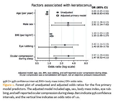
RESULTS: Participants with keratoconus had a higher BMI (29.4±6.4 vs. 26.3±5.1 kg/m²; p=0.005), larger neck circumference (40.1±4.6 vs. 37.6±4.6 cm; p=0.004), more frequent eye rubbing (71.6% vs. 51.0%; p=0.020), and more frequent ocular compression (43.2% vs. 20.4%; p=0.009) than controls. High-risk classifications did not differ significantly between groups according to either the Berlin Questionnaire (p=0.206) or STOP-BANG (p=0.139). In exploratory models adjusted for age and sex, positive associations were observed for the Berlin Questionnaire (OR, 2.58; 95% CI, 0.91-7.28) and STOP-BANG (OR, 3.30; 95% CI, 0.83-13.05). In the primary model, younger age (OR, 0.88; 95% CI, 0.83-0.93), male sex (OR, 2.91; 95% CI, 1.14-7.42), and higher BMI (OR, 1.15; 95% CI, 1.05-1.27) remained associated with keratoconus, whereas the estimates for behavioral factors were attenuated. No association with disease severity was detected.
CONCLUSIONS: Participants with keratoconus had higher anthropometric measurements and more frequent mechanical behaviors; however, the prevalence of high-risk obstructive sleep apnea classifications did not differ significantly between groups. Questionnaire-based and anthropometric findings are not independent because BMI contributes to both screening instruments, whereas neck circumference contributes to the STOP-BANG score. Ocular compression was assessed using a nonvalidated self-report measure rather than objective pressure measurements. Severity analyses were underpowered, and causality cannot be inferred from the study design.
Keywords: Keratoconus; Sleep apnea, obstructive; Risk factors; Body mass index; Sleep; Logistic models; Patient acuity; Tomography; Surveys and questionnaires
INTRODUCTION
Keratoconus (KC) is a progressive, bilateral, and often asymmetric ectatic corneal disorder characterized by central or paracentral thinning, biomechanical instability, and protrusion, resulting in irregular astigmatism and visual impairment. Traditionally considered a noninflammatory condition, KC may involve chronic low-grade inflammation, as evidenced by elevated cytokines, matrix metalloproteinases (MMPs), and oxidative stress markers in the tear film(1,2).
The etiology of KC is multifactorial, with genetic predisposition, environmental exposure, allergy, and mechanical trauma all implicated(3-8). Repetitive eye rubbing may disrupt the stromal collagen architecture and trigger inflammatory cascades(9,10).
An association between keratoconus and obstructive sleep apnea (OSA) has been proposed. OSA is characterized by recurrent upper-airway obstruction, intermittent hypoxia, and systemic inflammation and is strongly associated with increased body mass index (BMI) and neck circumference(11,12). A higher prevalence of OSA or its risk factors has been reported among individuals with keratoconus, suggesting potentially shared anatomic and inflammatory mechanisms(13,14). Proposed mechanisms include systemic oxidative stress and direct mechanical stress on the cornea during sleep. Prone or lateral decubitus sleeping positions may result in sustained pressure on the globe against the pillow, and greater corneal steepening has been observed in eyes on the preferred sleeping side in patients with asymmetric disease(15,16).
Validated screening tools, such as the Berlin Questionnaire and the Snoring, Tiredness, Observed apnea, high blood Pressure, Body mass index, Age, Neck circumference, and Gender (STOP-BANG) questionnaire, identify individuals at increased risk of OSA but differ in their emphasis. The Berlin Questionnaire is more symptom-oriented and incorporates obesity, whereas STOP-BANG prioritizes sensitivity and includes age, sex, BMI, and neck circumference(17-20). Because these instruments contain overlapping anthropometric variables, concordant results should not be considered independent validation. The associations of anthropometric and sleep-related variables with keratoconus severity also remain unclear; tear-film mediators and sleep posture have been associated with disease severity or tomographic asymmetry(15,16,21).
The present study compared questionnaire-estimated OSA risk, anthropometric characteristics, and mechanical behaviors between participants with keratoconus and controls and explored their associations with tomographic disease severity.
METHODS
Study design and ethical approval
This was a prospective, cross-sectional observational study. The study protocol was approved by the Research Ethics Committee of Hospital de Olhos Paulista (CEP/HOLHOS SP) on June 4, 2024 (Approval No. 6,866,697; CAAE 78456924.7.0000.9867). Recruitment commenced only after ethical approval and was conducted during the approved period from June 2024 to January 2025. All participants provided written informed consent before undergoing any study procedures. The study was conducted in accordance with the Declaration of Helsinki.
Study population
A total of 123 adults aged 18-60 years were included. Keratoconus was confirmed in 74 participants based on clinical and tomographic criteria(22). Both groups were prospectively enrolled during the same approved recruitment period at Hospital de Olhos Paulista, São Paulo. The 49 controls were recruited at the same institution and had no clinical or tomographic evidence of keratoconus according to the Rabinowitz-McDonnell topographic criteria(23). The available study records did not specify the clinical reason for the controls’ attendance.
Exclusion criteria included other ocular diseases affecting visual acuity or corneal measurements, systemic conditions known to affect the cornea (i.e., diabetes or connective tissue disease), and a previous diagnosis of or treatment for obstructive sleep apnea.
Previous corneal surgery was not an exclusion criterion for participants with keratoconus, who were recruited from a tertiary corneal service. Among the 74 patients, 33 (44.6%) had undergone corneal cross-linking, intrastromal corneal ring segment implantation, or keratoplasty in at least one eye, and 23 had undergone surgery in the more affected eye. Because these procedures can alter the parameters used for Amsler-Krumeich staging, the primary severity analysis was restricted to the 51 patients whose more affected eye was surgically naive. The entire cohort was analyzed separately as a sensitivity analysis.
Ophthalmologic evaluation
All participants underwent measurement of best-corrected visual acuity (BCVA), slit-lamp biomicroscopy, fundus examination, and corneal tomography using the Pentacam rotating Scheimpflug system (Oculus Optikgeräte GmbH, Wetzlar, Germany). Controls had no clinical or tomographic evidence of keratoconus according to the Rabinowitz-McDonnell criteria(23). Their quantitative tomographic parameters were used for eligibility assessment but were not entered into the analytical database.
For participants with keratoconus, the responsible researcher reviewed Pentacam data available in the medical records and assigned the Amsler-Krumeich stage. The recorded tomographic parameters included maximum keratometry (Kmax), mean keratometry, and thinnest corneal thickness(24). The eye with the highest Kmax was selected for severity analyses. BCVA was determined using the best spectacle correction; visual acuity with rigid contact lenses was not analyzed.
Anthropometric evaluation
Weight and height were self-reported by participants in both groups, and BMI was calculated as weight in kilograms divided by height in meters squared. Neck circumference was measured by the responsible researcher using a flexible measuring tape at the level of the thyroid cartilage. The thresholds used (≥43cm for men and ≥41cm for women) were printed on the administered form and corresponded to those specified in the Brazilian Portuguese adaptation of the STOP-BANG questionnaire(25). A sensitivity analysis using a threshold of >40 cm for both sexes is reported in the Statistical Model Output file(19).
OSA risk assessment
Paper-based Berlin and STOP-BANG questionnaires were completed at reception before the clinical consultation in a face-to-face session under the medical guidance of the same responsible researcher. No electronic platform was used. Individual responses, including information regarding hypertension, were recorded on the paper forms, and risk categories were calculated manually.
High risk according to the Berlin Questionnaire was defined as having at least two positive categories. STOP-BANG risk was classified as low (0-2 positive items), intermediate (3-4 positive items), or high (5-8 positive items or at least two positive STOP items plus male sex, BMI >35 kg/m², or neck circumference ≥43cm for men or ≥41cm for women), according to the administered Brazilian version of the questionnaire(17-19,25). The two instruments were used to provide complementary screening profiles and were not considered independent confirmation of OSA(20). Neither instrument is diagnostic of OSA.
Behavioral and allergic history
A study-specific, nonvalidated questionnaire was administered face-to-face by the same researcher. Eye rubbing was assessed using the question, “Coça os olhos?” (“Do you rub your eyes?”), with a yes/no response and without standardized assessment of frequency, intensity, or laterality.
Preferred sleeping position was assessed using the question, “Tem posição preferida para dormir ou lado? Qual?” (“Do you have a preferred sleeping position or side? Which one?”), with an open-ended response. In addition, a separate structured field in the study database, labeled “Dorme com os olhos afundados no travesseiro” (“Sleeps with the eyes pressed into the pillow”), recorded whether participants reported sleeping with neither eye, the right eye, the left eye, or both eyes pressed against the pillow. For participant-level analyses, this variable was dichotomized as any ocular compression versus none.
Because this variable was nonvalidated and did not quantify pressure, duration, or frequency, it is referred to as self-reported ocular compression during sleep. The questionnaire was based on behavioral factors previously reported in studies of keratoconus(7,15,16).
Separate yes/no items assessed allergic rhinitis and allergic conjunctivitis. A positive response could have reflected a participant’s self-report, a previous medical diagnosis, information documented in the medical record, current symptoms, or ophthalmologic findings. The source of ascertainment and disease severity were not recorded separately. Asthma, dermatitis, medication use, and broader atopic conditions were not assessed.
Sample size
The sample comprised all eligible participants enrolled during the approved recruitment period. No formal a priori sample size calculation was performed. Post hoc power calculations were not performed because power derived from an observed nonsignificant effect would be circular. Therefore, the nonsignificant STOP-BANG comparison remains inconclusive, and the possibility of a type II error cannot be excluded.
Statistical analysis
Statistical analyses were performed using SPSS Statistics version 26.0 (IBM Corp., Armonk, New York), Minitab version 21.2 (Minitab LLC, State College, Pennsylvania), and Microsoft Excel (Microsoft Corp., Redmond, Washington). All regression, sensitivity, and severity analyses were independently recomputed using Python version 3.12 with statsmodels version 0.14.6 directly from the study database. The complete coefficient-level output is provided in the text-only Statistical Model Output file, which contains no additional tables or figures.
Normality was assessed using the Shapiro-Wilk test. Continuous variables were compared using the Student’s t test. Because several distributions deviated from normality, sensitivity analyses using the Mann-Whitney U test were also performed and yielded the same conclusions (Statistical Model Output file). Categorical variables were compared using the chi-square test when all expected cell counts were ≥5 and Fisher’s exact test when expected cell counts were sparse. BMI categories of ≥25 and ≥30 kg/m² were summarized descriptively to characterize the study population; continuous BMI was retained in the regression models.
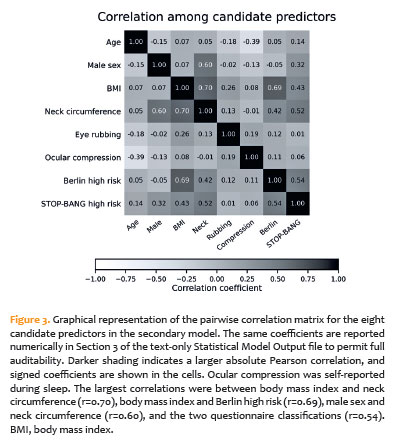
Predictors of keratoconus status were initially examined using separate univariable logistic regression models. The primary multivariable model included age, sex, BMI, eye rubbing, and self-reported ocular compression during sleep. Neck circumference was excluded because of its strong correlation with BMI (r=0.70). Questionnaire-based classifications were also excluded because BMI contributes to both instruments, whereas neck circumference contributes to the STOP-BANG score.
The primary model included five predictors for the 49 participants in the smaller group, corresponding to approximately 10 participants per predictor. This contrasted with approximately six participants per predictor in the secondary eight-predictor model. Variance inflation factors in the primary model ranged from 1.09 to 1.30. Additional exploratory models adjusted each questionnaire classification for age and sex, replaced BMI with neck circumference, and incorporated allergic disease. Because age and sex are themselves components of the STOP-BANG score, the age- and sex-adjusted STOP-BANG estimate was considered a partial-adjustment sensitivity analysis rather than a conventional confounder-adjusted effect estimate.
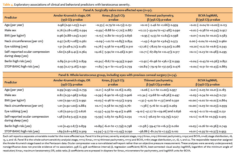
Severity analyses were exploratory and restricted to participants with keratoconus. The responsible researcher assigned the Amsler-Krumeich stage based on Pentacam data documented in the medical records. Because previous corneal procedures can alter the parameters used for staging, the primary severity analysis included the 51 patients whose more affected eye was surgically naive. The complete cohort of 74 patients was analyzed separately as a sensitivity analysis.
Disease stage was analyzed using ordinal logistic regression, whereas Kmax, thinnest corneal thickness, and BCVA were analyzed using separate univariable linear regression models. Given the inclusion of only 51 patients across four disease stages, the statistical power for the severity analyses was very limited.
RESULTS
Demographic and anthropometric characteristics
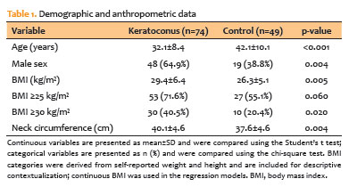
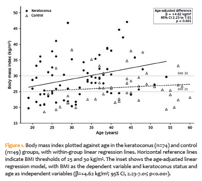
Mean age was 32.1±8.4 years in the keratoconus group and 42.1±10.1 years in controls (p<0.001), and 64.9% versus 38.8% were male (p=0.004). BMI was higher in the keratoconus group (29.4±6.4 vs. 26.3±5.1kg/m²; crude difference, 3.13 kg/m²; p=0.005). BMI was ≥25kg/m² in 71.6% versus 55.1% (p=0.060) and ≥30kg/m² in 40.5% versus 20.4% (p=0.020). Because weight and height were self-reported, these comparisons should be interpreted with caution. Neck circumference, the directly measured anthropometric variable, was also greater in the keratoconus group (40.1±4.6 vs. 37.6±4.6 cm; p=0.004; Table 1). After adjustment for age, the estimated BMI difference increased to 4.62 kg/m² (95% CI, 2.23-7.01; p<0.001; Figure 1), indicating that the older age of controls attenuated rather than explained the crude association; additional adjustment for sex yielded consistent results.
Behavioral, allergic, and sleep-related characteristics
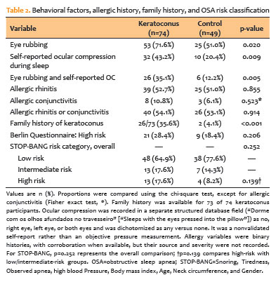
Eye rubbing was reported by 71.6% of keratoconus patients and 51.0% of controls (p=0.020). Self-reported ocular compression during sleep was present in 43.2% and 20.4%, respectively (p=0.009), and both behaviors were reported in 35.1% and 12.2%, respectively (p=0.005).
Allergic rhinitis (52.7% vs. 51.0%), allergic conjunctivitis (10.8% vs. 6.1%), and either condition (54.1% vs. 53.1%) were similarly frequent. Allergy was associated with eye rubbing across the sample (p=0.021). Allergy variables were based on binary histories, with corroboration when available; however, the source and severity were not recorded separately. A family history of keratoconus was reported by 35.6% and 4.1% of participants, respectively (p<0.001; Table 2).
OSA risk assessment
Berlin high-risk classification was present in 28.4% of keratoconus participants and 18.4% of controls (p=0.206). STOP-BANG low-, intermediate-, and high-risk categories comprised 64.9%, 17.6%, and 17.6% in the keratoconus group and 77.6%, 14.3%, and 8.2% in controls, respectively (overall p=0.252). When high risk was compared with low or intermediate risk combined, the difference remained nonsignificant (p=0.139; Table 2).
Tomographic parameters, BCVA, and disease staging
Among the 51 surgically naive eyes included in the primary severity analysis, stages 1-4 comprised 16, 15, 2, and 18 patients, respectively. Corresponding Kmax, thinnest corneal thickness, and BCVA summaries are presented with the severity results.
Logistic regression for keratoconus presence
In unadjusted analyses, younger age, male sex, higher BMI, larger neck circumference, eye rubbing, and self-reported ocular compression during sleep were associated with keratoconus. In the primary model, younger age (OR, 0.88; 95% CI, 0.83-0.93), male sex (OR, 2.91; 95% CI, 1.14-7.42), and higher BMI (OR, 1.15; 95% CI, 1.05-1.27) remained associated, whereas eye rubbing (OR, 1.43; 95% CI, 0.55-3.73) and self-reported ocular compression (OR, 1.47; 95% CI, 0.52-4.11) were attenuated (Table 3; Figure 2). Replacing BMI with neck circumference produced an OR of 1.17 per cm (95% CI, 1.03-1.33; Table 3, Panel C). Adding allergy left the estimates unchanged, and allergy was not associated with keratoconus (OR, 0.62; 95% CI, 0.24-1.58; Table 3, Panel C).
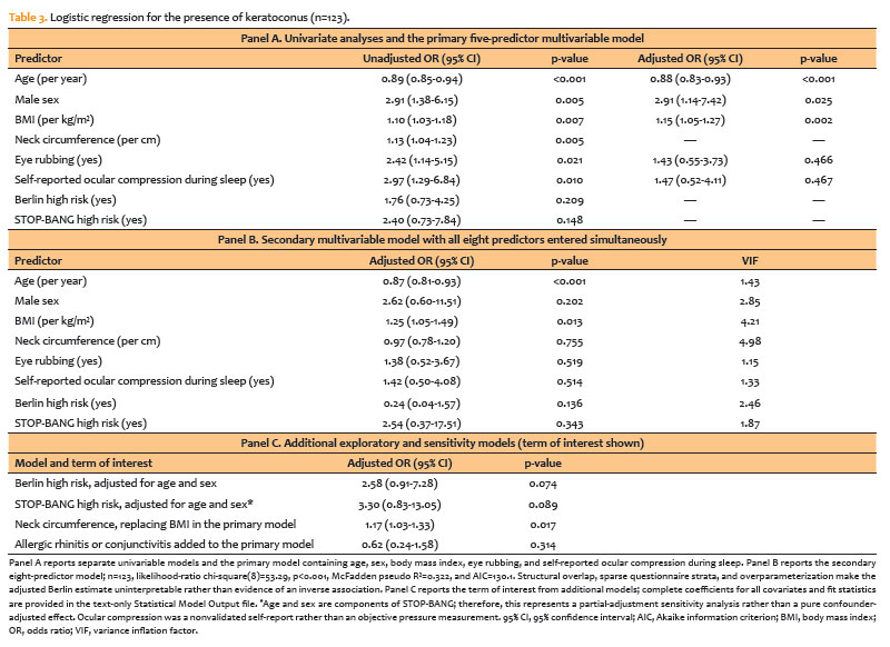
In the secondary eight-predictor model (Table 3, Panel B), only age and BMI retained significance. The predictor correlation matrix illustrates the structural overlap among BMI, neck circumference, and the two questionnaire classifications (Figure 3). In that model, the Berlin classification yielded an OR of 0.24 (95% CI, 0.04-1.57), a reversal of its unadjusted direction that reflects the small number of high-risk controls (9 of 49) and adjustment for overlapping anthropometric content; it should therefore be regarded as uninterpretable rather than as evidence of an inverse association.
Adjusted for age and sex only, the Berlin (OR, 2.58; 95% CI, 0.91-7.28; p=0.074) and STOP-BANG (OR, 3.30; 95% CI, 0.83-13.05; p=0.089) classifications showed positive but nonsignificant associations. These estimates are exploratory; for STOP-BANG, age and sex are also score components, so this represents a partial-adjustment sensitivity analysis rather than a pure confounder-adjusted effect.
Exploratory analyses of keratoconus severity
In the 51 patients whose more affected eye had not been operated on, no predictor was associated with severity. Neither eye rubbing (stage OR, 1.51; 95% CI, 0.48-4.72), self-reported ocular compression during sleep (OR, 1.60; 95% CI, 0.58-4.42), nor high-risk STOP-BANG classification (OR, 0.95; 95% CI, 0.27-3.26) was associated with Amsler-Krumeich stage, and none was associated with Kmax, thinnest corneal thickness, or BCVA (Table 4). Results were similar in all 74 patients (Table 4, Panel B). These analyses were severely underpowered; therefore, the absence of statistical significance does not constitute evidence of the absence of an association.
DISCUSSION
Patients with keratoconus had higher BMI and neck circumference than controls, representing an anthropometric profile associated with increased OSA risk, and reported eye rubbing more frequently. They also more frequently reported sleeping with one or both eyes pressed into the pillow; however, questionnaire-based OSA classifications did not differ significantly between the groups.
A methodological point warrants emphasis. Anthropometric variables are incorporated into the screening tools: the Berlin questionnaire scores for obesity, whereas STOP-BANG includes both BMI above 35 kg/m² and increased neck circumference. Therefore, the anthropometric differences and numerically higher questionnaire scores cannot be considered independent observations, and the contribution of sleep-specific items cannot be isolated. The older control group was structurally more likely to receive the age point in the STOP-BANG score. In contrast, the Berlin questionnaire incorporates hypertension but not age itself. Thus, for STOP-BANG, the age imbalance works against, rather than explains, the observed numerical direction. Most importantly, screening classifications do not constitute OSA diagnoses; without polysomnography, clinically valid “keratoconus with OSA” and “keratoconus without OSA” groups cannot be established.
The Brazilian National Health Survey 2019, which used measured anthropometry, estimated that 60.3% of adults had a BMI ≥25 kg/m² and 25.9% had a BMI ≥30 kg/m²(26). The corresponding proportions based on self-reported data were 71.6% and 40.5% in the keratoconus group and 55.1% and 20.4% in controls. Thus, the control group was somewhat below the national estimates, whereas the keratoconus group was above them. However, these external comparisons are contextual only because the national sample differed in age and sex and used measured rather than self-reported anthropometry. The crude BMI difference was 3.13 kg/m² and increased to 4.62kg/m² after adjustment for age, indicating that the older age of controls masked part of, rather than explained, the association. This adjusted finding was consistent with a population-based association reported in more than half a million adolescents(27). Because neck circumference was measured directly, its between-group difference is less susceptible to self-report error. Nevertheless, its strong correlation with BMI and sex limits interpretation of its independent contribution.
Eye rubbing was more frequent among patients with keratoconus in the unadjusted analysis, consistent with its recognized mechanical and inflammatory effects(1,10,21). Allergy is an important potential confounder because itching promotes eye rubbing, and keratoconus shares inflammatory features with allergic conjunctivitis(9,28). Binary histories of rhinitis and conjunctivitis were similar between the groups, and adding allergy to the model did not materially alter the estimates. However, the source, subtype, severity, medication use, asthma, and dermatitis were not systematically recorded; therefore, residual confounding cannot be excluded.
Self-reported ocular compression during sleep was more frequent among patients with keratoconus, consistent with reports linking preferred sleeping side and unfavorable sleeping posture to asymmetric disease(7,15,16). However, this variable should not be interpreted as a measure of observed or quantified pressure because it was derived from a single nonvalidated structured field and did not capture frequency, duration, or force. No association with disease severity was detected in the exploratory analyses, which had limited statistical power. Objective sleep studies remain necessary to establish the burden of OSA among patients with keratoconus(14,29,30).
A strength of this study is the joint evaluation of keratoconus and OSA screening risk using two validated questionnaires while integrating anthropometric, behavioral, and tomographic data(7,13,15).
Several limitations should be considered. These include the cross-sectional design, unmatched age and sex, and absence of polysomnography(12). Participants with a previous diagnosis or treatment of OSA were excluded; therefore, the screening comparisons apply only to individuals without recognized OSA. This exclusion may have attenuated high-risk classification rates and between-group contrasts and may limit the generalizability of the findings. Controls were a convenience sample from the same institution, and the clinical reason for their attendance was not retained, allowing the possibility of selection bias and limiting population representativeness. Paper questionnaires were administered and scored manually by a single researcher, which standardized their application but may have introduced interviewer or classification bias. Weight and height were self-reported in both groups, creating potential reporting errors that cannot be eliminated by age adjustment; neck circumference was directly measured and was therefore less susceptible to self-report error. The ocular compression variable was based on a single nonvalidated self-report field and did not quantify pressure, duration, or frequency. Allergy was assessed as a binary variable and was incompletely characterized, leaving the possibility of residual confounding(9,28). Control tomography was unavailable for post hoc quantitative comparison. Prior surgery affected staging variables, and the timing of tomography relative to surgery was undocumented for operated eyes. BCVA was assessed using spectacle correction, and seven participants lacked numeric logMAR data, five of whom were among the 51 surgically naive patients.
In summary, patients with keratoconus had higher BMI and neck circumference, more frequent eye rubbing, and more frequent self-reported ocular compression during sleep, but OSA screening classifications did not differ significantly between the groups. Behavioral estimates were attenuated after adjustment, while ocular compression represented a nonvalidated self-report rather than an objective measure of pressure, and the severity analyses were inconclusive. Prospective studies should incorporate polysomnography, measured anthropometry, prespecified assessments of sleep position and ocular compression, and standardized evaluation of allergic conditions.
ACKNOWLEDGMENTS
The authors thank Rafael Balsalobre for his valuable contribution to this study. This paper underwent Paperpal Preflight. The study received the award for Best Scientific Paper Presentation at the Brazilian Congress of Cataract and Refractive Surgery (BRASCRS 2025).
AUTHORS’ CONTRIBUTIONS
Significant contribution to conception and design: Jaqueline Fernandes Ruiz; Talita Gambogi Araújo; Thaís Cândida Borges; Leonardo Ugulino de Araújo Neto; Felipe Leal de Moraes Brito; Pablo Felipe Rodrigues; Luiz Antonio de Brito Martins; Myrna Serapião dos Santos; Bernardo Kaplan Moscovici. Data acquisition: Jaqueline Fernandes Ruiz; Talita Gambogi Araújo; Thaís Cândida Borges; Leonardo Ugulino de Araújo Neto; Felipe Leal de Moraes Brito. Data analysis and interpretation: Pablo Felipe Rodrigues; Luiz Antonio de Brito Martins; Myrna Serapião dos Santos; Bernardo Kaplan Moscovici. Manuscript drafting: Jaqueline Fernandes Ruiz; Talita Gambogi Araújo; Thaís Cândida Borges; Leonardo Ugulino de Araújo Neto; Felipe Leal de Moraes Brito. Significant intellectual content revision of the manuscript: Pablo Felipe Rodrigues; Luiz Antonio de Brito Martins; Myrna Serapião dos Santos; Bernardo Kaplan Moscovici. Final approval of the submitted manuscript: Jaqueline Fernandes Ruiz; Talita Gambogi Araújo; Thaís Cândida Borges; Leonardo Ugulino de Araújo Neto; Felipe Leal de Moraes Brito; Pablo Felipe Rodrigues; Luiz Antonio de Brito Martins; Myrna Serapião dos Santos; Bernardo Kaplan Moscovici. Statistical analysis: Pablo Felipe Rodrigues; Myrna Serapião dos Santos. Obtaining funding: not applicable. Supervision of administrative, technical, or material support: Luiz Antonio de Brito Martins; Myrna Serapião dos Santos. Research group leadership:Myrna Serapião dos Santos; Bernardo Kaplan Moscovici.
REFERENCES
1. Santodomingo-Rubido J, Carracedo G, Suzaki A, Villa-Collar C, Vincent SJ, Wolffsohn JS. Keratoconus: an updated review. Cont Lens Anterior Eye. 2022;45(3):101559.
2. Debourdeau E, Planells G, Chamard C, Touboul D, Villain M, Demoly P, et al. New keratoconus risk factors: a cross-sectional case-control study. J Ophthalmol. 2022;2022(1):6605771.
3. Rabinowitz YS. The genetics of keratoconus. Ophthalmol Clin North Am. 2003;16(4):607-20.
4. Gordon-Shaag A, Millodot M, Shneor E, Liu Y. The genetic and environmental factors for keratoconus. BioMed Res Int. 2015;2015(1):795738.
5. Gajecka M. The role of epigenetic changes in stability and progression of the disease phenotype in keratoconus. Adv Exp Med Biol. 2015;882:15-23.
6. de Azevedo Magalhães O, Gonçalves MC, Gatinel D. The role of environment in the pathogenesis of keratoconus. Curr Opin Ophthalmol. 2021;32(4):379-84.
7. Santos RT, Moscovici BK, Hirai FE, Benício CM, Nakano EM, Nosé W. Association between keratoconus, ocular allergy, and sleeping behavior. Arq Bras Oftalmol. 2021;84(1):17-21.
8. Moran S, Gomez L, Zuber K, Gatinel D. A case-control study of keratoconus risk factors. Cornea. 2020;39(6):697-701.
9. Hage A, Knoeri J, Leveziel L, Majoulet A, Buffault J, Labbé A, et al. [From ocular itching to eye rubbing: a review of the literature]. J Fr Ophtalmol. 2023;46(2):173-84.
10. Lema I, Sobrino T, Durán JA, Brea D, Díez-Feijoo E. Subclinical keratoconus and inflammatory molecules from tears. Br J Ophthalmol. 2009;93(6):820-4.
11. Arriola-Villalobos P, Benito-Pascual B, Peraza-Nieves J, Perucho-González L, Sastre-Ibañez M, Dupré-Peláez MG, et al. Corneal topographic, anatomic, and biomechanical properties in severe obstructive sleep apnea-hypopnea syndrome. Cornea. 2020;39(1):88-91.
12. Senaratna CV, Perret JL, Lodge CJ, Lowe AJ, Campbell BE, Matheson MC, et al. Prevalence of obstructive sleep apnea in the general population: A systematic review. Sleep Med Rev. 2017;34:70-81.
13. Gupta PK, Stinnett SS, Carlson AN. Prevalence of sleep apnea in patients with keratoconus. Cornea. 2012;31(6):595-9.
14. Saidel MA, Paik JY, Garcia C, Russo P, Cao D, Bouchard C. Prevalence of sleep apnea syndrome and high-risk characteristics among keratoconus patients. Cornea. 2012;31(6):600-3.
15. Mazharian A, Panthier C, Courtin R, Jung C, Rampat R, Saad A, et al. Incorrect sleeping position and eye rubbing in patients with unilateral or highly asymmetric keratoconus: a case-control study. Graefes Arch Clin Exp Ophthalmol. 2020;258(11):2431-9.
16. Corrêa DC, Dantas DO, Amaral DC, Moreira H, Louzada RN, Alves MR. Evaluation of the preferred sleeping position as a risk factor for keratoconus asymmetry. Optom Vis Sci. 2025;102(6):400-5.
17. Netzer NC, Stoohs RA, Netzer CM, Clark K, Strohl KP. Using the Berlin Questionnaire to identify patients at risk for the sleep apnea syndrome. Ann Intern Med. 1999;131(7):485-91.
18. Chung F, Yegneswaran B, Liao P, Chung SA, Vairavanathan S, Islam S, et al. STOP questionnaire: a tool to screen patients for obstructive sleep apnea. Anesthesiology. 2008;108(5):812-21.
19. Chung F, Yang Y, Brown R, Liao P. Alternative scoring models of STOP-bang questionnaire improve specificity to detect undiagnosed obstructive sleep apnea. J Clin Sleep Med. 2014;10(9):951-8.
20. Chiu HY, Chen PY, Chuang LP, Chen NH, Tu YK, Hsieh YJ, et al. Diagnostic accuracy of the Berlin questionnaire, STOP-BANG, STOP, and Epworth sleepiness scale in detecting obstructive sleep apnea: A bivariate meta-analysis. Sleep Med Rev. 2017;36:57-70.
21. Kolozsvári BL, Petrovski G, Gogolák P, Rajnavölgyi É, Tóth F, Berta A, et al. Association between mediators in the tear fluid and the severity of keratoconus. Ophthalmic Res. 2014;51(1):46-51.
22. Gomes JA, Tan D, Rapuano CJ, Belin MW, Ambrósio R Jr, Guell JL, et al.; Group of Panelists for the Global Delphi Panel of Keratoconus and Ectatic Diseases. Global consensus on keratoconus and ectatic diseases. Cornea. 2015;34(4):359-69.
23. Rabinowitz YS, McDonnell PJ. Computer-assisted corneal topography in keratoconus. Refract Corneal Surg. 1989;5(6):400-8.
24. Kamiya K, Ishii R, Shimizu K, Igarashi A. Evaluation of corneal elevation, pachymetry and keratometry in keratoconic eyes with respect to the stage of Amsler-Krumeich classification. Br J Ophthalmol. 2014;98(4):459-63.
25. Fonseca LB, Silveira EA, Lima NM, Rabahi MF. STOP-Bang questionnaire: translation to Portuguese and cross-cultural adaptation for use in Brazil. J Bras Pneumol. 2016;42(4):266-72.
26. Instituto Brasileiro de Geografia e Estatística (IBGE). Pesquisa nacional de saúde: 2019: atenção primária à saúde e informações antropométricas - Brasil. Rio de Janeiro: IBGE; 2020.
27. Eliasi E, Bez M, Megreli J, Avramovich E, Fischer N, Barak A, et al. The association between keratoconus and body mass index: a population-based cross-sectional study among half a million adolescents. Am J Ophthalmol. 2021;224:200-6.
28. Gijs M, Adelaar TI, Vergouwen DP, Visser N, Dickman MM, Ollivier RC, et al. Tear fluid inflammatory proteome analysis highlights similarities between keratoconus and allergic conjunctivitis. Invest Ophthalmol Vis Sci. 2023;64(15):9.
29. Pedrotti E, Demasi CL, Fasolo A, Bonacci E, Brighenti T, Gennaro N, et al. Obstructive sleep apnea assessed by overnight polysomnography in patients with keratoconus. Cornea. 2018;37(4):470-3.
30. Pellegrini M, Bernabei F, Friehmann A, Giannaccare G. Obstructive sleep apnea and keratoconus: a systematic review and meta-analysis. Optom Vis Sci. 2020;97(1):9-14.
Submitted for publication:
September 30, 2025.
Accepted for publication:
September 9, 2026.
Funding: This study received no specific financial support.
Disclosure of potential conflicts of interest: The authors declare no potential conflicts of interest.

How to cite this article:
 ABO is licensed under a Creative Commons Attribution-NonComercial 4.0 Internacional.