Arq. Bras. Oftalmol. 2025; 88 (3): 10.5935/0004-2749.2024-0358
Total: 1268
J. William Harbour1; Zélia Maria Corrêa2
DOI: 10.5935/0004-2749.2024-0358
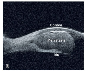
A 32-year-old woman with multiorgan metastatic cervix adenocarcinoma reported pain and blurred vision in the right eye (OD). Her vision was counts fingers with an intraocular pressure of 30 mmHg OD. Figure 1A depicts the fleshy vascularized iris mass with layered hyphema and pseudohypopyon. The iris mass measuring 6.9 × 6.8 × 3.3 mm on 50 MHz ultrasonography is shown in Figure 1B. Choroid OD and left eye were normal. She expired prematurely despite systemic chemotherapy. Cervix cancer is the second most prevalent malignancy in women rarely metastasizing to the iris(1,2).

REFERENCES
1. Nair AG, Asnani HT, Mehta VC, Mehta SV, Pathak RS. Metastatic adenocarcinoma of the cervix presenting as a choroidal mass: A case report and review of the literature on cervical metastases to the eye. Indian J Ophthalmol. 2015;63(8):674-8.
2. Kurosawa A, Sawaguchi S. Iris metastasis from squamous cell carcinoma of the uterine cervix. Case report. Arch Ophthalmol. 1987;105(5):618.
Submitted for publication:
November 19, 2024.
Accepted for publication:
December 11, 2024.
Informed consent was obtained from all patients included in this study.
Funding: This study received no specific financial support.
Disclosure of potential conflicts of interest: The authors declare no potential conflicts of interest.

How to cite this article:
 ABO is licensed under a Creative Commons Attribution-NonComercial 4.0 Internacional.