Arq. Bras. Oftalmol. 2024; 87 (5): 10.5935/0004-2749.2024-0155
Total: 1978
Giulia Aragão; Nicole B. M. Almeida; Newton Kara-Junior
DOI: 10.5935/0004-2749.2024-0155
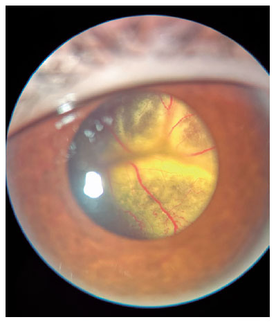
Coats’ disease is an idiopathic exudative retinopathy that is characterized by retinal telangiectasias, aneurysms, and capillary nonperfusion. It is associated with intraretinal and subretinal exudations, which frequently progress to exudative retinal detachment(1). Coats’ disease is mostly unilateral and progressive and predominantly affects males during childhood. Although the average age at the time of diagnosis is 8-16 years, adult cases have also been described(2). The most commonly used classification was proposed by Shields et al., and it is based on funduscopic findings. Although this classification can aid in the disease diagnosis(1), in a majority of the patients, some form of ancillary testing, such as fluorescein angiography, ultrasound, computerized tomography, or magnetic resonance imaging, is required(2). The most common manifestations of the disease are decreased visual acuity, strabismus and leukocoria(3). The differential diagnoses of Coats’ disease include retinoblastoma, retinal vasoproliferative tumor, familial exudative retinopathy, retinal capillary hemangioblastoma, and familial retinal arterial macroaneurysm(1). The aim of treatment in Coats’ disease is the ablation of abnormal retinal vasculature, preservation of vision, and prevention of disease progression to retinal detachment(3). Thus, the treatment options include photocoagulation, cryotherapy, and surgery(1). Antivascular endothelial growth factor or corticosteroids may also be injected intravitreally as adjuvant therapy(1).
AUTHOR CONTRIBUTIONS:
Substantial contribution to conception and design: Nicole B. M. Almeida, Giulia Aragão, Newton Kara-Junior. Acquisition of data: Nicole B. M. Almeida, Giulia Aragão, Newton Kara-Junior. Analysis and interpretation of data: not applicable. Drafting of the manuscript: Nicole B. M. Almeida. Critical revision of the manuscript for important intellectual content: Newton Kara-Junior. Have given final approval of the submitted manuscript: Newton Kara-Junior, Nicole B. M. Almeida, Giulia Aragão. Statistical analysis: not applicable. Obtaining funding: not applicable. Administrative, technical, or material support supervision: not applicable. Research group leadership: not applicable.
REFERENCES
1. Dorado AM, De-Pablo-Cabrera A, Muñoz-Gallego A, Moreno-García L, Barceló-Mendiguchía A, Tejada-Palacios P. Dexamethasone intravitreal implant as an adjuvant treatment for pediatric patients with Coats’ disease. Arq Bras Oftalmol [Internet]. 2023;86(4):380-3.
2. Shields JA, Shields CL, Honavar SG, Demirci H: Clinical variations and complications of Coats disease in 150 cases: the 2000 Sanford Gifford Memorial Lecture. Am J Ophthalmol. 2001;131:561-71.
3. Ghorbanian S, Jaulim A, Chatziralli IP. Diagnosis and treatment of Coats’ Disease: a review of the literature. Ophthalmologica [Internet]. 2012 [cited 2024 May 24];227(4):175-82.
Submitted for publication:
May 16, 2024.
Accepted for publication:
May 21, 2024.
Funding: This study received no specific financial support.
Disclosure of potential conflicts of interest: None of the authors have any potential conflicts of interest to disclose.

How to cite this article:
 ABO is licensed under a Creative Commons Attribution-NonComercial 4.0 Internacional.