Arq. Bras. Oftalmol. 2019; 82 (3): 10.5935/0004-2749.20190034
Total: 2279
Anita Zimmermann; Keila Miriam Monteiro de Carvalho; Camila Atihe; Sara Martins Vieira Zimmermann; Valeriana Leme de Moura Ribeiro
DOI: 10.5935/0004-2749.20190034
ABSTRACT
Purposes: This study aimed to present the characteristics of visual development from a clinical viewpoint in infants and preschool children aged 0-6 years who were born at term with no pregnancy or childbirth complications.
Methods: We conducted a bibliographic review on visual development in infants and preschool children.
Results: We described visual development in children according to age groups: 0-1 month, 1-3 months, 3-6 months, 6-10 months, 10 months-1 year and
4 months, 1 year and 4 months-2 years, 2-4 years, and 4-6 years.
Conclusion: Visual responses in infants and preschool children born at term and with normal development were observed to occur in an integrated manner with neuromotor functions in addition to cognitive and psycho-emotional sensory, behavioral, and visual capacity.
Keywords: Vision, ocular; Child; Infant; Visual acuity
RESUMO
Objetivos: Este estudo teve como objetivo apresentar as características do desenvolvimento visual do ponto de vista clínico em bebês e pré-escolares de 0 a 6 anos que nasceram a termo sem complicações na gravidez ou no parto.
Métodos: Foi realizada uma revisão bibliográfica sobre o desenvolvimento visual em lactentes e pré-escolares.
Resultados: Descrevemos o desenvolvimento visual em crianças de acordo com as faixas etárias: 0-1 mês, 1 a 3 meses, 3 a 6 meses, 6 a 10 meses, 10 meses a 1 ano e 4 meses, 1 ano e 4 meses a 2 anos, 2 a 4 anos e 4 a 6 anos.
Conclusão: Observou-se que as respostas visuais em lactentes e pré-escolares nascidos a termo e com desenvolvimento normal ocorrem de forma integrada às funções neuromotoras, além da capacidade sensorial, comportamental e visual cognitiva e psicoemocional.
Descritores: Visão ocular; Criança; Lactente; Acuidade visual
INTRODUCTION
Visual capacity of the central nervous system in humans progressively develops from the birth. The occipital region of the human brain has a specific area for receiving and interpreting images captured by eyes(1). Optic nerve fiber myelination progresses to completion in the tenth week after birth and consequently rapidly increases the synaptic density of the visual cortex from birth to four months of extrauterine life, reflecting in improved visual perception, fixation, and functional coordination of accompanying motivators of visual stimuli(1,2).
Development of the visual system immediately starts after birth via visual stimuli and interactions with the environment, which concomitantly occur with the child's global development, i.e., neuropsychomotor development, visual-motor coordination, cognitive abilities, and behavioral, environmental, and sociocultural adaptation(2-4). The anatomical and neurophysiological integrity of this system is essential for the occurrence of the maturation process, which differs with age and is interdependent of genetic, cognitive, and environmental aspects(5,6).
After birth, the visual system undergoes a continuous maturation process involving the eyeball and pathways and neural networks of cortical areas and cortical association areas that integrating different parts. In early life, development of the still immature retinas is accelerated by the fovea and macula, the optical pathways are partially myelinated, and the visual cortex is rudimentary(1,7-9).
Many anatomical changes occur in the visual maturation process, such as the increase in central cone density and elongated outer photoreceptor segments, which develop slowly until age 7 years(3,10), enabling progressive improvement in functional vision and development(11,12).
The light stimuli received by the retina are transmitted to the occipital cortex as specific stimuli formed from the photochemical reactions by the retina after the light capture(1,12,13). The occipital cortex integrates the stimuli received from both eyes into a unique visual perception.
Completing its functional maturity between the ages of 5 and 6 years, this sensorineural mechanism is called binocular fusion. The perceived and unified images are evaluated regarding their form, color, light, and relative location based on the surroundings(9,14), raising awareness of the object's spatial location, i.e., stereopsis(7,11,15).
At 15 days after birth, visual acuity is estimated at 20/400. At this stage, the child shows interest in objects >10 cm in diameter(1,16,17).
Considering normal visual development and cognitive development, children up to age 18 months have similar vision to adults, and a fully developed visual system is perceived until the child becomes 10 years old(10).
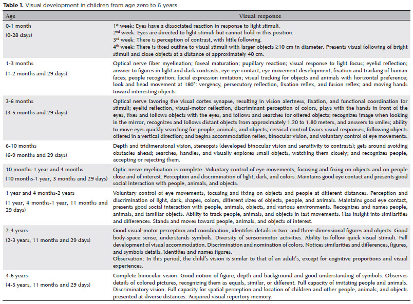
Visual development in children
Information about the child's vision can be obtained through objective ophthalmological exams, including electrophysiological tests(10), which evaluate maturation of the cortical visual processes independent of the child's active participation.
Psychophysical tests(13,14,18,19) identify visual responses in children from age zero to 6 years and relate them to normal visual development expected in children born at term without compromising development, with intact visual, sensory, and motor neuron abilities(11,16,20-22). These tests are standardized with figures, followed with or without the use of recreational objects that encourage visual responses observed through the child's spontaneous behavior(4,10,17).
Normal vision development follows a child's global development in sensorineural nuances; vision maturation is obtained by the visual stimuli provided by both the environment in which the child lives and the child's caregivers. Table 1 shows the maturation of the child's normal visual development, with no developmental compromises, according to each age group.
Considerations
After birth, body cells of the infant are still in full development, forming continuous connections and neural communications. The maturation of the eyes and optic pathways is directly related to the child's visual and neuromotor development(23,24).
The visual system's ability to interpret perceived images is developed following cognitive development together with other skills related to child development, forming and organizing the visual repertory. Stimuli, motivations, and visual experiences are important for maturation of the visual system and its developmental functions(18,25-28).
Each visual function has a specific profile depending on functional potentiality at birth, child development, and the child's visual neuroperception capabilities(6).
Visual system development in children occurs concomitantly with child development. Visual system integrity as well as the neurophysiological structures enable the vision to progress in its functional maturation steps after childbirth(2,23-25).
Objective and psychophysical evaluation of vision must be specific for children and results must be consistent with the child's age(20). From the beginning of extrauterine development, the child must receive continuous stimuli and visual experiences to develop the optical system in order to present expected visual responses in each age group.
CONCLUSION
Visual responses in infants and preschool children born at term and with normal development occur in an integrated manner to neuromotor functions in addition to cognitive and psycho-emotional sensory and behavioral and visual capacity.
REFERENCES
1. Bicas HE. Physiology of binocular vision. Arq Bras Oftalmol. 2004; 67(1):172-80.
2. Braddick O, Atkinson J. Development of human visual function. Vision Res. 2011;51(13):1588-609.
3. Kozma P, Kovacs I, Benedek G. Normal and abnormal development of visual functions in children. Acta Biol Szeged. 2001;45(1-4):23-42.
4. Gagliardo HG, Gonçalves VMG, Lima MCMP. Método para Avaliação da conduta visual de lactentes. Arq Neuropsiquiatr. 2004; 62(2A):300-6.
5. Larsson E, Martin L, Holmström G. Peripheral and central visual fields in 11-year-old children who had been born prematurely and at term. J Pediatr Ophthalmol Strabismus. 2004;41(1):39-45.
6. Dutton G. Visual problems in children with damage to the brain. Vis Impair Res. 2002;4(2).113-21. https://doi.org/10.1076/vimr.4.2.113.15640
7. Huttenlocher PR. Morphometric study of human cerebral cortex development. Neuropsychologia. 1990;28(6):517-27.
8. Ruas TC, Ravanini SG, Martinez CS, Gagliardo HR, Françoso MF, Rim PH. Avaliação do comportamento visual de lactentes no primeiro e segundo meses de vida. J Hum Growth Develop. 2006;16(3):1-8.
9. Salomão SR. Desenvolvimento da acuidade visual de grades. Psicol USP. 2007;18(2):63-81.
10. Coelho AC, Marta DC, Dias MA, Salvador M, Reis VN, Pacheco ZM. Olho vivo: analisando a acuidade visual das crianças e o emprego do lúdico no cuidado de enfermagem. Esc Anna Nery Rev Enferm. 2010;42(2):318-23.
11. Berezovsky A. Maturação funcional da retina em bebês prematuros. Psicol USP. 2007;18(2):35-45.
12. Snir M, Friling R, Weinberger D, Sherf I, Axer-Siegel R. Refraction and keratometry in 40 week old premature (corrected age) and term infants. Br J Ophthalmol. 2004;88(7):900-4.
13. Oda JY, Santana DM, Carvalho J. Plasticidade e regeneração funcional do sistema nervoso: contribuição ao estudo de revisão. Arq Ciênc Saúde Unipar. 2002;6(2):171-6.
14. Pallagrosi M. Neuroplasticity of the developing brain and child cortical visual impairment. Ann Ist Super Sanita. 1993;29(1):163-5.
15. O'Connor AR, Stephenson TJ, Johnson A, Tobin MJ, Ratib S, Moseley M, et al. Visual function in low birthweight children. Br J Ophthalmol. 2004;88(9):1149-53.
16. Kutzbach BR, Summers CG, Holleschau AM, MacDonald JT. Neurodevelopment in children with albinism. Ophthalmology. 2008; 115(10):1805-8, 1808.e1-2.
17. Zimmermann A, Silva SV, Zimmermann SM, Lira RP, Carvalho KM. Teller test with functional vision evaluation in children with low vision. Rev Bras Oftalmol. 2015;74(6):362-5.
18. Kronbauer AL, Schor P, Carvalho LA. Medida da visão e testes psicofísicos. Arq Bras Oftalmol. 2008;71(1):122-7.
19. Salomão SR, Ejzenbaum F, Berezovsky A, Sacai PY, Pereira JM. Age norms for monocular grating acuity measured by sweep-VEP in the first three years of age. Arq Bras Oftalmol. 2008;71(4):475-9.
20. Graziano RM, Leone CR. Frequent ophthalmologic problems and visual development of extremely preterm newborn infants. J Pediatr (Rio J). 2005;81(1 Suppl):S95-100. Review. Portuguese.
21. Olitsky SE, Nelson BA, Brooks S. The sensitive period of visual development in humans. J Pediatr Ophthalmol Strabismus. 2002;39(2): 69-72; quiz 105-6. Review.
22. Colenbrander A. The historical evolution of visual acuity measurement. S.l. Volume 10. Vis Impair Res. 2008;10(2-3):57-66.
23. Wright JD Jr, Boger WP 3rd. Visual complaints from healthy children. Surv Ophthalmol. 1999;44(2):113-21. Review.
24. Ferreira AP, Albuquerque RC, Rabelo AR, Farias FC, Correia RC, Gagliardo HG, et al. Comportamento visual e desenvolvimento motor de recém-nascidos prematuros no primeiro mês de vida. Rev Bras Crescimento Desenvolv Hum. 2011;21(2):335-43.
25. Ventura DF. Visão de cores no primeiro ano de vida. Psicol USP. 2007;18(2):83-97.
26. Macchi M, Rossi LN, Cortinovis I, Menegazzo L, Burri SM, Piller M, et al. Development of visual perception and attention, assessed by backward masking and application in children with epilepsy. Dev Med Child Neurol. 2003;45(8):562-7.
27. Manoni AS, Campos D, Borges G. Gonçalves VM. O conceito do sistema neurofuncional aplica-se à percepção de faces. Rev Neurocienc. 2008;16(4):316-21.
28. Holmström G, Larsson E. Long-term follow-up of visual functions in prematurely born children-a prospective population-based study up to 10 years of age. J AAPOS. 2008;12(2):157-62.
Submitted for publication:
April 9, 2018.
Accepted for publication:
August 14, 2018.
Funding: No specific financial support was available for this study.
Disclosure of potential conflicts of interest: The authors have no potential conflicts of interest to disclose.

How to cite this article:
 ABO is licensed under a Creative Commons Attribution-NonComercial 4.0 Internacional.