Arq. Bras. Oftalmol. 2018; 81 (5): 10.5935/0004-2749.20180085
Total: 1541
Alexis Galeno Matos1; Viviane Pinho Gurgel1; Pedro Javier Yugar1; Alejandro Sebastian Yugar2
DOI: 10.5935/0004-2749.20180085
ABSTRACT
We report on a case of two sisters, daughters of consanguineous parents, presenting with a similar condition of low visual acuity associated with retinal dystrophy in both eyes associated with alopecia and bone alterations or syndactyly.
Keywords: Ectodermal dysplasia/genetics; Syndactyly; Alopecia; Retina/abnormalities; Retinal dystrophy; Hand deformities, congenital/genetics; Syndrome
RESUMO
Relatamos um caso de duas irmãs, filhas de pais consanguíneos, apresentando uma condição semelhante de baixa acuidade visual associado à distrofia retiniana em ambos os olhos associado à alopecia e alterações ósseas ou sindactilia.
Descritores: Displasia ectodérmica/genética; Sindactilia; Alopecia; Retina/anormalidades; Distrofia retiniana; Deformidades congênitas da mão/genética; Síndrome
INTRODUCTION
In 1956, Albrectsen and Svendsen described two siblings of consanguineous parents presenting with syndactyly, sparse hair, and retinal degeneration with normal psychomotor development(1).
Ectodermal dysplasia, ectrodactyly, and macular dystrophy (EEM) syndrome results from mutation of the CDH3 gene (MIM 114021), which decodes the classical P-cadherin molecule(2). Cadherins are integral membrane glycoproteins responsible for calcium-dependent intercellular adhesion wherein the CDH3 protein manifests in a variety of tissues, including follicular capillaries, retinal pigment epithelium, and limb development(3,4).
EEM syndrome is characterized by scarce hair at birth with little growth during the patients’ life, and some patients may have dental anomalies. Limb malformations may present significant phenotypic variability among previously reported cases(5,6). Macular dystrophy develops with significant progressive loss of visual acuity between the ages of 16 and 20 years(6).
Fundoscopy may not show clinically visible changes early in the course of EEM syndrome, yet it progresses with deterioration of the retinal pigment epithelium and atrophic areas in the macular region. In addition, middle periphery hyperpigmentation, white deposits, and an orange peel appearance may be observed. Electroretinographic scans may show reduced wave amplitude, revealing retinal dysfunction, and electrooculogram demonstrates normal values. In addition, the vessels, optic disc, and peripheral retina(7,8).
CASE REPORTS
Case 1 (#1 heredogram)
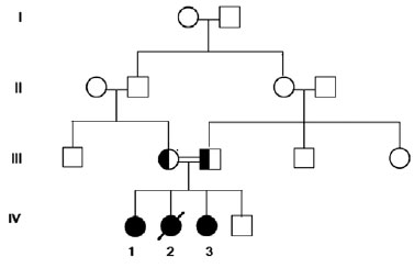
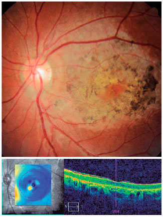
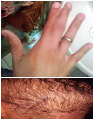
A 15-year-old girl with consanguineous parents presented with low visual acuity. An ophthalmologic examination demonstrated acuity 20/100 (-5.00 sph) and 20/150 (-5.75 sph). Dry eye was diagnosed with positive Schirmer test (8 mm) along with decreased BUT in both eyes. Fundoscopy demonstrated pigmentary alteration in the macular region of both eyes. During anamnesis, the patient reported hair loss since childhood, for which she wore a wig, in addition to a procedure for corrective repair of syndactyly between 2° and 3° for right chirodactyl (Figures 1 and 2).
Case 2 (#3 heredogram)
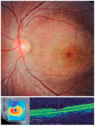
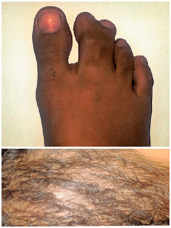
A female child aged 9 years, the sister of patient 1, presented with low visual acuity of 20/80 (LP) in the right and 20/100 (LP) in the left eye. We performed biomicroscopy and tonometry with no observed changes, and no signs of dry eye were diagnosed. Fundoscopy revealed hypopigmented macular lesions in both eyes with capillary rarefaction and syndactyly between the second and third pododactyls (Figures 3 and 4).
DISCUSSION
EEM syndrome is an autosomal recessive disease requiring a copy of the defect among the parents of the affected individual with a high incidence in blood relatives(9).
The CDH3 gene encoding the P-cadherin protein contains 16 exons on human chromosome 16q22.1(2), and it is known that the mutation contains the intragenic deletion of exons 12 and 13, resulting in continuous transcription of exon 11 to exon 14 and, while maintaining the structure, loss of functionality occurs in the future protein(10). In addition, CDH3 gene mutations are also responsible for hypotrichosis associated with juvenile macular dystrophy and may represent phenotypic heterogeneity of the same syndrome(11,12). Both conditions are similarly associated with thin and sparse hair accompanied with macular dystrophy; however, individuals with EEM develop malformations of the hands and feet(11,13).
In the cases presented, the changes in patient 1, such as hypotrichosis and macular dystrophy, are more evident. Optical coherence tomography revealed macular alterations. The patient underwent corrective surgery for syndactyly. Patient 2, who was younger, presented a milder but evolving condition after 2-year follow-up assessment where the syndactyly was not corrected. In addition, electroretinogram is not available in our city, but the retinal findings were characteristic of the dystrophy reported in the literature.
Only the older sister was diagnosed with signs of dry eye, perhaps because her alopecia and retinal symptoms were more severe or her younger sister had not yet presented.
The patients reported similar alterations in their deceased sister (#2 heredogram), who died because of unknown causes; their brother did not have alterations. Both of our patients were referred for dermatological follow-up. This ophthalmologic pathology, for which no available treatment can control the evolution of low visual acuity, may lead to bilateral blindness.
REFERENCES
1. Albrectsen B, Svendsen IB. 1956. Hypotrichosis, syndactyly, and retinal degeneration in two siblings. Acta Derm Venereol. 1956; 36(2):96-101.
2. Sprecher E, Bergman R, Richard G, Lurie R, Shalev S, Petronius D, et al. Hypotrichosis with juvenile macular dystrophy is caused by a mutation in CDH3, encoding P-cadherin. Nat Genet. 2001; 29(2):134-6.
3. Shimoyama Y, Yoshida T, Terada M, Shimosato Y, Abe O, Hirohashi S. Molecular cloning of a human Ca2+-dependent cell-cell adhesion molecule homologous to mouse placental cadherin: its low expression in human placental tissues. J Cell Biol. 1989;109(4 Pt 1): 1787-94.
4. Müller-Röver S, Tokura Y, Welker P, Furukawa F, Wakita H, Takigawa M, et al. E- and P-cadherin expression during murine hair follicle morphogenesis and cycling. Exp Dermatol. 1999;8(4):237-46.
5. Xu L, Overbeek PA, Reneker LW. Systematic analysis of E-, N- and P-cadherin expression in mouse eye development. Exp Eye Res. 2002;74(6):753-60.
6. Naqvi SK. Identification of genes involved in human hereditary skin disorders [thesis]. Islamabad: Quaid-i-Azam University; 2012
7. Genderen MM, Dixon MJ, Riemslag FC, Stilma JS, Meire FM. The EEM syndrome, a ten-year follow-up of two new patients. In: Genderen MM. Electrophysiology in visually impaired children. Utrecht. Quasi Grafische; 2006. p. 83-93
8. Karti O, Abali S, Ayhan Z, Gokmeydan E, Nalcaci S, Yaman A, et al. CDH3 gene related hypotrichosis and juvenile macular dystrophy - A case with a novel mutation. Am J Ophthalmol Case Rep. 2017; 7:129-33.
9. Ohdo S, Hirayama K, Terawaki T. Association of ectodermal dysplasia, ectrodactyly, and macular dystrophy: the EEM syndrome. J Med Genet. 1983;20(1):52-7.
10. Halford S, Holt R, Németh AH, Downes SM. Homozygous deletion in CDH3 and hypotrichosis with juvenile macular dystrophy. Arch Ophthalmol. 2012;130(11):1490-2.
11. Kjaer KW, Hansen L, Schwabe GC, Marques-de-Faria AP, Eiberg H, Mundlos S, et al. Distinct CDH3 mutations cause ectodermal dysplasia, ectrodactyly, macular dystrophy (EEM syndrome). J Med Genet. 2005;42(4):292-8.
12. Vicente LP, Finzi S, Susanna R Jr, Young TL. Hypotrichosis with juvenile macular dystrophy: a case report with molecular study. Arq Bras Oftalmol. 2017;80(1):49-51.
13. Shimomura Y, Wajid M, Shapiro L, Christiano AM. P-cadherin is a p63 target gene with a crucial role in the developing human limb bud and hair follicle. Development. 2008 Feb;135(4):743-53.
Submitted for publication:
January 29, 2018.
Accepted for publication:
May 27, 2018.
Funding: No specific financial support was available for this study
Disclosure of potential conflicts of interest: None of the authors have any potential conflicts of interest to disclose

How to cite this article:
 ABO is licensed under a Creative Commons Attribution-NonComercial 4.0 Internacional.