
Selam Yekta Sendul1; Burcu Dirim1; Cemile Ucgul Atılgan2; Mehmet Demir1; Ali Olgun1; Semra Tiryaki Demir1; Saniye Uke Uzun1; Gurcan Dogukan Arsalan1; Dilek Guven1
DOI: 10.5935/0004-2749.20180011
ABSTRACT
Purpose: This study aimed to share the results of patients who underwent anterior tarsal flap rotation combined with anterior lamellar reposition because of cicatricial upper eyelid entropion, and to determine the effectiveness and reliability of this surgical technique.
Methods: Fifteen eyes of 11 patients (2 right eyes; 5 left eyes; and 4 bilateral eyes) on whom we performed anterior tarsal flap rotation surgery combined with anterior lamellar reposition because of cicatricial entropion were included in this study. The medical records of the patients were analyzed retrospectively, and the causes of cicatricial entropion as well as the preoperative and postoperative ophthalmic examination findings were recorded. Normal anatomical appearance and function of eyelid were considered to have been achieved.
Results: The mean age was 59.81 ± 18 years. The mean follow-up period was 21.72 ± 14 months (range, 5-43 months). The causes of cicatricial entropion were postoperative cicatrices development due to multiple electrolyzes for trichiasis and/or distichiasis in 7 eyes, trachoma in 6 eyes, and trauma in 2 eyes. Irritation and watering were detected in all patients preoperatively, whereas corneal opacity and erosion were detected in 10 patients and epithelial erosion was detected in one patient. Full anatomical and functional success was achieved for all patients.
Conclusion: Anterior tarsal flap rotation combined with anterior lamellar reposition in the repair of cicatricial entropion was found to be an effective and reliable alternative surgical procedure.
Keywords: Trachoma/complications; Eyelids/surgery; Entropion/surgery; Cicatrix; Surgical flaps; Ophthalmologic surgical procedures/methods
RESUMO
Objetivo: Compartilhar os resultados dos pacientes submetidos à rotação de retalho tarsal anterior, combinados com a reposição lamelar anterior devido à entrópio cicatricial da pálpebra superior e determinar a eficácia e a confiabilidade desta técnica cirúrgica.
Métodos: Foram incluídos neste estudo quinze olhos de 11 pacientes em quem realizamos cirurgia de rotação de retalho tarsal anterior combinada com reposição lamelar anterior devido ao entrópio cicatricial. Os registros médicos dos pacientes foram analisados retrospectivamente e as causas da entrópio cicatricial, bem como os achados do exame oftalmológico pré-operatório e pós-operatório foram registrados. A integridade anatômica e funcional da pálpebra foi considerada como sucesso cirúrgico.
Resultados: A idade média foi de 59,81 ± 18 anos. O período médio de seguimento foi de 21,72 ± 14 meses (intervalo 5-43 meses). As causas da entrópio cicatricial foram o desenvolvimento de cicatrizes pós-operatórias devido a eletrólises múltiplas para triquíase e/ou distiquiase em 7 olhos, tracoma em 6 olhos e trauma em 2 olhos. Todos os pacientes foram tiveram irritação e lacrimejamento pré-operatório, enquanto que 10 pacientes apresentavam opacidade e erosão da córnea e 1 paciente apresentava apenas erosão epitelial. O sucesso total anatômico e funcional foi alcançado em todos os casos.
Conclusão: A rotação do retalho tarsal anterior combinada com a reposição lamelar anterior no reparo da entrópio cicatricial é um procedimento cirúrgico alternativo efetivo e confiável.
Descritores: Tracoma/complicações; Pálpebras/cirurgia; Entrópio/cirurgia; Cicatriz; Retalhos cirúrgicos; Procedimentos cirúrgicos oftalmológicos/métodos
INTRODUCTION
Upper eyelid cicatricial entropion is primarily caused by trachoma, especially in Eastern countries, whereas in Western countries it is caused by chronic blepharitis, previous surgeries, pemphigus, trauma, chronic drug use, and anophthalmic socket syndrome(1-3). Recurring infections, trauma, or multiple eyelid surgeries lead to scarring in the conjunctiva and/or tarsal tissue. This scarring results in the turning of the eyelid inwards and contact of the eyelashes with the corneal surface.
Erosion of the corneal surface and consequent watering are the initial outcomes of recurrent contact of the eyelashes, which in time leads to corneal opacification and consequent vision loss(4-6). The definitive treatment for cicatricial entropion is surgery. Although many surgical techniques have been devised, the majority of these techniques unfortunately fail to achieve the full success desired, both functionally and cosmetically, and recurrence of entropion has frequently been reported(7-13).
The purpose of this study was to share the results of patients referred to our clinic because of cicatricial upper eyelid entropion, who were treated with anterior tarsal flap rotation combined with anterior lamellar reposition (ATFR+ ALR) as an alternative surgical technique, and to determine its effectiveness and reliability.
METHODS
The medical records of 15 eyes of 11 patients for whom we performed ATFR + ALR to treat cicatricial entropion between April 2013 and June 2016 were analyzed retrospectively. Previous eye diseases, trauma, and eyelid surgeries of the patients were searched, and the causes of cicatricial entropion were determined and recorded. Preoperative full ophthalmologic examinations were made in which eyelid and tarsal diseases, corneal and conjunctival surface disorders, and the shape and position of the eyelashes were analyzed and recorded. Electrolysis or surgical treatments performed previously due to trichiasis or distichiasis were analyzed.
Perioperative and postoperative complications due to surgical techniques were noted. The patients were followed up postoperatively on the first day, first week, first month, third month, and biannually thereafter. The mean follow-up period was 21.72 ± 14 (range, 5-42 months). Although the functional success criteria were based on whether preoperative watering, irritation, and contact of the eyelashes with the cornea improved, the criteria for anatomical success were based on whether lagophthalmos, entropion, ectropion, and eyelid contour disorders improved. The study was conducted in accordance with the tenets of the Declaration of Helsinki by obtaining written informed consent from all patients, with the approval of the local ethics review board.
Surgical technique
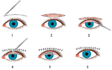
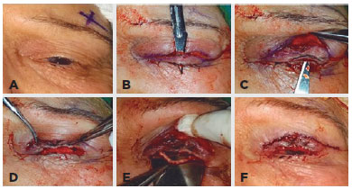
The surgical repair was performed with the patient under local anesthesia. The facial area was sterilized with antiseptic solution then the eyelid sulcus line was marked with a surgical marker. The skin and the orbicularis oculi muscle were passed by skin-crease incision. Then tarsus was reached through blunt dissection. Proceeding toward the anterior from the pretarsal area, the dissection was continued until the eyelid margin. Then, the anterior and posterior lamellae were separated both in the pretarsal direction and at the gray line by using a no. 11 scalpel blade. At this stage, in patients with posterior lamellar deficits, the tarsal tissue also was sectioned at horizontal full thickness to create an area for hard palate graft or auricular cartilage graft, and the graft was placed and sutured to the tarsus by using 6-0 Vicryl® suture. Then, the anterior lamella was reposed backward and sutured to the tarsus by using three 6-0 Vicryl sutures, and the anterior lamellar reposition was completed. Thereafter, the posterior lamella was sectioned at full horizontal thickness to be 2 mm back from the eyelid margin, thereby creating a tarsal flap. The tarsal flap was rotated 90º toward the anterior lamella and sutured to the previously reposed anterior lamella by using a 7-0 Vicryl suture (Figure 1). Blepharoplasty surgery was performed in patients whom upper eyelid skin excess was detected perioperatively. Then the eyelid skin was sutured by using 6-0 Prolene® suture (Figure 2). All patients used 2 × 1 ciprofloxacin pomade and 4 × 1 loteprednol etabonate + tobramycin eye drops for a week.


RESULTS
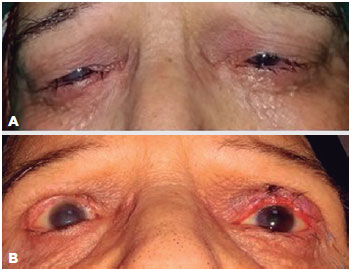
A total of 11 patients (6 males and 5 females; mean age, 59.81 ± 18 years; range, 20-80 years) were included in the study. Cicatricial entropion was present in a total of 15 of the eyes: 2 right eyes; 5 left eyes; and 4 bilateral eyes (Figure 3). The causes of cicatricial entropion were multiple electrolyzes and surgical treatment for trichiasis and/or distichiasis, upon which cicatrices developed postoperatively in 7 eyes, trachoma in 6 eyes, and trauma in 2 eyes. Preoperatively, all patients experienced irritation, watering, light sensitivity, and foreign body sensation, 10 patients had corneal opacity and erosion, and 1 patient had only epithelial erosion.

In the surgical repair, all patients were given ATFR + ALR, and 9 eyes were additionally given blepharoplasty in the same session or in a separate session. One of the 2 patients with posterior lamellar deficit underwent tarsal reconstruction with a hard palate graft in the same session, whereas the other patient underwent tarsal repair with auricular cartilage and tarsal fracture.
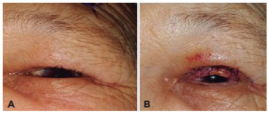
Postoperatively, all patients stated that their complaints of irritation, watering, light sensitivity and foreign body sensation had been resolved. None of the patients experienced problems, such as eyelid contour disorders, ectropion, or recurring entropion. Although 2 patients developed early-stage minimal lagophthalmos postoperatively, they had recovered by the 1-month follow-up. No other perioperative or postoperative serious complications were detected in any of the patients. Full anatomical and functional success was achieved for all patients (Table 1, Figure 4).

DISCUSSION
Entropion or trichiasis is a serious eye health problem, and surgical repair enables patients to lead a comfortable life. Considering that approximately 7-8 million people worldwide have trichiasis, 1-1.5 million of whom are irreversibly blind, the problem is serious(4,5). Even though the most important cause worldwide of cicatricial entropion is trachoma, chronic conjunctivitis, trauma, and recurring surgeries are more common causes in developed or developing countries(1,2). In our study, the major cause of cicatricial entropion was cicatrices caused by damage and/or burns in the upper eyelid tarsal tissue following previous multiple electrolyzes to treat trichiasis and/or distichiasis, with trachoma as a secondary cause.
In the literature, many surgical techniques have been devised and performed for treatment of cicatricial upper eyelid entropion(7-10,13-19). Some of these surgical techniques pertain to the anterior lamella, some to the posterior lamella, and some to both lamellae. Examples of these surgical techniques are anterior lamellar repositioning, bilamellar tarsal rotation, posterior lamellar tarsal rotation and tarsal fracture. Obviously each method has specific advantages, yet trichiasis or recurrence are quite frequent. Studies have reported that the postoperative trichiasis rate was 10% in the first three months and reached 60% within 3 years(7,8,15,20-27). Of course, many factors have a role in recurrence, such as progression of the entropion, surgical procedure, and skill of the surgeon.
In the literature, some studies have found that surgical techniques of posterior lamellar rotation have been more successful surgical procedure in terms of both recurrence and metaplastic eyelashes. The Cuenod Nataf procedure, described in the 1930s, was a posterior lamellar rotation with anterior lamellar resection and recession, and many different variations of this technique are presently used by many surgeons(19,28,29). Also, different variations of the posterior lamellar rotation technique, described by Trabut(16) in the 1950s, were defined in the following years by different surgeons(9,13). Similar to Trabut's posterior lamellar rotation technique, the posterior tarsal margin rotation technique defined by Seiff et al.(9) has also been reported to be effective. Similarly, Yagci and Palamar(13), in a study of posterior lamellar advancement and tarsal rotation, reported quite successful long-term results. In our study, we performed combined surgery with an anterior approach involving both anterior and posterior lamellae that gave successful results. No recurrence has been detected to date in our patients The benefit of performing surgery using an anterior approach is that in event of there being a need for additional surgery to the anterior lamella, such as blepharoplasty, the same section can be used. Again, when posterior lamellar shortening due to cicatrices is present this technique provides an additional opportunity for lengthening the posterior lamella, such as perioperative hard palate graft, which is a significant advantage of this particular surgical technique.
The fact that there are so many different surgical techniques in the treatment of entropion and that new techniques or variations are still being defined corroborates the fact that desired success often cannot be achieved in this field currently(7-9,17,18,28,29). We have therefore sought to identify alternative techniques by drawing on our previous clinical experience and the literature. We have noticed from our experience that anterior lamellar reposition was especially successful for a short period of time when applied alone, but its long-term results were inadequate(15,21). The posterior lamellar rotation techniques of Cuenod, Nataf, and Trabut, or surgical variations similar to these techniques, drew our attention to the posterior lamella(9,13,16,17,28-30). We then began to ask the following questions: Can we provide a barrier to the front of lashes by rotating the posterior lamella onto the anterior lamella? Can we prevent the metaplastic eyelashes in particular from touching the cornea if we turn the direction of the posterior lamella outward? With these questions in mind, we decided to apply ATFR+ ALR. The results matched our expectations: prevention of the recurrence of entropion by rotating the posterior lamella by 90º; and prevention of the metaplastic eyelashes from touching the cornea by changing their direction.
What could be the possible complications of such a surgical technique? The first complication that comes to mind is of course lagophthalmos, because of upper eyelid shortening, problems of blood supply to the flap, and, most importantly, the probability of recurrence long term. Indeed, a certain amount of lagophthalmos was observed in the early postoperative term, but it lessened upon recovery of the eyelid. Another advantage of this surgical technique is that in eyelids with extremely advanced contracture, it is possible to perform additional horizontal tarsal fracture, and again, in cases with tarsal shortening, additional grafts, such as auricular cartilage or hard palate graft, may be placed on the intertarsal region. In our study, posterior lamellar shortening was noticed perioperatively in two cases and, accordingly, additional grafts were placed on the intertarsal region. In our study, we experienced no problems with the blood supply to the flap. The probable reason for this is that the flap is double sided, and the eyelid can be supplied by rich internal and external arterial arcades on both sides.In conclusion, we think that ATFR+ALR in the repair of cicatricial entropion is an effective and reliable alternative surgical procedure. However, we believe that the benefits of this technique should be supported by long-term results of large case series.
REFERENCES
1. Polack S, Brooker S, Kuper H, Mariotti S, Mabey D, Foster A. Mapping the global distribution of trachoma. Bull World Health Organ. 2005;83(12):913-9.
2. Kersten RC, Kleiner FP, Kulwin DR. Tarsotomy for the treatment of cicatricial entropion with trichiasis. Arch Ophthalmol. 1992;110(5):714-7.
3. Cruz AA, Akaishi PM, Al-Dufaileej M, Galindo-Ferreiro A. Upper eyelid crease approach for margin rotation in trachomatous cicatricial entropion without external sutures. Arq Bras Oftalmol. 2015;78(6):367-70.
4. Pascolini D, Mariotti SP. Global estimates of visual impairment: 2010. Br J Ophthalmol. 2012;96(5):614-18.
5. Global WHO Alliance for the elimination of blinding trachoma by 2020. Wkly Epidemiol Rec. 2012;87(17):161-8.
6. Habtamu E, Wondie T, Aweke S2, Tadesse Z, Zerihun M, Zewudie Z, et al. Posterior lamellar versus bilamellar tarsal rotation surgery for trachomatous trichiasis in Ethiopia: a randomised controlled trial. Lancet Glob Health. 2016;4(3):175-84. Comment in: Lancet Glob Health. 2016;4(3):e140-1.
7. Reacher MH, Muñoz B, Alghassany A, Daar AS, Elbualy M, Taylor HR. A controlled trial of surgery for trachomatous trichiasis of the upper eyelid. Arch Ophthalmol. 1992;110(5):667-74.
8. Reacher MH, Huber MJ, Canagaratnam R, Alghassany A. A trial of surgery for trichiasis of the upper eyelid from trachoma. Br J Ophthalmol. 1990;74(2):109-13.
9. Seiff SR, Carter SR, Tovilla y Canales JL, Choo PH. Tarsal margin rotation with posterior lamella superadvancement for the management of cicatricial entropion of the upper eyelid. Am J Ophthalmol. 199;127(1):67-71.
10. Kemp EG, Collin JR. Surgical management of upper eyelid entropion. Br J Ophthalmol. 1986;70(8):575-9.
11. Collin JR. A manual of systematic eyelid surgery. 2nd Ed. London, UK: Churchill-Livingstone; 1989.
12. Baylis HI, Silkiss RZ. A structurally oriented approach to the repair of cicatricial entropion. Ophthal Plast Reconstr Surg. 1987;3(1):17-20.
13. Yagci A, Palamar M. Long-term results of tarsal margin rotation and extended posterior lamellae advancement for end stage trachoma. Ophthal Plast Reconstr Surg. 2012;28(1):11-3.
14. Rajak SN, Collin JR, Burton MJ. Trachomatous trichiasis and its management in endemic countries. Surv Ophthalmol. 2012;57(2):105-35.
15. Ahmed RA, Abdelbaky SH. Short term outcome of anterior lamellar reposition in treating trachomatous trichiasis. J Ophthalmol. 2015;2015:568363.
16. Trabut G. Entropion-trichiasis en Afrique du Nord. Arch Ophthalmol. 1949;9:701-7.
17. Négrel AD, Chami-Khazraji Y, Arrache ML, Ottmani S, Mahjour J. The quality of trichiasis surgery in the kingdom of Morocco. Sante. 2000;10(2):81-92.
18. Bowman RJ, Faal H, Myatt M, Adegbola R, Foster A, Johnson GJ, et al. Longitudinal study of trachomatous trichiasis in the Gambia. Br J Ophthalmol. 2002;86(3):339-43.
19. Zerihun N. Trachoma in Jimma zone, south western Ethiopia. Trop Med Int Health. 1997;2(12):1115-21.
20. West SK, West ES, Alemayehu W, Melese M, Munoz B, Imeru A, at al. Single-dose azithromycin prevents trichiasis recurrence following surgery: randomized trial in Ethiopia. Arch Ophthalmol. 2006;124(3):309-14.
21. Burton MJ, Bowman RJ, Faal H, Aryee EA, Ikumapayi UN, Alexander ND, et al. Long term outcome of trichiasis surgery in the Gambia. Br J Ophthalmol. 2005;89(5):575-9.
22. Burton MJ, Kinteh F, Jallow O, Sillah A, Bah M, Faye M, et al. A randomised controlled trial of azithromycin following surgery for trachomatous trichiasis in the Gambia. Br J Ophthalmol. 2005;89(10): 1282-8. Comment in: Br J Ophthalmol. 2005;89(10): 1232-3.
23. Rajak SN, Habtamu E, Weiss HA, Kello AB, Gebre T, Genet A, et al. Absorbable versus silk sutures for surgical treatment of trachomatous trichiasis in Ethiopia: a randomised controlled trial. PLoS Med. 2011;8(12):e1001137.
24. Rajak SN, Habtamu E, Weiss HA, Kello AB, Gebre T, Genet A, et al. Surgery versus epilation for the treatment of minor trichiasis in Ethiopia: a randomised controlled noninferiority trial. PLoS Med. 2011;8(12):e1001136.
25. Gower EW, West SK, Harding JC, Cassard SD, Munoz BE, Othman MS, et al. Trachomatous trichiasis clamp vs standard bilamellar tarsal rotation instrumentation for trichiasis surgery: results of a randomized clinical trial. JAMA Ophthalmol. 2013;131(3):294-301.
26. Adamu Y, Alemayehu W. A randomized clinical trial of the success rates of bilamellar tarsal rotation and tarsotomy for upper eyelid trachomatous trichiasis. Ethiopian Med J. 2002;40(2):107-14.
27. Khandekar R, Mohammed AJ, Courtright P. Recurrence of trichiasis: a long-term follow-up study in the Sultanate of Oman. Ophthalmic Epidemiol. 2001;8(2-3):155-61.
28. Thanh TTK, Khandekar R, Luong VQ, Courtright P. One year recurrence of trachomatous trichiasis in routinely operated Cuenod Nataf procedure cases in Vietnam. Br J Ophthalmol. 2004; 88(9):1114-8.
29. Thommy CP. A modified technique for correction of trachomatous cicatricial entropion. Br J Ophthalmol. 1980;64(4):296-8.
30. Hu VH, Harding-Esch EM, Burton MJ, Bailey RL, Kadimpeul J, Mabey DC. Epidemiology and control of trachoma: systematic review. Trop Med Int Health. 2010;15(6):673-91.
Submitted for publication:
June 13, 2017.
Accepted for publication:
August 8, 2017.
Funding: No specific financial support was available for this study.
Disclosure of potential conflicts of interest: None of the authors have any potential conflict of interest to disclose.
Ethics approval: This study was approved by the research ethics committee of Sisli Hamidiye Etfal Training and Research Hospital (# 1379/2017).