

Michelle de Lima Farah1; Murillo Santinello2; Luis Eduardo Morato Rebouças de Carvalho1; Carlos Fumiaki Uesugui1; Ronaldo Boaventura Barcellos1
DOI: 10.5935/0004-2749.20180008
ABSTRACT
Purpose: To describe a new method for measuring anomalous head positions by using a cell phone.
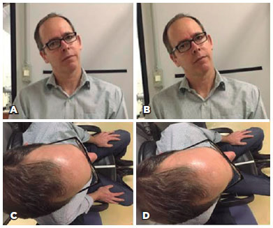
Methods: The photo rotation feature of the iPhone® PHOTOS application was used. With the patient seated on a chair, a horizontal stripe was fixed on the wall in the background and a sagittal stripe was fixed on the seat. Photographs were obtained in the following views: front view (photographs A and B; with the head tilted over one shoulder) and upper axial view (photographs C and D; viewing the forehead and nose) (A and C are without camera rotation, and B and D are with camera rotation). A blank sheet of paper with two straight lines making a 32-degree angle was also photographed. Thirty examiners were instructed to measure the rotation required to align the reference points with the orthogonal axes. In order to set benchmarks to be compared with the measurements obtained by the examiners, blue lines were digitally added to the front and upper view photographs.
Results: In the photograph of the sheet of paper (p=0.380 and a=5%), the observed values did not differ statistically from the known value of 32 degrees. Mean measurements were as follows: front view photograph A, 22.8 ± 2.77; front view B, 21.4 ± 1.61; upper view C, 19.6 ± 2.36; and upper view D, 20.1 ± 2.33 degrees. The mean difference in measurements for the front view photograph A was -1.88 (95% CI -2.88 to -0.88), front view B was -0.37 (95% CI -0.97 to 0.17), upper view C was 1.43 (95% CI 0.55 to 2.24), and upper view D was 1.87 (95% CI 1.02 to 2.77).
Conclusion: The method used in this study for measuring anomalous head position is reproducible, with maximum variations for AHPs of 2.88 degrees around the X-axis and 2.77 degrees around the Y-axis.
Keywords: Smartphone; Strabismus; Nystagmusm physiologic; Head; Torticollis
RESUMO
Objetivo: Descrever novo método de medida da posição anômala da cabeça (PAC) usando celular.
Métodos: Foi utilizado o recurso de rotação de fotografias do aplicativo fotos do iPhone®. Com paciente em uma cadeira, foram fixadas duas faixas, uma horizontal, na parede ao fundo e outra sagital sobre o assento. Fotografias: frontal (1 A e 1 B), com a cabeça inclinada sobre um ombro, e superior (1 C e 1 D), visualizando testa e nariz. Também fotografada uma folha sulfite com duas retas desenhadas formando um ângulo de 32º. Trinta examinadores foram orientados a mensurar a rotação necessária para alinhar os pontos de referência com os eixos ortogonais. Para estabelecer medidas de referência a serem comparadas com aquelas obtidas pelos examinadores, foram acrescentadas digitalmente linhas azuis nas fotos frontal e superior.
Resultados: Na foto da folha de papel (p=0,380 e a=5%), os valores observados não diferem estatisticamente do valor conhecido de 32º. Média das medidas: foto frontal 1A, 22,8 ± 2,77, frontal 1B, 21,4 ± 1,61, superior 1C, 19,6 ± 2,36 e superior 1D, 20,1±2,33. A média das diferenças das medidas na foto frontal 1A foi de -1,88 (IC 95% -2,88 a -0,88), frontal 1B de -0,37 (IC 95% -0,97 a 0,17), superior 1C de 1,43 (IC 95% 0,55 a 2,24) e superior 1D foi 1,87 (IC 95% 1,02 a 2,77).
Conclusões: O método utilizado neste estudo para medida da posição anômala da cabeça é reprodutível e apresenta variação máxima de 2,88º nas posições anômalas da cabeça ao redor do eixo X e 2,77º do Y.
Descritores: Smartphone; Estrabismo; Nistagmo fisiológico; Cabeça; Torcicolo
INTRODUCTION
The anomalous head position (AHP) is an important sign of various diseases. Some of these diseases are orthopedic, such as scoliosis and congenital torticollis, while others are ophthalmologic, such as some forms of strabismus, nystagmus, blepharoptosis, visual field defects, and refractive errors(1).
In an attempt to obtain the best visual acuity, patients with alterations in the visual field or those who have lost sight in one eye may assume an AHP. In cases of early-onset homonymous hemianopia in children, torticollis, which is ipsilateral to the visual field defect, was observed. In cases of central visual field defect in children with fixation loss, elevation of the mentum has been reported(1,2).
Refractive error-related AHP appears because of attempts to obtain the best visual acuity. Khawam et al.(3) observed astigmatism of 1-4D in most patients with refractive error-related AHP. High myopic individuals may lower the mentum or tilt the head in an attempt to create a pinhole effect with the nose or forehead.
Patients with strabismus can assume an AHP to maintain normal binocularity or obtain better visual acuity(1,4,5). In this condition, normal eye movements are limited. In order to focus on fixed objects within the visual field, the head is moved physiologically so as to supplement wider deviations(6). When patients with normal binocular vision experience duction limitation, they can turn the head in order to avoid the field of action of the affected muscle and thus eliminate diplopia, as in extraocular muscle pareses or restrictive syndromes (Graves' disease, Duane syndrome, Brown's syndrome, conditions secondary to trauma, or conditions associated with retinal detachment surgeries). Normally, these patients with restrictive strabismus exhibit a small field of binocular vision, and therefore, proposed treatments seek to increase the field. Amblyopic patients with restrictive syndromes of the fixing eye assume an AHP to maintain fixation irrespective of the absence of normal binocular vision(2,7).
In addition to strabismus, the presence of nystagmus is closely associated with the adoption of an AHP. When the intensity (amplitude and/or frequency) of nystagmus is smaller in the primary eye position (PEP), the patient can move the head in order to improve visual acuity. Regardless of the cause, the objective measure of this position relative to the PEP is important for surgical planning and follow-up, as well as for assessing the progression of acquired forms of AHP(8,9).
The variation in head position relative to the physiological position, as measured in degrees, can be converted into prismatic diopters when planning surgical correction, especially around the Y-axis, which involves intervening in the horizontal rectus muscles. In cases involving displacements around the Z-axis, the vertical rectus muscles are operated, whereas in cases involving displacements around the X-axis, the oblique muscles are operated(10).
In 1993, the orthopedists Garret, Youda, and Madson developed a device to measure the range of motion of the cervical spine called the cervical range of motion (CROM)(11). In 2000, Kushner(12) used this device to evaluate the field of binocular vision at a fixed distance, quantify the magnitude of AHPs, and assess duction limitation. This approach has a high cost and low availability for this type of assessment, and there is currently no method that is practical, precise, and simple for measuring AHP in routine ophthalmologic examination.
The present study aimed to describe a simple and novel method involving the use of an iPhone® photo editing application for measuring AHP and to establish the accuracy of this method.
METHODS
This is an empirical study to verify the accuracy of our method for AHP evaluation. The study complied with the guidelines set forth in the Declaration of Helsinki. The study protocol was approved by the research ethics committee at Irmandade da Santa Casa de Misericórdia de São Paulo (CAAE number: 54730316.2.0000.5479)
The iPhone®, which is manufactured by Apple®, has an iOS® operating system that is periodically updated by the company. Versions 9.0 and higher have an application called PHOTOS that allows the adjustment of brightness, contrast, exposure, and rotation of photographic records in the editing mode. In this study, 30 examiners evaluated five photographs of a patient in order to measure the degree of head tilt around the X- and Y-axes (tilt over the shoulder and lateral rotation, respectively) and assess the correlation of the values obtained by each examiner.
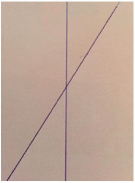
The patient participating in this study was seated in a chair with a wall in the background. With the patient seated in the chair, a horizontal stripe was fixed on the wall in the background and a sagittal stripe was fixed on the seat. Using the iPhone®, the patient was photographed in the following two views: front view (photograph A; Figure 1 A [without camera rotation] and photograph B; Figure 1 B [with camera rotation]), with the head tilted over the right shoulder, and upper axial view (photograph C; Figure 1 C [without camera rotation] and photograph D; Figure 1 D [with camera rotation]), where the forehead could be seen and the nose and head were turned to the left. Another photograph was taken of a sheet of paper with two straight lines that made a 32-degree angle (Figure 2).
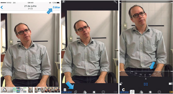
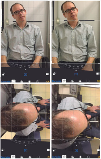
The examiners received the photographs through the WhatsApp® application and were instructed to save the images to their smartphones and open them with the PHOTOS program for editing. In the upper right corner of the screen, the examiner had to click on the word "editar" (Portuguese for "edit") (Figure 3 A). In the following screen, in the lower left corner, next to "cancelar" (Portuguese for "cancel"), the examiner had to click on the edit icon. After clicking, the photograph was framed within the compass in the lower portion of the screen (Figure 3 B). On placing fingers over the compass, a set of lines forming a grid was layered onto the image. The examiner can then rotate the image and observe in the compass the displacement angle obtained from editing the image (Figure 3 C). All images were aligned with the reference strips positioned on the seat and on the wall behind the patient's head (Figure 4). Each examiner then performed rotational adjustment of the patient's AHP using the grid superimposed on the image as a reference. The patient's front view photographs were edited so as to align the head with the 90-degree axis (Figure 5 A), and the upper view photographs were edited to align the head with the 180-degree axis (Figure 5 B). This same rotation feature was used to measure the angle between the two straight lines in the third photograph. The examiners were instructed to inform the researchers about any difficulty in obtaining the measurements. The mean values and standard deviations of all measurements obtained by the 30 examiners for the presented images were calculated.
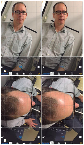
With regard to the photograph of the sheet of paper, Student's t-test was applied to compare the measurements found by the examiners with the actual value of the angle (32 degrees). The following hypotheses were considered: H0, the observed angles measure, on average, 32 degrees; and H1, they are different from the value of 32 degrees.
In order to set benchmarks for comparisons with the measurements obtained by the 30 examiners, blue lines were digitally added to the images. In front view photographs, the blue line was equidistant from the eyebrows and passed through the center of the nose and glasses, while in upper view photographs, the blue line was equidistant from the eyebrows and passed through the center of the nose (Figure 6). One of the examiners analyzed the edited images by using the method described, and the measurements obtained were compared with those recorded by the 30 examiners.
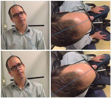
The mean of the measurements from the patient's upper view and front view photographs were compared with the reference value (measured value - reference value). A 95% confidence interval (95% CI) was calculated for the mean of the differences.
RESULTS
None of the 30 examiners reported difficulty in taking the measurements.
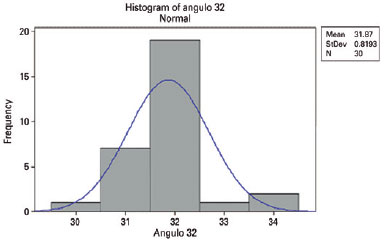
In the photograph of the straight lines on the sheet of paper, the mean of the measurements taken by the examiners was 31.87 ± 0.81 degrees. The Student's t-test (84% power) was used to compare the measurements obtained by the examiners with the real value of the angle (32 degrees). With a p-value of 0.380 and a of 5%, we accept the null hypothesis, where the observed values do not differ statistically from the known value of 32 degrees (Figure 7).
In the patient's front view photograph A, the mean of the examiners' measurements was 22.8 ± 2.77 degrees, and in the patient's front view photograph B, the mean was 21.4 ± 1.61 degrees.
In the patient's upper view photograph C, the mean was 19.6 ± 2.36 degrees, and in the patient's upper view photograph D, the mean was 20.1 ± 2.33 degrees.
The mean values found by the examiners for the angles from the patient's four photographs were compared with the value found from the same photographs with digital marking. The torticollis measurements from the patient's front view photographs A and B and from the patient's upper view photograph C were 21 degrees, whereas the measurement from the patient's upper view photograph D was 22 degrees.
The mean differences in the measurements for the front view photograph A was -1.88 (95% CI -2.88 to -0.88), front view photograph B was -0.37 (95% CI -0.97 to 0.17), upper view photograph C was 1.43 (95% CI 0.55 to 2.24), and upper view photograph D was 1.87 (95% CI 1.02 to 2.77).
DISCUSSION
The emergence of new applications and hardware has resulted in a substantial increase in the use of smartphones in the clinical routine of many physicians. The Apple® iPhone® with iOS 9 or higher operating system is equipped with an application to adjust the brightness, contrast, exposure, and rotation of photographic records. We describe a novel method involving the use of this program for measuring AHP, which is capable of making the clinical ophthalmologic practice of torticollis evaluation quick and simple. In our study, none of the 30 examiners reported difficulty in evaluating the photographs they were presented, which confirms the method's simplicity and reproducibility.
The first step in this study was to check whether the iPhone® photo editing program can accurately measure an angle between two lines. The values observed by the examiners in the photograph of the sheet of paper did not differ statistically from the real value. It can therefore be concluded that the examiners generally measured the angle correctly. With a mean of 31.87 ± 0.81 degrees, good accuracy was evident when measuring the angle between the two lines using the iPhone® photo editing program.
Proper positioning of the iPhone® when viewing the head reference points is essential to avoid alterations to the measurements. The nose and the forehead need to be seen when evaluating the rotation around the Y-axis, and the face needs to be seen when evaluating the rotation around the X-axis. Furthermore, if the phone is rotated, the images go out of alignment. To prevent poor image alignment from influencing the results, reference lines were placed for image rectification prior to the head rotation analysis step. The patient's photographs (both upper and front views) were taken with a cell phone at different tilts. As already explained, the examiners were instructed to align the images with the reference lines placed on the wall (behind the patient's head and sagittally oriented on the seat). Once this alignment was achieved, the examiners proceeded to measure the AHP. The value found was then subtracted from the value obtained from the alignment with the reference lines. We thus attempted to minimize the error induced by poor positioning of the iPhone®.
The mean angles obtained from the photographs were compared with the angles obtained from analyzing the digitally marked photographs. Considering the margin of error, the patient's front view photograph A had a maximum variation of 2.88 degrees when compared to the reference value (photograph with markings). Additionally, the maximum variations for the front view photograph B, upper view photograph C, and upper view photograph D were 0.97 degrees, 2.24 degrees, and 2.77 degrees, respectively. This analysis showed that the variations in AHP measurements are small from clinical and surgical standpoints.
The variations in the results obtained with the front and upper view photographs by the examiners are probably associated with the existence of many points that can be considered as reference points (the forehead, nose, ears, glasses, and eyebrows in the front view photographs, and the forehead and nose in the upper view photographs). When assessing so many asymmetric references, variations in the AHP measurements are expected, indicating the need to have a standardized reference point in order to make this method more precise. Adding artificial reference points that are either physical (lines or points on the patient's head) or digital (an edited line layered onto the image) could increase accuracy but would increase the complexity of the method.
Kushner(12) used CROM to evaluate the field of binocular vision at a fixed distance, quantify the magnitude of anomalous head positions, and assess duction limitation. The variations in values obtained from the mean measurements determined by Kushner(12) were 1.1 ± 2.6 degrees when assessing duction limitation and 1.0 ± 0.70 degrees when quantifying AHP. The use of CROM was confirmed to be a good approach for conducting such analyses, and the variations observed were not statistically significant. Although this approach is accurate, the equipment is expensive and has to be properly fixed to the patient's head, unlike the method presented herein.
Other studies aimed at developing clinically valid devices for measuring head position have been carried out(14-16). Hald et al.(14) have shown that the patient's head position could be accurately assessed using a motion sensor-based system (InterSense, Inc., Billerica, MA). The need for proper fixation of the sensor to the patient's head and its high cost are limitations for its use. Using an iPhone® does not necessarily imply acquisition of new equipment and adds further functions, other than those described in the study.
Kim et al.(15) developed an infrared optical head motion sensor using two Nintendo Wii® controls (WiiMote; Nintendo Co., Ltd., Kyoto, Japan). This system showed strong agreement with CROM, had good reliability in the comparison between tests, and was less expensive than the system by Hald. Nevertheless, patients were required to wear the device on their heads, and the examiners needed to set a 3D-virtual space before taking measurements.
Oh et al.(16) developed a digital system for measuring head position by incorporating Microsoft Kinect®, and they found agreement with measurements taken with the help of the CROM device. This approach does not rely on the use of any apparatus on the patient's head and requires good understanding of Microsoft Kinect®. However, the distance between the patient and the Microsoft Kinect Head Tracker® can influence head position measurements.
The literature shows a variety of treatments for torticollis. Studies by Wang and Parks et al.(13,17) Parks and Wang et al. have presented experience in this aspect. Parks proposed a standard operation for eliminating or reducing torticollis around the Y-axis (left- or right-hand turns), thereby correcting about 30 degrees for moderate and large AHPs, without different gradations for different AHPs. The values for this surgery were as follows: in the eye in abduction, 6 mm recession of the lateral rectus muscle and 7 mm resection of the medial rectus; in the eye in adduction, 5 mm recession of the medial rectus and 8 mm resection of the lateral rectus(10,17). On the other hand, there are tables and formulas for converting AHP measured in degrees into diopters, thus allowing different surgeries to be programmed for AHPs of different sizes(10).
Wang et al.(13) conducted a study to assess the efficacy of surgery for the correction of idiopathic horizontal nystagmus associated with AHC AHP and strabismus, defining three intensities of torticollis. The surgery was selected for each case according to head rotation values around the Y-axis. In patients with a rotation of 15 degrees or less, the Anderson procedure was performed. On the other hand, in patients with rotations of 15-25 degrees, Kestenbaum surgery (5-4-4-5) was the procedure of choice, while in patients with rotations of 25-35 degrees, the Parks technique (5-8-6-7) was performed. Following surgery, 84% of patients had a normal head position or showed a deviation of 8 diopters or less.(16) Surgeries for correcting torticollis aim to improve posture, have variable responses and, as shown by Wang et al.(13), yield good results. The establishment of reference ranges for 10-by-10 surgical programming in this study suggested that controlled attenuation parameter (CAP) measurement and variation over time for the same patient make it difficult to establish smaller ranges. The result of 8 diopters or less that is considered as successful suggests that, over this range, AHP is not noticed and that the precision observed with the proposed method would thus be suitable for verifying whether AHP correction has been successful. Considering that the surgical indication for operative correction of torticollis is based on deviation ranges and that AHP changes over time, the mean variations of 1.61 to 2.77 degrees found in the images evaluated by the examiners may be considered sufficient for good surgical planning, as well as for the follow-up of acquired forms of AHP. The imprecisions in obtaining measurements and the inherent variations of AHP throughout the day reinforce the idea that the introduction of a practical method can be of great importance.
The present study has some limitations. This study did not employ other existing methods, such as CROM, for the measurement of AHP in the patients evaluated. The asymmetry of the reference points used in the study may be a confounding factor in head position measurements, indicating the need for a fixed, possibly external, reference point for evaluating torticollis with better precision. A larger number of examiners and the establishment of reference points on images for analysis would increase measurement precision and decrease the confidence interval.
Based on the findings of the present study, we conclude that our method used for measuring AHP is reproducible and has a maximum variation of 2.88 degrees around the X-axis and 2.77 degrees around the Y-axis for AHPs.
REFERENCES
1. Davitt BV. Abnormal head postures: a review. Am Orthopt J. 2001; 51:137-43.
2. Kushner BJ. Ocular causes of abnormal head postures. Ophthalmology. 1979;86(12):2115-25.
3. Khawam E, el Baba F, Kaba F. Abnormal ocular head postures: Part IV. Ann Ophthalmol.1987;19(12):466-72.
4. Halachmi-Eyal O, Kowal L. Assessing abnormal head posture: a new paradigm. Curr Opin Ophthalmol. 2013;24(5):432-7.
5. Lim HW, Kim JH, Park SH, Oh SY. Clinical measurement of compensatory torsional eye movement during head tilt. Acta Ophthalmol. 2017;95(2):e101-e106.
6. Boricean ID, Barar A. Understanding ocular torticollis in children. Oftalmologia. 2011;55(1):10-26.
7. Kekunnaya R, Isenberg SJ. Effect of strabismus surgery on torticollis caused by congenital superior oblique palsy in young children. Indian J Ophthalmol. 2014;62(3):322-6.
8. Felius J, Muhanna ZA. Visual deprivation and foveation characteristics both underlie visual acuity deficits in idiopathic infantile nystagmus. Invest Ophthalmol Vis Sci. 2013;54(5):3520-5.
9. Teodorescu L. Anomalous head postures in strabismus and nystagmus diagnosis and management. Rom J Ophthalmol. 2015;59(3): 137-40.
10. Pietro-Díaz J, Souza-Dias C. Estrabismo. São Paulo: Santos Editora; 2002.
11. Garrett TR, Youdas JW, Madson TJ. Reliability of measuring forward head posture in a clinical setting. J Orthop Sports Phys Ther. 1993; 17(3):155-60.
12. Kushner BJ. The usefulness of the cervical range of motion device in the ocular motility examination. Arch Ophthalmol. 2000;118(7): 946-50.
13. Wang P, Lou L, Song L. Design and efficacy of surgery for horizontal idiopathic nystagmus with abnormal head posture and strabismus. J Huazhong Univ Sci Technolog Med Sci. 2011;31(5):678-81.
14. Hald ES, Hertle RW, Yang D. Development and validation of a digital head posture measuring system. Am J Ophthalmol. 2009; 147(6):1092-100,1100. e1-3.
15. Kim J, Nam KW, Jang IG, Yang HK, Kim KG, Hwang JM. Nintendo Wii remote controllers for head posture measurement: accuracy, validity, and reliability of the infrared optical head tracker. Invest Ophthalmol Vis Sci. 2012;53(3)1388-96.
16. Oh BL, Kim J, Kim J, Hwang JM, Lee J. Validity and reliability of head posture measurement using Microsoft Kinect. Br J Ophthalmol. 2014;98(11):1560-4.
17. Parks MM. Symposium: nystagmus. Congenital nystagmus surgery. Am Orthopt J. 1973;23:35-9.
Submitted for publication:
February 3, 2017.
Accepted for publication:
August 8, 2017.
Funding: No specific financial support was available for this study.
Disclosure of potential conflicts of interest: None of the authors have any potential conflict of interest to disclose.
Approved by the following research ethics committee: Irmandade da Santa Casa de Misericórdia de São Paulo (CAEE: 54730316.2.0000).