INTRODUCTION
The eyelids of Asian people have several well-established characteristics that distinguish them from eyelids of people of European descent, including their shapes and folds, the laterally inclined palpebral fissure, the several forms of the epicanthus tarsalis, and the frequently hidden caruncle(1-7). Structural features common to all eyelids, regardless of age and racial background, allow for a greater comprehension of the underlying anatomical basis of the characteristics that are distinctive, and of the surgical techniques essential for their maintenance. Failure to recognize and maintain inherent structures can lead to the loss of characteristic ethnic features and a cosmetically unacceptable result(8).
Racial characteristics may lead to differences in how aging affects the eyelids of people of Asian and European descent. The purpose of this study was to investigate differences in the position of the palpebral fissure in Japanese subjects and in Brazilian subjects of European descent over 50 years of age with the use of computerized measurements.
METHODS
A cross-sectional study evaluating the eyelid fissure position in 50 Japanese subjects (100 eyelids) and 50 Brazilians of European descent (100 eyelids) over 50 years of age was performed. The study was approved by the Regional Ethics Committee, and informed consent was obtained from all the participants. The Japanese subjects were living in Hamamatsu, Japan, and the Brazilian subjects in Botucatu, Brazil. The exclusion criteria were alteration of palpebral position, prior palpebral surgery, family tree with Asian progeny for controls, and refusal of authorization to record digital images.
Images were obtained with a Nikon® Coolpix 4100 digital camera and flash in frontal view 30 cm from the subject’s face. The subject was opposite to and at the same eye level as the examiner. The images were obtained with the eyes in the primary position and with the gaze at the center of the examiner’s nose to obtain clear photographs of both eyes. The images were then transferred to a computer running Windows and processed by Scion Imaging Software® (Scion Corporation, USA). The following parameters were measured and evaluated (Figure 1): fissure width: the distance between the medial canthus and the lateral canthus obtained by a line linking the lateral and medial corners of the eyelids; fissure height: the distance between the upper eyelid margin and the lower eyelid margin with open eyes obtained by a line linking the upper and lower eyelid margins and passing through the pupil; margin reflex distance (MRD): the perpendicular distance between the highest point of the open upper eyelid margin and the pupil reflex (MRD1), and the distance between the margin of the inferior eyelid margin to the pupil reflex (MRD2).
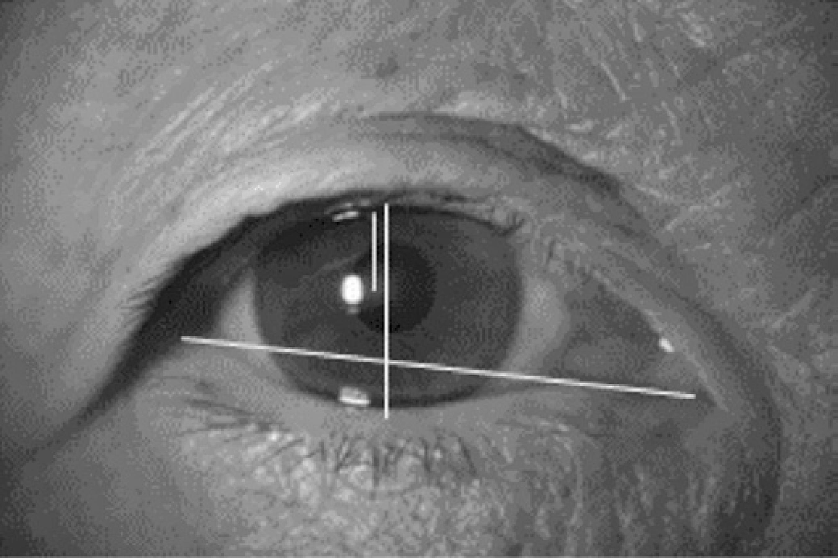
Figure 1 Representation of the width dimension of the palpebral fissure, obtained by a line linking the lateral and medial corners; the height dimension was obtained by a line linking the superior and inferior palpebral eyelid and passing through the pupil; MRD1 reflecting the distance between the open upper eyelid margin and the pupil reflex; MRD2 is the difference between the height and the MRD1
The measurement data were analyzed descriptively according to age. Differences between the Japanese and the Brazilian groups, and differences in laterality were analyzed by two-factor analysis of variance complemented with Tukey’s multiple comparisons test. Linear relationships between eyelid measurements and race were evaluated by the Pearson test.
RESULTS
The mean age of the participants was 63.9 ± 8.9 years (median 64.0-50.0; 80.0) in the Japanese group and 64.7 ± 8.5 years (median 64.0-50.0; 80.0) in the Brazilian group, with no significant difference (p>0.05). Thirty-two of the Japanese subjects were male and 18 were female, and 21 of the Brazilian subjects were male and 29 were female. The participants were divided into seven age groups. Among the Japanese subjects, there were 11 individuals aged 50-54 years (group 1), 8 aged 55-59 years (group 2), 6 aged 60-64 years (group 3), 10 aged 65-69 years (group 4), 8 aged 70-74 years (group 5), and 7 aged 75-80 years (group 6). Among the Brazilian subjects, there were 7 individuals aged 50-54 years (group 1), 7 aged 55-59 years (group 2), 12 aged 60-64 years (group 3), 10 aged 65-69 years (group 4), 5 aged 70-74 years (group 5), and 9 aged 75-80 years (group 6).
All the measurements were similar in the right and left eyelids of both groups (p>0.05) (Table 1), and therefore we considered only the right eyelid in the analyses.
Table 1 Mean and standard deviation of eyelid parameters according to laterality in Japanese subjects and Brazilian subjects of European descent
| Variable | Group | Eyelid | P value | |
|---|---|---|---|---|
| Right | Left | |||
| Fissure width | Japanese | 23.2 ± 2.0 | 22.9 ± 2.4 | p>0.05 |
| Brazilian | 21.4 ± 1.5 | 21.6 ± 1.4 | p>0.05 | |
| P value | p<0.001 | p<0.005 | ||
| Fissure height | Japanese | 7.6 ± 1.2 | 7.5 ± 1.4 | p>0.05 |
| Brazilian | 8.6 ± 1.5 | 8.6 ± 1.7 | p>0.05 | |
| P value | p<0.01 | p<0.01 | ||
| MRD1 | Japanese | 2.7 ± 0.9 | 2.7 ± 0.9 | p>0.05 |
| Brazilian | 3.6 ± 1.6 | 3.6 ± 1.7 | p>0.05 | |
| P value | p<0.01 | p<0.01 | ||
| MRD2 | Japanese | 4.9 ± 0.8 | 4.8 ± 0.7 | p>0.05 |
| Brazilian | 5.0 ± 1.5 | 5.0 ± 1.6 | p>0.05 | |
| P value | p>0.05 | p>0.05 | ||
The width of the eyelid fissure was significantly greater in Japanese than in Brazilian subjects (p<0.01), whereas the height and the MRD1 of the eyelid fissure were significantly greater in Brazilian than in Japanese subjects (p<0.01) (Tables 1 and 2).
Table 2 Mean and standard deviation of eyelid measures in Japanese subjects and Brazilian subjects of European descent according to age*
| Measure | Age group | |||||||
|---|---|---|---|---|---|---|---|---|
| 1 | 2 | 3 | 4 | 5 | 6 | 7 | All ages | |
| Width | ||||||||
| Brazilian | 21.91ab | 21.05ab | 21.57ab | 21.22ab | 20.83ab | 22.11ab | 22.74ba | p<0.05 |
| Japanese | 25.01ba | 24.10ba | 24.08ba | 21.93ab | 21.81ab | 20.95ab | 20.93aa | p<0.05 |
| P value | p<0.01 | p<0.01 | p<0.01 | p<0.05 | p<0.05 | p<0.05 | p<0.05 | |
| Height | ||||||||
| Brazilian | a9.13aa | 7.68 | 08.9200 | 08.81bb | 08.16ab | 08.57ab | 08.94ab | p>0.05 |
| Japanese | 08.73ba | 07.58ab | 08.21ba | 07.76ab | 07.18aa | 06.37ab | 06.37aa | p<0.05 |
| P value | p>0.05 | p>0.05 | p>0.05 | p<0.05 | p<0.05 | p<0.05 | p<0.05 | |
| MRD1 | ||||||||
| Brazilian | a3.54a | 03.60ab | 03.93ab | 04.06ab | 02.99ab | 03.12ab | 03.12ab | p>0.05 |
| Japanese | 03.36ba | 02.64ab | 03.12ab | 02.60ab | 02.46ab | 01.88ab | 02.34ab | p<0.05 |
| P value | p>0.05 | p>0.05 | p>0.05 | p<0.05 | p>0.05 | p<0.05 | p>0.05 | |
*values followed by different letters are significantly different.
There was a linear relationship between all of the studied variables (width, height, and MRD1) and age among Japanese subjects (p<0.05) (Figure 2), but only width was associated with age among Brazilian subjects (p<0.05) (Table 3).
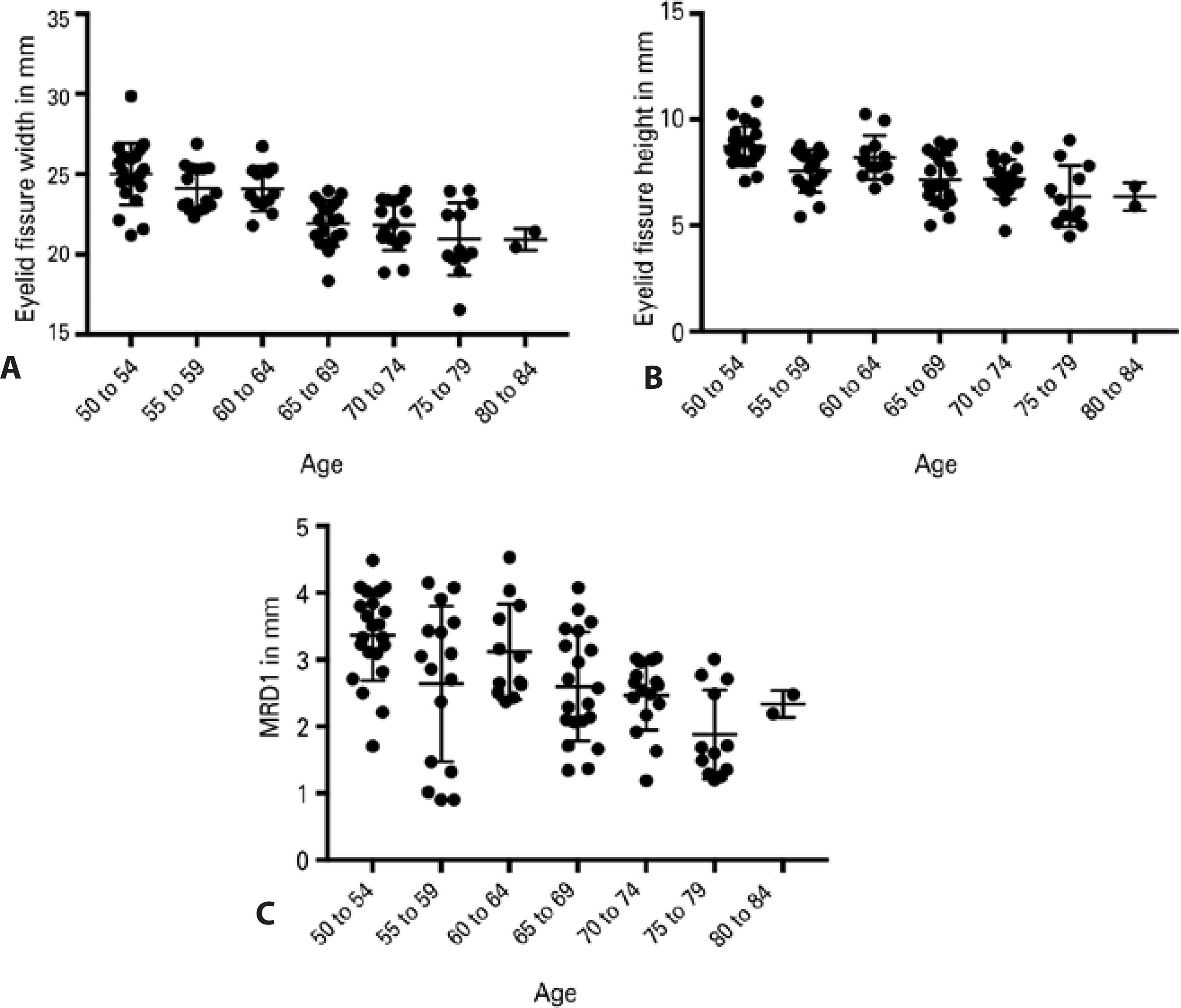
Table 3 Linear association between eyelid measure variables versus age in Japanese subjects and Brazilian subjects of European descent
| Association | Correlation coefficient (p value) | |
|---|---|---|
| Japanese | Brazilian | |
| Fissure width vs. age | -0.649 (<0.001) | -0.032 (0.826) |
| Fissure height vs. age | -0.468 (<0.001) | -0.010 (0.946) |
| MRD1 vs. age | -0.333 (<0.018) | -0.127 (0.380) |
| MRD2 vs. age | -0.368 (<0.009) | -0.142 (0.326) |
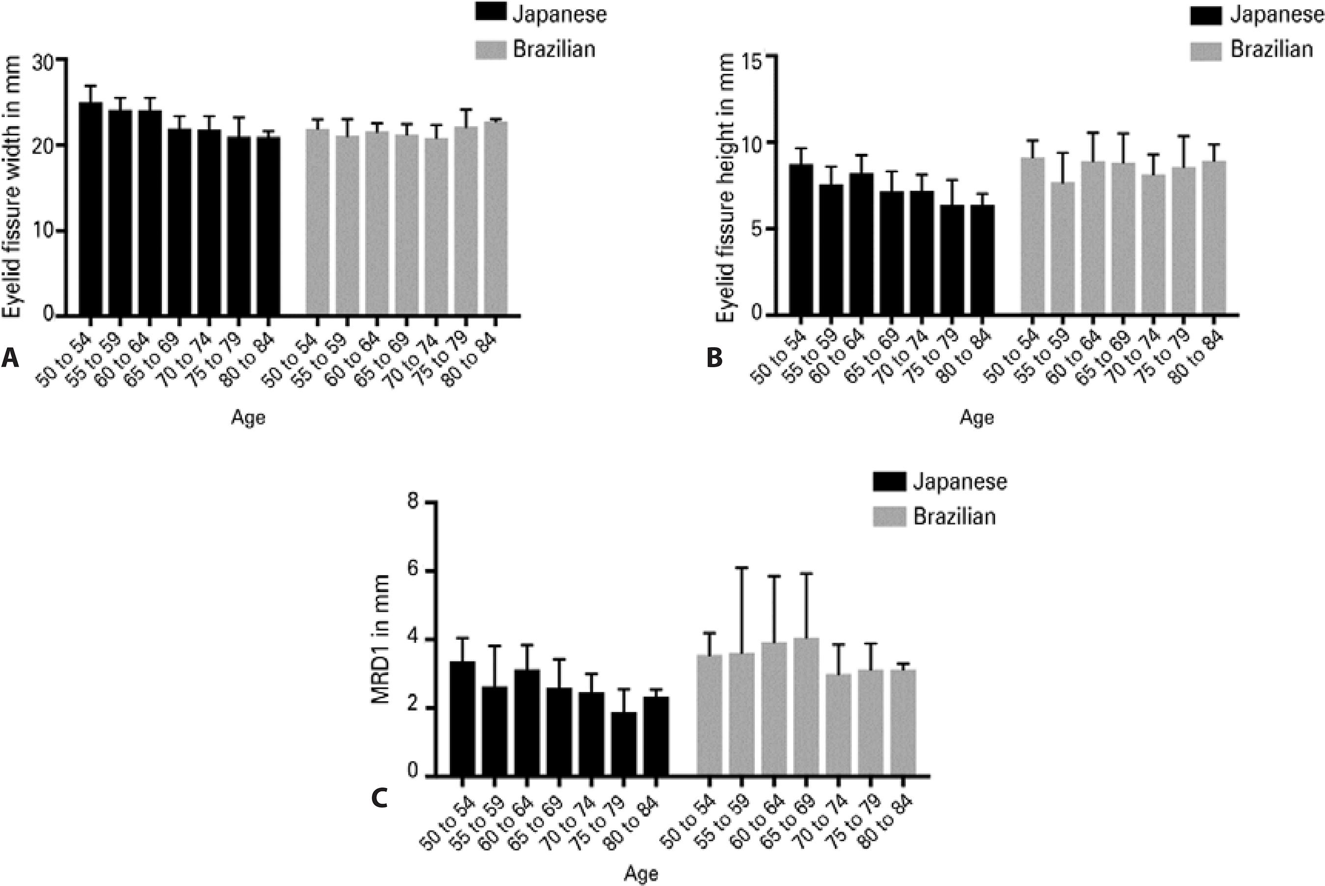
The width of the eyelid fissure varied more in Japanese than in Brazilian subjects. Among subjects between 50 and 64 years of age, the width of the eyelid fissure was greater in Japanese than in Brazilian subjects, after which both groups had similar values (Figure 3). The vertical fissure height was lower in Japanese than in Brazilian subjects and declined further with increasing age in Japanese subjects (p<0.05) (Figure 3). The values of MRD1 and their variability were higher in Brazilian than in Japanese subjects (p<0.05) (Figure 3). There were no significant differences in MRD2 between Brazilians and Japanese (p>0.05).
DISCUSSION
The majority of studies that involve evaluation and measurement of the eyelid in Asian people do not consider Japanese subjects(2,4,8). The present study confirmed variations in dimensions between eyelids of Japanese subjects and Brazilian subjects of European descent. Fissure height and MRD1 were larger in Brazilians, and fissure width was larger in Japanese.
The previously described specific anatomical features of eyelids of Asian people are responsible for some of these differences. The upper and lower eyelids of Asian people are typically characterized by a fuller appearance than the eyelids of people of European descent. The inferior extension of preaponeurotic fat and brow fat into the upper eyelids of Asian people accounts for their fullness and their difference from the upper eyelids of people of European descent9.
Although we did not compare upper and lower eyelids, alterations with age affect both due to superficial and deep tissue alterations. A comparison of age-related changes in facial skin wrinkles between women of European and Japanese descent living in the United States found that among younger women, those of European descent had greater wrinkle formation in all areas of the face than did women of Japanese descent(10,11).
Age-related changes in the lower eyelid are involved in the transformation of the three parts of the orbicularis oculi muscle, the relaxation of the suborbicularis retaining fasciae, the disappearance of the preseptal adipose tissue area due to orbital fat herniation, and the indistinctness of the palisade structure of the retinacula cutis(10,12).
Some superficial differences are directly affected by deep tissue modifications. The bony skeleton serves as the scaffolding for the soft tissues of the face. According to Lambros’s theory,(13) with aging, the facial skeleton rotates so that the frontal bone moves anteriorly and inferiorly, while the maxilla moves posteriorly and superiorly in a clockwise rotation relative to the cranial base. This rotation causes bony angles to become more acute and, will likely affect the position of the overlying soft tissue, with effects on eyelid position and the shape of the palpebral fissure.
The absence or reduced size of the crease and sulcus in the upper eyelids of Asian people is an important difference between people of Asian and European descent. This difference is due to the fact that Asians have fusion of the orbital septum to the levator aponeurosis at variable distances below the superior tarsal border, with a protruding preaponeurotic fat pad and a thick subcutaneous fat layer that prevent levator fibers from extending toward the skin near the superior tarsal border and the primary insertion of the levator aponeurosis into the orbicularis muscle and into the upper eyelid skin closer to the eyelid margin(2). Due to structural differences related to increased fat pad protrusion in the upper eyelids of Asian people and because the sulcus in elderly people can be covered by the eyelid crease, even among people of European descent, making it more difficult to visualize the upper lid crease, we decided not to analyze this parameter in the present study.
The greater effects of aging on eyelid height and MRD in Japanese people as compared with Europeans are most likely secondary to differences in the insertion of the orbital septum and prominent orbital fat protrusion. Changes in the nasal and central fat pads of the upper eyelid are also associated with aging. Other authors have shown that in the upper eyelids of people older than 70 years, the medial fat pad becomes prominent whereas the central fat pad atrophies, with implications for aesthetic and functional upper eyelid blepharoplasty(14).
The eyelids of Asian people have more subcutaneous and suborbicular fat than do the eyelids of people of European descent(2,15). This characteristic may be responsible for a heavier eyelid aspect among Asian people, making them more susceptible to age-related effects on fissure height and MRD1, as observed in the present study.
Facial sagging and reduced dermal elasticity are well-known morphological features associated with aging and gender. Although we did not compare men and women in this study, the morphology and areas of sagging in male faces are similar to those in females in the cheek, but sagging of the lower eyelid is more severe in men after middle age. Furthermore, the dermal elasticity of male facial skin decreases with age, similar to that of females, and may therefore be associated with sagging in the male face(16).
Aging changes eyelid position, primarily affecting the width of the eyelid fissure, which gradually shortens among people aged 50 years or older. This shortening is commonly observed in the eyelids of people of European descent and is likely due to progressive laxity of the medial and lateral canthal structures(17). We did not observe alterations of width in Japanese subjects, which may suggest that the disinsertion of the lateral canthus is more marked in Brazilian than in Japanese subjects or may be due to obliquity of the fissure, since oblique fissures are more commonly observed among Asians and Indians than among people of European descent(18).
This study found mean fissure heights of 7.65 ± 1.25 mm in the Japanese group and 8.62 ± 1.51 mm in the Brazilian group. Another study reported heights of 8.2 mm in Asian men and 8.5 mm in Asian women at 18 years of age(19). Our study evaluated only individuals over the age of 50 years, and the different results may be due to age-related disinsertion of the elevator muscle aponeurosis, causing involutional ptosis and deeper skin creases(17). According to other studies, the vertical dimension of the eyelid fissure reaches a specific level between 10 and 13 years of age, and decreases gradually as people approach their fiftie(7).
In conclusion, the width of the eyelids is reduced with increasing age, especially in Brazilians of European descent. The height of the eyelid fissure and MRD1 are more reduced with age in Japanese subjects than in Brazilians of European descent. Thus, there are racial differences in age-related changes in the eyelids, which are more marked in Japanese subjects.

