INTRODUCTION
Optical coherence tomography angiography (OCTA) is a novel technology that generates volumetric angiography images with applicability for the diagnosis and follow-up of a wide range of retinal diseases. However, OCTA image artifacts can alter vascular appearance, leading to false clinical interpretations1. Optic disc pit (ODP) is a rare clinical entity characterized by a congenital cavity of the optic nerve head2. The disease may be complicated by serous macular detachment, causing progressive visual loss. ODP may be rarely asso ciated with peripapillary choroidal neovascularization (CNV)3. We report a case of ODP maculopathy in which preoperative OCTA revea led artifactual subfoveal CNV and postoperative OCTA was normal.
CASE REPORT
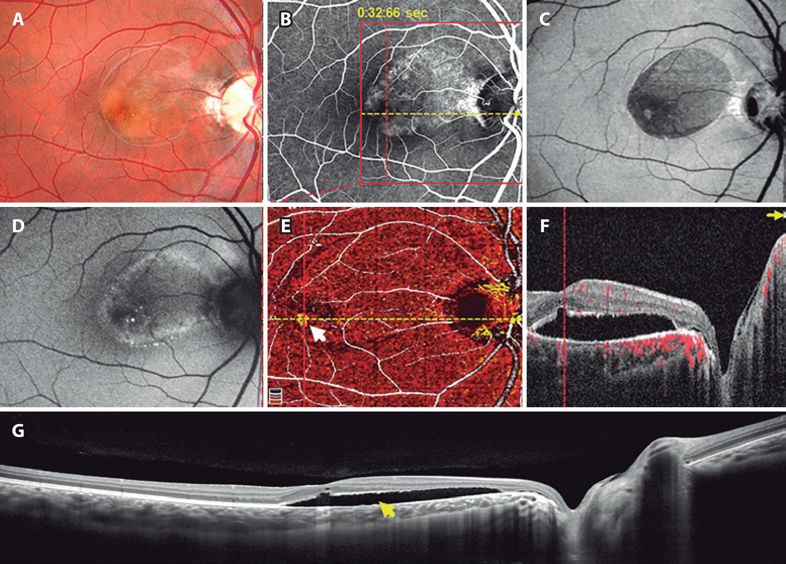
A 19-year-old caucasian, otherwise healthy man presented with decreased visual acuity in the right eye for 3 months. Best-corrected visual acuity (BCVA) was 20/60 in the right eye and 20/20 in the left eye. Intraocular pressures were 12 mmHg and 10 mmHg in the right and left eyes, respectively. Pupillary reflexes were normal in both eyes. Anterior segment slit-lamp exam was unremarkable in both eyes. Dilated funds exam revealed ODP associated with serous macular detachment in the right eye. Dilated funds exam of the left eye was normal. Multimodal imaging was performed (OCTA; RTVueXR Avanti device; Optovue Inc.; Fremont, CA, USA. Retinography; Topcon Retinal Camera TRC 50DX; Topcon Corp., Tokyo, Japan. Fluorescein angiography; Heidelberg HRA Spectralis/HRA2; Heidelberg Engi neering, Heidelberg, Germany) (Figure 1). OCT identified communication between the optic disc and the macular serous detachment. OCTA displayed a small, discrete subfoveal area suggestive of CNV surrounded by an area of relatively decreased choriocapillaris vessel density (Figure 1 E). OCTA signal superimposed on OCT B-scan de monstrated choriocapillaris detection underneath the center of the fovea, surrounded by areas of no detection on each side (Figure 1 F) There was no evidence of leakage in the fluorescein angiogram and no evidence of CNV on OCT in the area corresponding to the suspicious subfoveal CNV.

Figure 1 Preoperative multimodal imaging of the right eye: optic disc pit maculopathy in a 19-year-old man. A) Color image shows an optic disk pit associated with serous macular detachment. B) Fluorescein angiography. C) En-face OCT (superficial retina). D) Fundus autofluorescence. E) OCTA: image suggesting CNV (white arrow). F) OCT B-scan indicating the presence of communication between the optic nerve and serous retinal detachment. G) OCT showing subretinal fluid (yellow arrow).
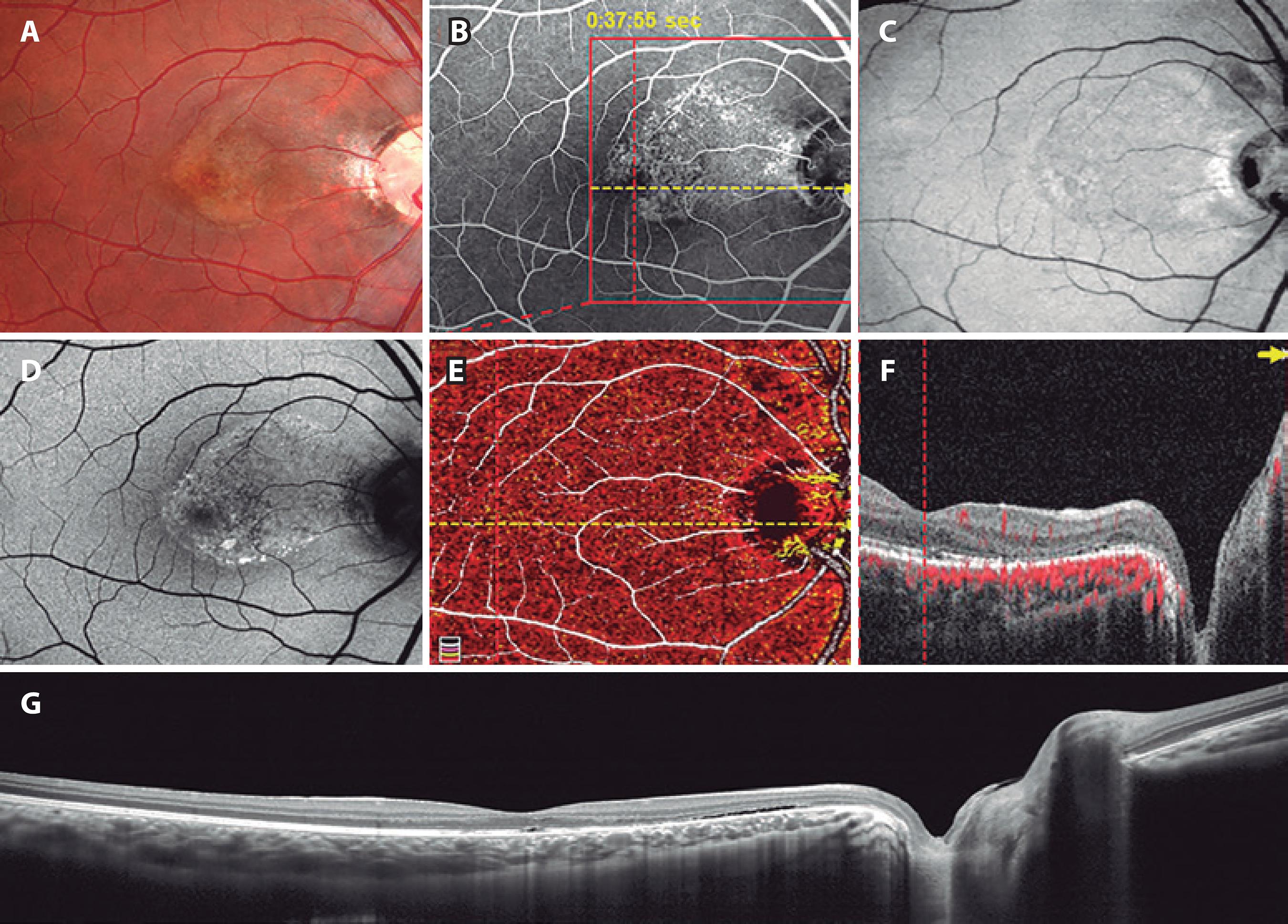
The patient underwent 23-gauge pars plana vitrectomy in the right eye. Triamcinolone-assisted posterior hyaloid detachment, as well as fluid-air exchange, was performed. No laser or peeling was performed during the surgery and air was used as the vitreous substitute. Six weeks after surgery, multimodal imaging was repeated (Figure 2). There was subtotal resorption of the subretinal fluid. BCVA improved to 20/20 in the right eye. OCTA demonstrated normal choriocapillaris signal throughout the macula (Figure 2 E). OCTA signal superimposed on OCT B-scan also demonstrated normal choriocapillaris signal throughout the macula (Figure 2 F).

Figure 2 Postoperative multimodal imaging of the right eye: resolution of optic disc pit maculopathy in a 19-year-old man. A) Color image. B) Fluorescein angiography. C) En-face OCT (superficial retina). D) Fundus autofluorescence. E) OCTA: disappearance of suspicious CNV image. F and G) OCT B-scan indicating near-complete absorption of subretinal fluid.
DISCUSSION
OCTA images can produce both positive and negative artifacts, which are important to recognize when interpreting clinical images. For instance, reflection artifacts may project superficial vessels into deeper layers and shadowing or eye movement artifacts may create areas devoid of vessels1.
We presented a case of a young patient diagnosed with ODP maculopathy whose preoperative OCTA suggested subfoveal CNV. Note that OCTA displayed a ring of relatively decreased choriocapillaris signal surrounding a small, subfoveal area of relatively increased choriocapillaris signal (Figure 1 E). The same phenomenon was present in the OCTA signal superimposed on the OCT B-scan (Figure 1 F). However, fluorescein angiogram and OCT did not demonstrate any signs of CNV in the same area, which suggests an artifactual lesion.
The patient was scheduled for PPV, which caused near-complete resolution of subretinal fluid 6 weeks postoperatively. Postoperative OCTA revealed normal choriocapillaris, confirming the artifactual na ture of the suspected subfoveal CNV. Possible mechanisms for the abovementioned artifactual CNV include light scattering by the elevated retina and/or presence of subretinal fluid.
In conclusion, optical coherence tomography angiography may produce artifacts in optic disc pit maculopathy that simulate choroidal neovascularization. Multimodal imaging is important to accurately interpret unusual optical coherence tomography angiography findings.


 English PDF
 Print
Print
 Send this article by email
 How to cite this article
 Submit a comment
 Mendeley
 Scielo
 Pocket
 Share on Linkedin

