INTRODUCTION
Sebaceous proliferations such as sebaceous hyperplasia and sebaceous adenoma, which mainly present after the age of 60 years, typically arise in the head and neck(1). Sebaceous adenomas were first reported in 1949 and characterized as benign skin tumors presenting as pink or yellow nodules approximately 5 mm in size(2). Sebaceous adenomas rarely develop in the conjunctiva and are mainly encountered on the caruncle(3,4). As an example of another rare localization, sebaceous adenoma of the palpebral conjunctiva combined with atypical adenomatous hyperplasia of the endometrium was reported in a 42-year-old female(5). Bulbar conjunctiva was reported in only one case in association with a multiple malignancy syndrome called Muir-Torre syndrome(6). We aimed to report a unique sebaceous adenoma primarily located at the nasal bulbar conjunctiva in an adult in the absence of systemic malignancy.
CASE REPORT
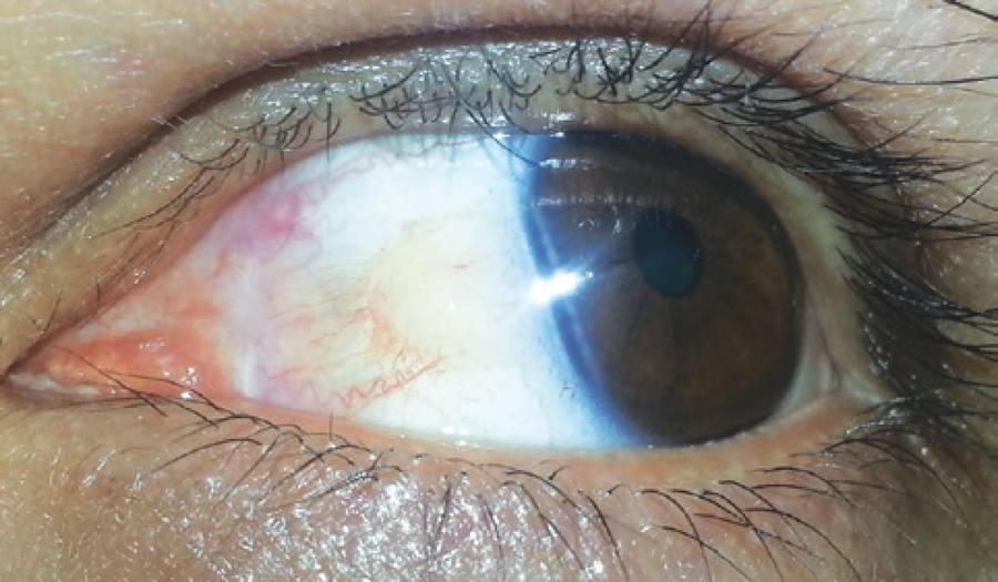
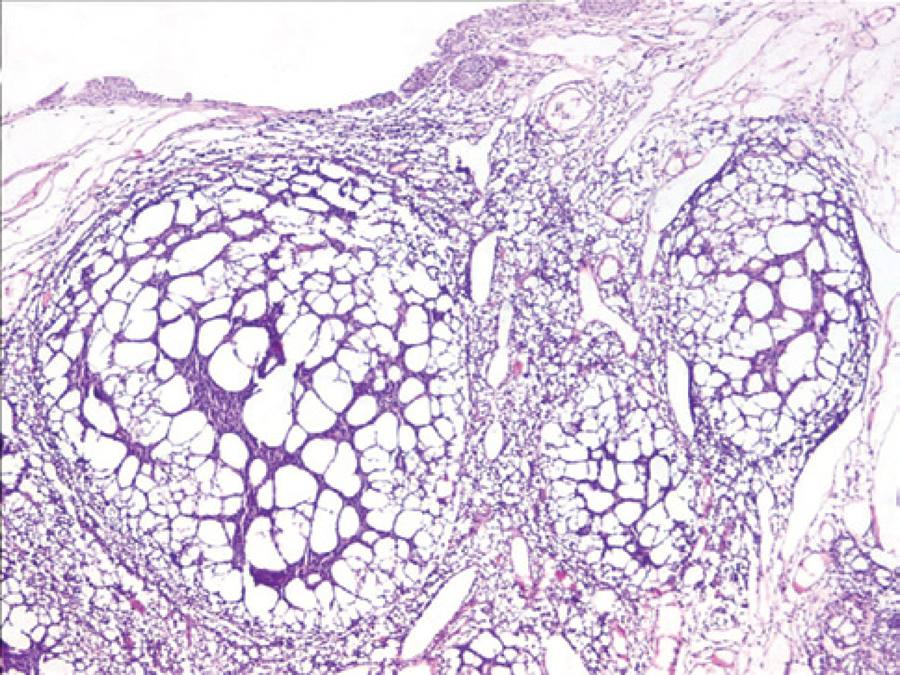
A 34-year-old male presented with a solid mobile mass that had developed three months before his first visit. There was no evidence of trauma or infection. A lobulated pink-yellowish, richly vascularized lesion on the surface of the nasal side of the bulbar conjunctiva in his left eye, measuring 7 × 11 mm in size, was detected on performing slit-lamp biomicroscopic examination. The anterior margin of the mass was superonasally located, 7 mm away from the limbus, and it posteriorly and nasally extended through the caruncle; however, the nasal margin of the mass did not pass the plica semilunaris (Figure 1). On ocular examination, best-corrected visual acuity was 20/20 in both eyes. The tarsal and fornical conjunctival surface and caruncles were normal. The remaining ophthalmic examination was noncontributory. Because of recent enlargement and cosmetic concerns, the tumor with a 2-mm strip of normal-appearing conjunction was excised under the presumptive diagnosis of papilloma. The margin of the excised lesion was tumor-negative. A subconjunctival tumor with enlarged and lobulated sebaceous glands consisting of well-differentiated cells with low mitotic activity (1 mitosis/mm2) was identified, and no atypia was observed on histopathological examination (Figure 2); sebaceous adenoma was diagnosed. Because of the unusual diagnosis and localization, a further work-up was offered for detecting any possible malignancies. Systemic examinations, blood tests, and radiological examinations were performed. There was no history of malignancy in the family. The patient has been followed up for 24 months. There were no malignancies and recurrence of the lesion in the excised area.

Figure 1 Yellow-pinkish lobulated vascularized tumor with a gel-like appearance lying on the surface of the bulbar conjunctiva was clearly observed during down gaze.
DISCUSSION
The meibomian glands and glands of Zeiss are sebaceous glands in the eyelids, and the brow cilia and the caruncle are other areas in which sebaceous glands are located. However, normally, sebaceous glands are not present in the conjunctiva(3,4,6,7). In the present case, the tumor was present on the bulbar conjunctiva with no connection to the caruncle.
It is unclear how a tumor of sebaceous origin can primarily form on the bulbar conjunctiva. There may have been a faulty migration of sebaceous gland cells from the caruncle through the nasal bulbar conjunctiva or it may have derived from pluripotent basal cells of the conjunctival epithelium.
The best-known association between sebaceous tumors including sebaceous hyperplasia, adenoma or carcinoma, and visceral malignancy is represented by the Muir-Torre syndrome. Previously recorded cases associated with the Muir-Torre syndrome were located on the eyelids, palpebral conjunctiva, or caruncle, except for a single case located at the bulbar conjunctiva associated with glioblastoma multiforme and chronic lymphocytic leukemia(8). Our patient was examined for any potential malignancies; however, all systemic examinations were normal. No recurrence of the lesion or other malignancies was found at the 2-year follow-up. With regard to the risk of potential malignancy, we have decided to continue annual examination of the patient.
To the best of our knowledge, this case is the first isolated sebaceous adenoma of the bulbar conjunctiva in the absence of systemic malignancy. A vast majority of benign and malign lesions from ectodermal, mesodermal, and endodermal origins can be detected in the presence of primary conjunctival tumors. Furthermore, sebaceous adenoma should be considered in the diagnosis of benign conjunctival tumors, and histopathologic evaluation must be performed to reach a definite diagnosis.


 English PDF
 Print
Print
 Send this article by email
 How to cite this article
 Submit a comment
 Mendeley
 Scielo
 Pocket
 Share on Linkedin

