
Felipe Placeres Borges1; Denny Marcos Garcia1; Antonio Augusto Velasco e Cruz1
DOI: 10.1590/S0004-27492010000400005
ABSTRACT
PURPOSE: To determine if the distribution of inter-blink time intervals is constant with repeated measurements with and without topical ocular anesthesia. METHODS: Inter-blink time was measured in 15 normal subjects ranging from 19 to 32 years (mean ± SD= 23.9 ± 3.20) with the magnetic search coil technique on 3 different occasions, the last one with topical ocular anesthesia. RESULTS: One-way analysis of variance for repeated measurements showed that topical anesthesia significantly reduced the blink rate (blinks per minute), which was constant in the first two measurements (F=8.27, p=0.0015. First measurement: mean ± SD= 13.7 ± 7.8; second measurement: 13.1 ± 8.5 SD; with topical anesthesia: = 7.2 ± 4.6). However, distributions shape was not affected when the blink rate was reduced. The three distributions followed a Log Normal pattern, which means that the time interval between blinks was symmetrical when the time logarithm was considered. CONCLUSIONS: Topical ocular anesthesia reduces the rate of spontaneous blinking, but does not change the distribution of inter-blink time interval.
Keywords: Blinking; Blinking; Administration, topical; Anesthesia; Ophthalmic solutions; Eyelids
RESUMO
OBJETIVOS: Determinar se a distribuição dos intervalos do piscar espontâneo se mantém em medidas repetidas com e sem anesthesia tópica ocular. MÉTODOS: Os intervalos entre movimentos de piscar da pálpebra superior foram medidos com rastreamento magnético (Magnetic Search Coil) em 15 sujeitos (11 do sexo masculino) normais com idades entre 19 a 32 anos (média 23,86 ± 3,20 dp anos). RESULTADOS: Análise de variância unifatorial para medidas repetidas mostrou que a anesthesia tópica ocular diminuiu significativamente a frequência média (número de blinks/minuto) do piscar espontâneo, a qual se manteve constante nas duas primeiras medidas (F=8,27, p=0,0015. Primeira medida 13,7 ± 7,8 DP; segunda medida 13,1 ± 8,5; com anestesia tópica 7,2 ± 4,6). No entanto, a forma da distribuição nas 3 medidas obedeceu uma distribuição do tipo Log Normal, de modo que os intervalos de piscar foram normalmente distribuídos quando o logaritmo do intervalo foi considerado. CONCLUSÕES: A anesthesia tópica ocular diminui significativamente a frequência de piscar, mas não altera a distribuição dos intervalos do piscar espontâneo.
Descritores: Piscadela; Piscadela; Administração tópica; Anestesia; Soluções oftálmicas; Pálpebras
ORIGINAL ARTICLES
Distribution of spontaneous inter-blink interval in repeated measurements with and without topical ocular anesthesia
Distribuição dos intervalos do piscar espontâneo em medidas repetidas com e sem anestesia tópica ocular
Felipe Placeres Borges; Denny Marcos Garcia; Antonio Augusto Velasco e Cruz
Physician, Ophthalmology, Otorrinolaringology and Head and Neck Surgery Department, Faculdade de Medicina de Ribeirão Preto, Universidade de São Paulo - USP - Ribeirão Preto (SP), Brazil
ABSTRACT
PURPOSE: To determine if the distribution of inter-blink time intervals is constant with repeated measurements with and without topical ocular anesthesia.
METHODS: Inter-blink time was measured in 15 normal subjects ranging from 19 to 32 years (mean ± SD= 23.9 ± 3.20) with the magnetic search coil technique on 3 different occasions, the last one with topical ocular anesthesia.
RESULTS: One-way analysis of variance for repeated measurements showed that topical anesthesia significantly reduced the blink rate (blinks per minute), which was constant in the first two measurements (F=8.27, p=0.0015. First measurement: mean ± SD= 13.7 ± 7.8; second measurement: 13.1 ± 8.5 SD; with topical anesthesia: = 7.2 ± 4.6). However, distributions shape was not affected when the blink rate was reduced. The three distributions followed a Log Normal pattern, which means that the time interval between blinks was symmetrical when the time logarithm was considered.
CONCLUSIONS: Topical ocular anesthesia reduces the rate of spontaneous blinking, but does not change the distribution of inter-blink time interval.
Keywords: Blinking; Blinking/physiology; Administration, topical; Anesthesia; Ophthalmic solutions/pharmacology; Eyelids
RESUMO
OBJETIVOS: Determinar se a distribuição dos intervalos do piscar espontâneo se mantém em medidas repetidas com e sem anesthesia tópica ocular.
MÉTODOS: Os intervalos entre movimentos de piscar da pálpebra superior foram medidos com rastreamento magnético (Magnetic Search Coil) em 15 sujeitos (11 do sexo masculino) normais com idades entre 19 a 32 anos (média 23,86 ± 3,20 dp anos).
RESULTADOS: Análise de variância unifatorial para medidas repetidas mostrou que a anesthesia tópica ocular diminuiu significativamente a frequência média (número de blinks/minuto) do piscar espontâneo, a qual se manteve constante nas duas primeiras medidas (F=8,27, p=0,0015. Primeira medida 13,7 ± 7,8 DP; segunda medida 13,1 ± 8,5; com anestesia tópica 7,2 ± 4,6). No entanto, a forma da distribuição nas 3 medidas obedeceu uma distribuição do tipo Log Normal, de modo que os intervalos de piscar foram normalmente distribuídos quando o logaritmo do intervalo foi considerado.
CONCLUSÕES: A anesthesia tópica ocular diminui significativamente a frequência de piscar, mas não altera a distribuição dos intervalos do piscar espontâneo.
Descritores: Piscadela; Piscadela/fisiologia; Administração tópica; Anestesia; Soluções oftálmicas/farmacologia; Pálpebras
INTRODUCTION
Spontaneous blinking activity is essential for the maintenance of ocular surface integrity(1). The so-called blinking rhythm is defined by the frequency of blinks occurring within a given time interval(2). Typically, normal persons are considered to blink 10 to 20 times per minute, depending on their mental activity at any given time(3).
An issue rarely mentioned in the literature but of theoretical relevance for the elucidation of neural mechanisms underlying the blinking rate is the distribution of consecutive inter-blink intervals. This topic was first considered by Ponder and Kennedy(2) who described four patterns of interblink distribution: "J" pattern, symmetrical or normal patterns, irregular patterns, and bimodal. These patterns may be the expression of an intrinsic feature of each individual determined by mesencephalic dopaminergic activity. Although the hypothesis of blinking rate central regulation is well accepted in neuropsychology(4-9), there are several lines of ophthalmologic evidence showing that the condition of the ocular surface modulates the blink rate(1,10-11). A classical example is the reduced blink rate that occurs after topical ocular anesthesia(12).
The purpose of the present study was to investigate whether the pattern of distribution of blinking frequency changes after repeated measurements and after ocular surface anesthesia.
METHODS
The study was conducted on 15 subjects (11 males) aged from 19 to 32 years (mean ± SD: 23.86 ± 3.20 years) with no systemic diseases or oculo-palpebral problems or procedures, including previous surgeries, that might alter the palpebral dynamics.
Blinking activity was recorded with a capturing system called Magnetic Search Coil. This is a cubic metal structure that produces a weak magnetic field, where the subject is placed while wearing a small coil on his upper eyelid (4.85 mm in diameter, 20 volts, 5 mg, a copper wire 0.16 mm in diameter). As the eyelid slides over the ocular surface, the coil produces an electric signal proportional to the angle relative to the field. This signal is preprocessed with a 10 kHz low-pass filter which is amplified 20,000X and digitized by a analogue-to-digital converter (National Instruments PCI-6220) sampled at 200 Hz, with 12 bit precision. The angular position of the eyelid, in degrees, is obtained with a spatial resolution of approximately 0.1º (equivalent to a linear dislocation of 0.02 mm) by means of a calibration factor measured with the aid of a radius transfer device with a radius similar to the mean radius of the human eye.
For the recording of spontaneous blinking, the subject was instructed to watch a film during 5 minutes on a monitor located at the distance of 1 m. This experiment was repeated twice with an interval of at least 3 days between the experiments. The third acquisition was carried out after the instillation of a drop of anesthetic eye-drops (Visonest®, 5 mg proxymetacaine hydrochloride, vehicle qsp. 1 ml) in the conjunctival sac of both eyes, with one eye being randomly chosen for examination. Thus, three distributions of blinking intervals were obtained for each subject. The first minute of each acquisition was considered to be a period of adaptation and was discarded.
Data were analyzed statistically using the Microcal Origin 8.0® and SAS® JMP 7.0® software. Descriptive statistical techniques were used (dispersal graphs and histograms) and groups were compared by repeated-measures unifactorial ANOVA and by the Kolmogorov and Shapiro-Wilk distribution tests.
RESULTS
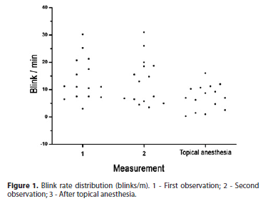
A total of 2042 blinking movements were recorded and distributed as follows: 803 in the first measurement, 771 in the second, and 468 in the third (with anesthesia). Figure 1 illustrates the blink rates distribution obtained for the studied subjects within a 4 minute interval.

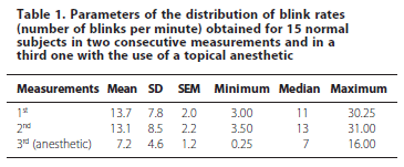
Table 1 lists the parameters of the obtained distributions.

Unifactorial analysis of variance for repeated measures (ANOVA) showed that there was a difference between the rates (F=8.27, p=0.0015). The Tukey test revealed that the rates obtained in the 1st and 2nd measurements did not differ significantly one from another but both differed from the 3rd measurement obtained with the use of a topical anesthetic.
Figure 2 illustrates the distribution of the inter-blink time intervals obtained for 2 study subjects under the three different conditions. The data for the sample as a whole were grouped in order to analyze the anesthetic effect on the inter-blink rate (Table 2 and Figure 3). The Kolmogorov test showed that the 3 conditions followed a Log Normal distribution (J pattern).
DISCUSSION
The results of the present experiment unequivocally show that the conditions of the ocular surface modulate the spontaneous blink rate, as suggested by some authors(1). While in repeated measurements the blink rate was quite constant, after the topical anesthetic administration, the number of blinks per minute decreased significantly.
These data can be interpreted as opposite evidence to the notion that spontaneous blinking activity is not regulated by a central mechanism. However, analysis of inter-blink intervals indicated that the most common pattern observed in the present sample was a Log Normal or J-shaped distribution. In other words, most of the time the subjects blinked at short intervals and only a few times at increasing intervals. The number of blinks separated by a longer time interval was smaller than the number of blinks separated by short intervals. Thus, the histogram representing the distribution of inter-blink intervals was J-shaped.
An interesting property of J-shaped distributions is that this shape becomes symmetrical or normal when the values on the abscissa are log transformed. This characteristic was not affected by the reduced blink rate induced by topical anesthesia.
Apparently, the conditions of the ocular surface modulate the number of blinks but not the inter-blink interval. The neural control of blinking involves a complex set of supranuclear mesencephalic structures (superior colliculus, periaqueductal gray matter, substantia nigra, pyramidal tract, and medial longitudinal fasciculus) that control the orbicular musculature activation, as well as the concomitant inhibition of the upper eyelid levator muscle tonus(4). The inter-blink time intervals temporarily reflect the joint action of these inhibitory (levator muscle) and activating (orbicular muscle) processes. The nervous system regulates logarithmically the inter-blink interval in a symmetrical manner. This characteristic seems to be invariable and it would be interesting to determine whether the inter-blink time intervals also follow a Log Normal distribution in the presence of conditions that affect blink rate such as psychosis(13), Parkinson's disease(5), sleep deprivation(14), drug use(8), and dry eye, as well as the rates induced by refractive surgery such as LASIK(15).
CONCLUSIONS
Topical ocular anesthesia reduces the rate of spontaneous blinking, but does not change the pattern of inter-blink time interval distributions.
REFERENCES
1. Nakamori K, Odawara M, Nakajima T, Mizutani T, Tsubota K. Blinking is controlled primarily by ocular surface conditions. Am J Ophthalmol. 1997;124(1):24-30.
2. Ponder E, Kennedy W. On the act of blinking. Q J Exp Physiol. 1927;18:89-110.
3. Karson CN, Berman KF, Donnelly EF, Mendelson WB, Kleinman JE, Wyatt RJ. Speaking, thinking, and blinking. Psychiatry Res. 1981;5(3):243-6.
4. Esteban A, Traba A, Prieto J. Eyelid movements in health and disease. The supranuclear impairment of the palpebral motility. Neurophysiol Clin. 2004;34(1):3-15.
5. Agostino R, Bologna M, Dinapoli L, Gregori B, Fabbrini G, Accornero N, et al. Voluntary, spontaneous, and reflex blinking in Parkinson's disease. Mov Disord. 2008;23(5):669-75.
6. Blin O, Masson G, Azulay JP, Fondarai J, Serratrice G. Apomorphine-induced blinking and yawning in healthy volunteers. Br J Clin Pharmacol. 1990;30(5):769-73.
7. Colzato LS, Slagter HA, Spapé MM, Hommel B. Blinks of the eye predict blinks of the mind. Neuropsychologia. 2008;46(13):3179-83.
8. Colzato LS, van den Wildenberg WP, Hommel B. Reduced spontaneous eye blink rates in recreational cocaine users: evidence for dopaminergic hypoactivity. PLoS One. 2008;3(10):e3461.
9. Colzato LS, van den Wildenberg WP, van Wouwe NC, Pannebakker MM, Hommel B. Dopamine and inhibitory action control: evidence from spontaneous eye blink rates. Exp Brain Res. 2009;196(3):467-74.
10. Tsubota K, Hata S, Mori A, Nakamori K, Fujishima H. Decreased blinking in dry saunas. Cornea. 1997;16(2):242-3.
11. Al-Abdulmunem M. Relation between tear breakup time and spontaneous blink rate. Int Contact Lens Clin. 1999;26(5):117-20.
12. Naase T, Doughty MJ, Button NF. An assessment of the pattern of spontaneous eyeblink activity under the influence of topical ocular anaesthesia. Graefes Arch Clin Exp Ophthalmol. 2005;243(4):306-12.
13. Caplan R, Guthrie D. Blink rate in childhood schizophrenia spectrum disorder. Biol Psychiatry. 1994;35(4):228-34.
14. Barbato G, Ficca G, Beatrice M, Casiello M, Muscettola G, Rinaldi F. Effects of sleep deprivation on spontaneous eye blink rate and alpha EEG power. Biol Psychiatry. 1995;38(5):340-1.
15. Mian SI, Li AY, Dutta S, Musch DC, Shtein RM. Dry eyes and corneal sensation after laser in situ keratomileusis with femtosecond laser flap creation. Effect of hinge position, hinge angle, and flap thickness. J Cataract Refract Surg. 2009;35(12):2092-8.
 Correspondense address:
Antonio Augusto Velasco e Cruz
Departamento de Oftalmologia, Otorrinolaringologia e Cirurgia de Cabeça e Pescoço, Faculdade de Medicina de Ribeirão Preto - USP - Hospital das Clínicas-Campus
Av. Bandeirantes, 3.900
CEP 14049-900 - Ribeirão Preto (SP)
E-mail: [email protected]
Recebido para publicação em 21.12.2009
Última versão recebida em 05.04.2010
Aprovação em 15.07.2010
Study carried out at Ophthalmology, Otorrinolaringology and Head and Neck Surgery Department, Faculdade de Medicina de Ribeirão Preto, Universidade de São Paulo - USP -Ribeirão Preto (SP), Brazil.
Research support: Scientific Initiation scholarship - Process number 2008/08743-4
Nota Editorial: Depois de concluída a análise do artigo sob sigilo editorial e com a anuência dos Drs. Marcos Carvalho da Cunha e Sérgio Burnier sobre a divulgação de seus nomes como revisores, agradecemos suas participações neste processo.