
Tiago Eugênio Faria e Arantes1; Claudio Renato Garcia2; Janaína Jamile Ferreira Saraceno1; Cristina Muccioli2
DOI: 10.1590/S0004-27492010000100003
ABSTRACT
PURPOSE: To describe the features and outcomes of patients with AIDS-related cytomegalovirus retinitis after highly active antiretroviral therapy availability. METHODS: Retrospective chart review of 30 consecutive patients (44 eyes) with AIDS and newly diagnosed, active AIDS-related cytomegalovirus retinitis, examined from January 2005 to December 2007. RESULTS: The mean age was 34.8 years, 18 patients (60.0%) were male and median duration of AIDS was 90 months. Nineteen patients (63.3%) had evidence of highly active antiretroviral therapy failure and median CD4+ lymphocyte count was 12.5 cells/µl. Visual acuity at presentation was 20/40 or better in 27 eyes (61.4%). Retinitis involved Zone 1 in 13 eyes (39.5%). Despite specific anti-AIDSrelated cytomegalovirus therapy, 16 eyes (36.4%) presented relapse of retinitis and 10 eyes (22.7%) lost at least three lines of vision. When compared to highly active antiretroviral therapy responsive patients, eyes of highly active antiretroviral therapy failure patients were more likely to develop relapse of retinitis (p=0.03) and loss of at least three lines of vision (p=0.03). CONCLUSION: The patients in this series are essentially young men with longstanding AIDS, non-responsive to highly active antiretroviral therapy and with a similar immunological profile as noted before highly active antiretroviral therapy era. These findings have implications for the management of the disease and confirm the magnitude of rational periodic screening after diagnosis of AIDS.
Keywords: Cytomegalovirus; Retinitis; Acquired immunodeficiency syndrome; Antiretroviral therapy, highly active; AIDS-related opportunistic infections; Uveitis, posterior; Eye infections; HIV infections
RESUMO
OBJETIVO: Descrever as características e evolução clínica de pacientes com retinite por citomegalovírus relacionada à AIDS após o advento da terapia antirretroviral potente. MÉTODOS: Estudo retrospectivo dos prontuários de 30 pacientes consecutivos (44 olhos) com AIDS e retinite por citomegalovírus ativa recém-diagnosticada, atendidos entre janeiro de 2005 e dezembro de 2007. RESULTADOS: A idade média dos pacientes foi de 34,8 anos, 18 pacientes (60,0%) eram do sexo masculino e a mediana do tempo de diagnóstico de AIDS era 90 meses. Dezenove pacientes (63,3%) apresentavam evidência de falência da terapia antirretroviral potente e a mediana da contagem de linfócitos T CD4+ era 12,5 células/µl. A acuidade visual inicial era melhor ou igual a 20/40 em 27 olhos (61,4%). A retinite acometia a Zona 1 em 13 olhos (39,5%). Apesar da terapia antirretinite por citomegalovírus específica, 16 olhos (36,4%) apresentaram recidiva da retinite e 10 olhos (22,7%) perderam pelo menos três linhas de visão. Quando comparado aos de pacientes com boa resposta à terapia antirretroviral potente, olhos de pacientes com falência à terapia antirretroviral potente apresentaram mais recidiva da retinite (p=0,03) e perda de pelo menos três linhas de visão (p=0,03). CONCLUSÃO: Os pacientes nesta série são essencialmente homens jovens com longo tempo de diagnóstico de AIDS, má resposta à terapia antirretroviral potente e com um perfil imunológico semelhante ao encontrado antes do advento da terapia antirretroviral potente. Estes achados têm implicações no manejo da doença e confirmam a importância da triagem periódica e racional após o diagnóstico de AIDS.
Descritores: Citomegalovírus; Retinite; Síndrome de imunodeficiência adquirida; Terapia antirretroviral de alta atividade; Infecções oportunistas relacionadas com a AIDS; Uveíte posterior; Infecções oculares; Infecções por HIV
ORIGINAL ARTICLE
Clinical features and outcomes of AIDS-related cytomegalovirus retinitis in the era of highly active antiretroviral therapy
Características e evolução clínica da retinite por citomegalovírus em pacientes com AIDS na era da terapia antirretroviral potente
Tiago Eugênio Faria e ArantesI; Claudio Renato GarciaII; Janaína Jamile Ferreira SaracenoIII; Cristina MuccioliIV
IDoctoral Student - Department of Ophthalmology - Federal University of São Paulo - UNIFESP - Brazil
IIMD and preceptor of Uveitis and HIV Service - Department of Ophthalmology - Federal University of São Paulo - UNIFESP - Brazil
IIIDoctoral Student - Department of Ophthalmology - Federal University of São Paulo - UNIFESP - Brazil
IVAssociate Professor, head of Uveitis and HIV Service - Department of Ophthalmology - Federal University of São Paulo - UNIFESP - Brazil
ABSTRACT
PURPOSE: To describe the features and outcomes of patients with AIDS-related cytomegalovirus retinitis after highly active antiretroviral therapy availability.
METHODS: Retrospective chart review of 30 consecutive patients (44 eyes) with AIDS and newly diagnosed, active AIDS-related cytomegalovirus retinitis, examined from January 2005 to December 2007.
RESULTS: The mean age was 34.8 years, 18 patients (60.0%) were male and median duration of AIDS was 90 months. Nineteen patients (63.3%) had evidence of highly active antiretroviral therapy failure and median CD4+ lymphocyte count was 12.5 cells/µl. Visual acuity at presentation was 20/40 or better in 27 eyes (61.4%). Retinitis involved Zone 1 in 13 eyes (39.5%). Despite specific anti-AIDSrelated cytomegalovirus therapy, 16 eyes (36.4%) presented relapse of retinitis and 10 eyes (22.7%) lost at least three lines of vision. When compared to highly active antiretroviral therapy responsive patients, eyes of highly active antiretroviral therapy failure patients were more likely to develop relapse of retinitis (p=0.03) and loss of at least three lines of vision (p=0.03).
CONCLUSION: The patients in this series are essentially young men with longstanding AIDS, non-responsive to highly active antiretroviral therapy and with a similar immunological profile as noted before highly active antiretroviral therapy era. These findings have implications for the management of the disease and confirm the magnitude of rational periodic screening after diagnosis of AIDS.
Keywords: Cytomegalovirus; Retinitis; Acquired immunodeficiency syndrome; Antiretroviral therapy, highly active; AIDS-related opportunistic infections; Uveitis, posterior; Eye infections/etiology; HIV infections
RESUMO
OBJETIVO: Descrever as características e evolução clínica de pacientes com retinite por citomegalovírus relacionada à AIDS após o advento da terapia antirretroviral potente.
MÉTODOS: Estudo retrospectivo dos prontuários de 30 pacientes consecutivos (44 olhos) com AIDS e retinite por citomegalovírus ativa recém-diagnosticada, atendidos entre janeiro de 2005 e dezembro de 2007.
RESULTADOS: A idade média dos pacientes foi de 34,8 anos, 18 pacientes (60,0%) eram do sexo masculino e a mediana do tempo de diagnóstico de AIDS era 90 meses. Dezenove pacientes (63,3%) apresentavam evidência de falência da terapia antirretroviral potente e a mediana da contagem de linfócitos T CD4+ era 12,5 células/µl. A acuidade visual inicial era melhor ou igual a 20/40 em 27 olhos (61,4%). A retinite acometia a Zona 1 em 13 olhos (39,5%). Apesar da terapia antirretinite por citomegalovírus específica, 16 olhos (36,4%) apresentaram recidiva da retinite e 10 olhos (22,7%) perderam pelo menos três linhas de visão. Quando comparado aos de pacientes com boa resposta à terapia antirretroviral potente, olhos de pacientes com falência à terapia antirretroviral potente apresentaram mais recidiva da retinite (p=0,03) e perda de pelo menos três linhas de visão (p=0,03).
CONCLUSÃO: Os pacientes nesta série são essencialmente homens jovens com longo tempo de diagnóstico de AIDS, má resposta à terapia antirretroviral potente e com um perfil imunológico semelhante ao encontrado antes do advento da terapia antirretroviral potente. Estes achados têm implicações no manejo da doença e confirmam a importância da triagem periódica e racional após o diagnóstico de AIDS.
Descritores: Citomegalovírus; Retinite; Síndrome de imunodeficiência adquirida; Terapia antirretroviral de alta atividade; Infecções oportunistas relacionadas com a AIDS; Uveíte posterior; Infecções oculares/etiologia; Infecções por HIV
INTRODUCTION
Since the introduction of highly active antiretroviral therapy (HAART) in 1996, patients with acquired immunodeficiency syndrome (AIDS) have been experiencing changes in morbidity and mortality(1-3). The incidence of systemic complications and opportunistic infections, including cytomegalovirus (CMV) retinitis declined dramatically compared to the rates from the pre-HAART era(1-7). Cytomegalovirus retinitis is still one of the most common ocular manifestations and the major cause of blindness in patients with AIDS and, despite all the advances in AIDS treatment, it continues to be diagnosed(4-8).
The estimated lifetime probability of a patient with AIDS developing CMV retinitis in the pre-HAART era was about 30% (9) but its incidence has declined 55 to 85% after the introduction of HAART(2-6). In Brazil this drop in the incidence may be explained by the fact that HAART therapy is distributed with no cost by the National AIDS Programme from the National Ministry of Health(10). Patients with CD4+ lymphocytes count below 100 cells/µl and especially patients with CD4+ count 50 cells/µl or less have a proven increased risk of developing the retinitis(11).
Before HAART, CMV disease was characteristically a relapsing retinitis associated with progressive visual loss despite the repeated specific anti-CMV therapy(7,12). As a consequence of HAART, safe discontinuation of maintenance treatment with anti-CMV medication is possible in patients who experience immune reconstitution(13-14).
The purpose of this study is to describe the features and outcomes of Brazilian patients with AIDS-related CMV retinitis infected after availability of HAART.
METHODS
From January 2005 to December 2007, active untreated CMV retinitis was diagnosed in 30 (4.34%) of the 691 consecutive patients examined in Uveitis and AIDS Service of the Federal University of São Paulo - UNIFESP. Medical records, retinal drawings and fundus photographs were reviewed retrospectively. The patients who met the inclusion criteria had the diagnosis of AIDS according to the 1993 Center for Disease Control and Prevention Revised Surveillance Case Definition(15).
Data collection included demographic information, medical and ophthalmic history and a complete ophthalmologic examination that included pinhole visual acuity measurement using the Early Treatment Diabetic Retinopathy Study (ETDRS) chart, external examination, slit-lamp biomicroscopy, intraocular pressure measurement, and indirect ophthalmoscopy through a dilated pupil.
The diagnosis of CMV retinitis was based on the clinical exam and its progression was assessed by the presence of new lesions or border advancement of existing lesions(16). Retinitis lesions were described based on size and location. The location of each lesion was divided into three zones (1, 2 and 3) according to an already established standard classification system(17).
Patients on HAART therapy were receiving a combination of antiviral drugs with three or more drugs including at least one protease inhibitor or non-nucleoside reverse transcriptase inhibitor. Patients were considered to be HAART-naive if they were not under this drug regimen.
Newly diagnosed patients had less than 3 months of AIDS diagnosis. HAART-failure was defined as the presence of immunologic failure, virologic failure, or both, in patients who had been receiving HAART for at least 12 weeks before the diagnosis of CMV retinitis. Immunologic failure was defined as a CD4+ T lymphocyte count of 50 cells/µl or less. Virologic failure was defined as HIV RNA blood levels of 1,000 copies/ml or more(4). An immunologic response to HAART was defined as an increase in the CD4+ T cell count by at least 50 cells/µl from the nadir CD4+ T cell count to a level of 100 cells/µl or more.
Immune recovery uveitis was diagnosed as the presence of intraocular inflammation in a patient with healed CMV retinitis who had an immune response to HAART and was characterized by vitritis, macular edema, or epiretinal membrane formation in association with specific cytokine profiles(18).
Statistical analysis
Statistical analysis was performed using SigmaStat 3.11 (SPSS Inc, Chicago, IL). The distribution of continuous variable is expressed as mean ± standard deviation and categorical data are presented as frequencies. Relationships between two categorical variables were assessed using Fisher's exact test. Student t test was used for analysis of continuous variables and unpaired t test with Welch correction was used when applicable.
RESULTS
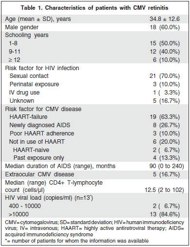
Thirty patients with newly diagnosed active CMV retinitis were included. The average age was 34.8 ± 12.6 years (median: 36 years) and 18 patients (60.0%) were male. The most frequent risk factor for HIV infection was sexual contact with HIV infected individual (21 patients - 70.0%) and the most frequent risk factor for CMV disease was HAART-failure (19 patients - 63.3%).
The median duration of AIDS (defined as the time from diagnosis of AIDS to the first ocular evaluation) was 90 months (mean ± SD: 93.2 ± 79.6 months); in two patients (6.7%), CMV retinitis was an initial manifestation of AIDS. The median CD4+ lymphocyte count was 12.5 cells/µl (mean ± SD: 26.8 ± 29.9 cells/µl). The median follow-up of the patients was 6.0 months (mean ± SD: 9.0 ± 7.0 months). Demographic and clinical characteristics are presented in table 1.

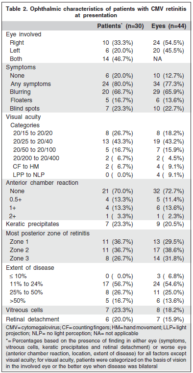
Fourteen patients (46.7%) had bilateral disease at the first visit; one patient with unilateral disease had involvement of the fellow eye during the follow-up. Most of the patients (24 patients - 80.0%) presented visual symptoms in the first evaluation and visual blurring was the most frequent (65.9% of the eyes). The visual acuity at baseline was 20/40 or better in 27 eyes (61.4%). Anterior segment findings included 1+ anterior chamber cells or more on 7 eyes (15.9%) and fine keratic precipates on 9 eyes (20.5%). The retinitis involved Zone 1 in 13 eyes (39.5%) and the area involved was greater than 50% in 6 eyes (13.6%). Retina was detached in 7 eyes (15.9%) of 6 patients (20.0%) at the time of the first appointment, and 7 eyes of 5 patients detached the retina during the follow-up period. Information regarding ophthalmologic findings is presented in table 2.

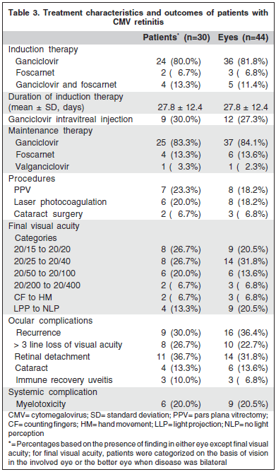
Induction and maintenance therapy doses consisted of a standard regimen(16) with the induction treatment extended until retinitis healing was achieved, despite the classically 2-3 week course of induction. Twenty-four patients (80.0%) received induction therapy with intravenous ganciclovir, two (6.7%) received intravenous foscarnet regimen and four patients (13.3%) that started treatment with ganciclovir switched to foscarnet later on due to inappropriate response or adverse effects. Ganciclovir was the elected drug for maintenance therapy in 25 patients (83.3%), foscarnet was elected in 4 (13.3%) and one patient (3.3%) was treated with valganciclovir. The average time of induction therapy was 27.8 ± 12.4 days (median: 21 days). Nine patients (12 eyes) underwent intravitreal injection of ganciclovir (2.0 mg in 0.1 ml - 2.6 ± 1.3 injection per eye). Six patients (21.4%) treated with ganciclovir experienced myelotoxicity. Patients with HAART-failure required longer duration of induction therapy when compared to HAART responsive patients (respectively, 31.3 ± 13.6 and 21.9 ± 7.4 days - p=0.021). Patients with more than 3 month of AIDS diagnosis also required longer induction treatment than patients with newly diagnosed AIDS (respectively, 30.3 ± 13.1 and 21.1 ± 7.4 days - p=0.025). Intravitreal injection of ganciclovir was necessary when time of induction therapy was extended due to lack of response to the systemic treatment (mean duration of induction therapy was 34.8 ±11.8 days inpatients who underwent intravitreal ganciclovir and 24.9 ± 11.7 days in patients that did not require combined local treatment - p=0.043). No complications such as retinal detachment, cataract or immune recovery uveitis were significantly associated with this procedure.
Sixteen eyes (36.4%) of nine patients (30.0%) experienced relapse of the retinitis while on maintenance therapy. The mean duration of the first remission in these patients was 41.2 ± 42.7 days (median: 22 days). Retinitis relapse was more likely to occur in eyes of patients with HAART-failure (p=0.030) and extraocular CMV disease (p=0.002).
During follow-up, 10 eyes (22.7%) of 8 patients (26.7%) had at least three lines of visual acuity loss (doubling of the visual angle) and this was associated with HAART-failure (p=0.031) and retinal detachment (p=0.006). At the final visit, 15 eyes (34.1%) were legally blind (20/200 or worse), with a significant association with zone 1 involvement (p=0.004).
Seven patients (23.3%) presented immune recovery during follow-up allowing anti-CMV maintenance therapy discontinuation; six of these patients had newly diagnosed AIDS and only one had more than three months of diagnosis of AIDS. The mean duration of maintenance therapy was 7.0 ± 4.1 months.
Eight eyes (18.2%) with retinal detachment underwent pars plana vitrectomy, eight eyes (18.2%) underwent retinal argon laser photocoagulation and three eyes (6.8%) had phacoemulsification with intraocular lens implantation performed. Treatment and clinical outcomes are summarized in table 3.

DISCUSSION
Cytomegalovirus retinitis accounts for 75 to 85% of CMVrelated diseases in AIDS(19-20) and even in the HAART era it still is the major cause of visual impairment in these patients(4,8,19-20)
In a previous study from May 2000 to February 2001(1), 4.5% of 200 of our patients were diagnosed with active CMV retinitis in the first visit. In our series, 5.6% of 691 consecutive examined patients from January 2005 to February 2007 met the criteria for newly diagnosed CMV retinitis, suggesting that its incidence has been stable in the last decade.
New cases of CMV retinitis continue to be diagnosed due to reasons such as late diagnosis of HIV infection or AIDS, poor adherence to HAART treatment or viral resistance to one or more components of HAART therapy(4,21). In developing countries, CMV retinitis is largely undiagnosed and the scale of the problem is still not known as there is no strategy for screening and management of the problem(21).
Patients with CMV retinitis presented a similar immunological profile as observed in the era before HAART. At that time patients had low CD4+ T-cell counts and elevated HIV viral loads(4,12), although patients receiving HAART showed a broader range of CD4+ count, including values of more than 50 cells/µl(4).
As in other studies, most of these patients have longstanding AIDS, had received HAART and are intolerant or non responsive to this therapy(4-6). Visual symptoms were frequent at the time of diagnosis and blurring was the most common presenting symptom, as previously described(22). Patients with more severe disease, including HAART-failure and extraocular CMV disease, had a worse prognosis, evolving with visual loss and CMV retinitis relapses.
The median time from the diagnosis of AIDS to the first visit was longer than the median time in previous studies(4,6); rates of retinal detachment and visual acuity of 20/200 or worse at presentation were also higher than previously described(4,23). This could be explained by the fact that not always the patients are submitted to a screening performed by an ophthalmologist that is familiar with the ocular complications of the disease. These difficulties could be diminished by specific training programs for general ophthalmologists and the use of telemedicine for evaluation and second opinion.
Treatment with specific anti-CMV therapy successfully controlled the disease with minimal complications. In patients unresponsive to HAART and with long standing AIDS the duration of induction treatment needed to be prolonged from the standard 2-3 weeks for adequate control of the retinitis. Local treatment with intraocular injection of ganciclovir 2.0 mg was considered an option for patients with inappropriate response to therapy and play a role in patients with systemic contraindications to anti-CMV drugs. The procedure was not associated with complications like retinal detachment or lens opacification.
Despite treatment, doubling of the visual angle occurred in 22.7% of the eyes and retinitis recurred in 30% of the patients. The most common cause of visual loss of 20/200 or worse was zone 1 involvement(23). Retinal detachment was not significantly associated with visual loss of 20/200 or worse in our series; surgical reattachment of the retina (pars plana vitrectomy with silicone oil tamponade) was successful in the majority of cases (5 of 8 eyes had visual acuity of 20/40 or better at the end of the follow-up). The mean duration of first retinitis remission in eyes with recurrences was shorter than the period described in the HAART era(5) and similar to the period before HAART availability(16). Probably the poor immunologic status of these patients and increasing rates of CMV resistance to ganciclovir and foscarnet is the reason for this. Unfortunately, CMV resistance testing was only available for a minority of patients.
CONCLUSION
This study is retrospective, but the results represent the heterogeneity of host profiles and clinical features of AIDSrelated CMV retinitis in the HAART era agreeing with the already published data. These findings have implications for evaluation and management of disease and confirm the importance of ophthalmic evaluation after diagnosis of AIDS, rational periodic screening of CMV retinitis and prompt treatment. These strategies are crucial for successful outcomes and preservation of vision.
REFERENCES
1. Arruda RF, Muccioli C, Belfort R Jr. [Ophthalmological findings in HIV infected patients in the post-HAART (Highly Active Anti-retroviral Therapy) era, compared to the pre-HAART era]. Rev Assoc Med Bras. 2004;50(2):148-52. Portuguese.
2. Goldberg DE, Smithen LM, Angelilli A, Freeman WR. HIV-associated retinopathy in the HAART era. Retina. 2005;25(5):633-49; quiz 682-3. Review.
3. Matos KT, Santos MC, Muccioli C. [Ocular manifestations in HIV infected patients attending the department of ophthalmology of Universidade Federal de Sao Paulo]. Rev Assoc Med Bras. 1999;45(4):323-6. Portuguese.
4. Holland GN, Vaudaux JD, Shiramizu KM, Yu F, Goldenberg DT, Gupta A, Carlson M, Read RW, Novack RD, Kuppermann BD; Southern California HIV/Eye Consortium. Characteristics of untreated AIDS-related cytomegalovirus retinitis. II. Findings in the era of highly active antiretroviral therapy (1997 to 2000). Am J Ophthalmol. 2008;145(1):12-22.
5. Lin DY, Warren JF, Lazzeroni LC, Wolitz RA, Mansour SE. Cytomegalovirus retinitis after initiation of highly active antiretroviral therapy in HIV infected patients: natural history and clinical predictors. Retina. 2002;22(3):268-77.
6. Jabs DA, Van Natta ML, Kempen JH, Reed Pavan P, Lim JI, Murphy RL, et al. Characteristics of patients with cytomegalovirus retinitis in the era of highly active antiretroviral therapy. Am J Ophthalmol. 2002;133(1):48-61.
7. Deayton JR, Wilson P, Sabin CA, Davey CC, Johnson MA, Emery VC, et al. Changes in the natural history of cytomegalovirus retinitis following the introduction of highly active antiretroviral therapy. AIDS. 2000;14(9):1163-70.
8. Jabs DA, Van Natta ML, Thorne JE, Weinberg DV, Meredith TA, Kuppermann BD, Spekowitz K, Li HK; Studies of Ocular Complications of AIDS Research Group. Course of cytomegalovirus retinitis in the era of highly active antiretroviral therapy: 1. Retinitis progression. Ophthalmology. 2004;111(12):2224-31.
9. Hoover DR, Peng Y, Saah A, Semba R, Detels RR, Rinaldo CR, Jr. et al. Occurrence of cytomegalovirus retinitis after human immunodeficiency virus immunosuppression. Arch Ophthalmol. 1996;114(7):821-7.
10. Brazil. Ministry of Health. National STD and AIDS Programme from Brazilian. Brasilia (DF): Ministry of Health; 2007.
11. Kuppermann BD, Petty JG, Richman DD, Mathews WC, Fullerton SC, Rickman LS, et al. Correlation between CD4+ counts and prevalence of cytomegalovirus retinitis and human immunodeficiency virus-related noninfectious retinal vasculopathy in patients with acquired immunodeficiency syndrome. Am J Ophthalmol. 1993;115(5):575-82.
12. Holland GN, Vaudaux JD, Jeng SM, Yu F, Goldenberg DT, Folz IC, Cumberland WG, McCannel CA, Helm CJ, Hardy WD; UCLA CMV Retinitis Study Group. Characteristics of untreated AIDS-related cytomegalovirus retinitis. I. Findings before the era of highly active antiretroviral therapy (1988 to 1994). Am J Ophthalmol. 2008;145(1):5-11.
13. Waib LF, Bonon SH, Salles AC, Benard G, de Oliveira AC, Pannuti CS, et al. Withdrawal of maintenance therapy for cytomegalovirus retinitis in AIDS patients exhibiting immunological response to HAART. Rev Inst Med Trop Sao Paulo. 2007;49(4):215-9.
14. Wohl DA, Kendall MA, Owens S, Holland G, Nokta M, Spector SA, Schrier R, Fiscus S, Davis M, Jacobson MA, Currier JS, Squires K, Alston-Smith B, Andersen J, Freeman WR, Higgins M, Torriani FJ; ACTG 379 Study Team. The safety of discontinuation of maintenance therapy for cytomegalovirus (CMV) retinitis and incidence of immune recovery uveitis following potent antiretroviral therapy. HIV Clin Trials. 2005;6(3):136-46.
15. 1993 revised classification system for HIV infection and expanded surveillance case definition for AIDS among adolescents and adults. MMWR Recomm Rep. 1992;41(RR-17):1-19.
16. Foscarnet-Ganciclovir Cytomegalovirus Retinitis Trial. 4. Visual outcomes. Studies of Ocular Complications of AIDS Research Group in collaboration with the AIDS Clinical Trials Group. Ophthalmology. 1994;101(7):1250-61.
17. Holland GN, Buhles WC Jr, Mastre B, Kaplan HJ. A controlled retrospective study of ganciclovir treatment for cytomegalovirus retinopathy. Use of a standardized system for the assessment of disease outcome. UCLA CMV Retinopathy. Study Group. Arch Ophthalmol. 1989;107(12):1759-66.
18. Schrier RD, Song MK, Smith IL, Karavellas MP, Bartsch DU, Torriani FJ, et al. Intraocular viral and immune pathogenesis of immune recovery uveitis in patients with healed cytomegalovirus retinitis. Retina. 2006;26(2):165-9.
19. Gallant JE, Moore RD, Richman DD, Keruly J, Chaisson RE. Incidence and natural history of cytomegalovirus disease in patients with advanced human immunodeficiency virus disease treated with zidovudine. The Zidovudine Epidemiology Study Group. J Infect Dis. 1992;166(6):1223-7. Comment in: J infect Dis. 1993;168(4):1071-2.
20. Pertel P, Hirschtick R, Phair J, Chmiel J, Poggensee L, Murphy R. Risk of developing cytomegalovirus retinitis in persons infected with the human immunodeficiency virus. J Acquir Immune Defic Syndr. 1992;5(11):1069-74.
21. Mahadevia PJ, Gebo KA, Pettit K, Dunn JP, Covington MT. The epidemiology, treatment patterns, and costs of cytomegalovirus retinitis in the post-haart era among a national managed-care population. J Acquir Immune Defic Syndr. 2004;36(4):972-7. Erratum in: J Acquir Immune Defic Syndr. 2004;37(1):1220.
22. Wei LL, Park SS, Skiest DJ. Prevalence of visual symptoms among patients with newly diagnosed cytomegalovirus retinitis. Retina. 2002;22(3):278-82.
23. Thorne JE, Jabs DA, Kempen JH, Holbrook JT, Nichols C, Meinert CL; Studies of Ocular Complications of AIDS Research Group. Causes of visual acuity loss among patients with AIDS and cytomegalovirus retinitis in the era of highly active antiretroviral therapy. Ophthalmology. 2006;113(8):1441-5.
 Endereço para correspondência:
Tiago Eugênio Faria e Arantes
Rua Botucatu, 822
São Paulo (SP) CEP 04023-062
E-mail: [email protected]
Recebido para publicação em 19.08.2009
Aprovação em 12.01.2010
Nota Editorial: Depois de concluída a análise do artigo sob sigilo editorial e com a anuência do Dr. Carlos Roberto Neufeld sobre a divulgação de seu nome como revisor, agradecemos sua participação neste processo.