Arq. Bras. Oftalmol. 2009; 72 (5): 10.1590/S0004-27492009000500028
Total: 1592
Paulo José Martins Bispo1; Ana Luisa Höfling-Lima2; Antonio Carlos Campos Pignatari3
DOI: 10.1590/S0004-27492009000500028
ABSTRACT
Bacterial endophthalmitis is a serious but uncommon intraocular infection which frequently results in vision loss. Early diagnosis and appropriate therapy are associated with better visual outcome. Conventional microbiological methods are currently used for microbiological characterization of eyes with suspected endophthalmitis. However, the sensitivity of bacterial detection from aqueous and vitreous humor using microbiology techniques is poor, and time-consuming to confirm the results. The application of molecular methods enhances significantly laboratory confirmation of bacterial endophthalmitis, demanding a shorter time to draw a definitive result and thereby promoting the early initiation of a more specific therapy to limit the empirical or unnecessary use of broad-spectrum antibiotics. PCR-based techniques, including post-PCR methods such RFLP, DNA probe hybridization and DNA sequencing have been successfully used for the diagnostic elucidation of clinically suspected bacterial endophthalmitis cases, showing promising application in the routine practice of ocular microbiology laboratories.
Keywords: Endophthalmitis; Eye infections, bacterial; Molecular diagnostic techniques; Polymerase chain reaction
RESUMO
Endoftalmite bacteriana é uma infecção intraocular grave, mas de baixa frequência, podendo resultar em grande prejuízo visual. O diagnóstico precoce e a rápida instituição de terapia adequada estão associadas a um melhor prognóstico da doença. Os métodos microbiológicos convencionais são utilizados rotineiramente para caracterização microbiológica de olhos com suspeita de endoftalmite. No entanto, a sensibilidade de detecção bacteriana em amostras de humor aquoso e vítreo utilizando técnicas microbiológicas é baixa, além de demandar um maior tempo para a confirmação dos resultados. A utilização de métodos moleculares aumenta significativamente os casos de endoftalmite bacteriana confirmados laboratorialmente, com tempo menor para a liberação de um resultado definitivo, auxiliando assim a instituição precoce de uma terapia mais específica, limitando o uso empírico ou desnecessário de antibióticos de amplo espectro. A técnica de PCR e outras metodologias para análises pós-PCR como, RFLP, hibridização com sondas e sequenciamento do DNA, tem sido utilizadas com sucesso para elucidação diagnóstica em casos com suspeita clínica de endoftalmite bacteriana, demonstrando promissora aplicação para a rotina dos laboratórios de microbiologia ocular.
Descritores: Endoftalmite; Infecções oculares bacterianas; Técnicas de diagnóstico molecular; Reação em cadeia da polimerase
ATUALIZAÇÃO CONTINUADA
Molecular biology applied to the laboratory diagnosis of bacterial endophthalmitis
Biologia molecular aplicada ao diagnóstico laboratorial de endoftalmite bacteriana
Paulo José Martins BispoI; Ana Luisa Höfling-LimaII; Antonio Carlos Campos PignatariIII
IBiomédico, Pós-graduando (Mestrado) do Laboratório Especial de Microbiologia Clínica - LEMC e Laboratório ALERTA, Disciplina de Infectologia, Departamento de Medicina, Universidade Federal de São Paulo - UNIFESP - São Paulo (SP) - Brazil
IIProfessora Titular do Departamento de Oftalmologia da UNIFESP - São Paulo (SP) - Brazil
IIIProfessor Titular da Disciplina de Infectologia do Departamento de Medicina e Diretor do Laboratório Especial de Microbiologia Clínica - LEMC - UNIFESP- São Paulo (SP) - Brazil
ABSTRACT
Bacterial endophthalmitis is a serious but uncommon intraocular infection which frequently results in vision loss. Early diagnosis and appropriate therapy are associated with better visual outcome. Conventional microbiological methods are currently used for microbiological characterization of eyes with suspected endophthalmitis. However, the sensitivity of bacterial detection from aqueous and vitreous humor using microbiology techniques is poor, and time-consuming to confirm the results. The application of molecular methods enhances significantly laboratory confirmation of bacterial endophthalmitis, demanding a shorter time to draw a definitive result and thereby promoting the early initiation of a more specific therapy to limit the empirical or unnecessary use of broad-spectrum antibiotics. PCR-based techniques, including post-PCR methods such RFLP, DNA probe hybridization and DNA sequencing have been successfully used for the diagnostic elucidation of clinically suspected bacterial endophthalmitis cases, showing promising application in the routine practice of ocular microbiology laboratories.
Keywords: Endophthalmitis/etiology; Eye infections, bacterial; Molecular diagnostic techniques; Polymerase chain reaction
RESUMO
Endoftalmite bacteriana é uma infecção intraocular grave, mas de baixa frequência, podendo resultar em grande prejuízo visual. O diagnóstico precoce e a rápida instituição de terapia adequada estão associadas a um melhor prognóstico da doença. Os métodos microbiológicos convencionais são utilizados rotineiramente para caracterização microbiológica de olhos com suspeita de endoftalmite. No entanto, a sensibilidade de detecção bacteriana em amostras de humor aquoso e vítreo utilizando técnicas microbiológicas é baixa, além de demandar um maior tempo para a confirmação dos resultados. A utilização de métodos moleculares aumenta significativamente os casos de endoftalmite bacteriana confirmados laboratorialmente, com tempo menor para a liberação de um resultado definitivo, auxiliando assim a instituição precoce de uma terapia mais específica, limitando o uso empírico ou desnecessário de antibióticos de amplo espectro. A técnica de PCR e outras metodologias para análises pós-PCR como, RFLP, hibridização com sondas e sequenciamento do DNA, tem sido utilizadas com sucesso para elucidação diagnóstica em casos com suspeita clínica de endoftalmite bacteriana, demonstrando promissora aplicação para a rotina dos laboratórios de microbiologia ocular.
Descritores: Endoftalmite/etiologia; Infecções oculares bacterianas; Técnicas de diagnóstico molecular; Reação em cadeia da polimerase
INTRODUCTION
Endophthalmitis is an inflammation of intraocular tissues, which can result from the introduction of a bacterial agent in the posterior segment of the eye. It requires urgent medical attention because it is a potentially destructive condition for the eye, and even with therapeutic and surgical intervention, it can lead to partial or complete vision loss after a few days of inoculation(1-2). Regarding the acquisition mechanism, endophthalmitis is classified as: (1) postoperative (acute or delayed-onset), (2) posttraumatic, (3) endogenous and (4) miscellaneous (e.g., secondary to keratitis)(3). Postoperative endophthalmitis is the most common presentation and it is frequently associated with cataract surgery. Causative pathogens generally originate from the normal conjunctival and eyelid flora. In the Endophthalmitis Vitrectomy Study (EVS), the recovery of gram-positive bacteria (94.2%) was far greater than gram-negative (6.5%) in acute postoperative endophthalmitis cases, where staphylococci, streptococci and enterococci were more frequently isolated(4). Posttraumatic endophthalmitis is mainly caused by normal ocular flora and environmental isolates. Staphylococci and B. cereus are the most common isolates(5-6). Endogenous endophthalmitis is a less common category, corresponding to 2 to 8% of all endophthalmitis cases. As a consequence of hematogenous dissemination, specially in compromised hosts or intravenous drug abusers, any pathogen causing bacteremia/sepsis could gain access to the posterior segment of the eye resulting in infection. However, it has been shown that the most common bacterial isolates are Staphylococcus aureus, group B streptococci, Streptococcus pneumoniae, Listeria monocytogene, Klebsiella spp., Escherichia coli, Pseudomonas aeruginosa, and Neisseria meningitidis(7).
The correct and early identification of the etiological agent by analysis of vitreous and/or aqueous humor is important for the institution of an early and effective antibiotic therapy, and for the prevention of the inappropriate use of broad-spectrum antibiotics, thereby minimizing the potential emergence of resistant bacterial strains. However, the institution of treatment for patients with clinical signs of intraocular infection, presenting negative cultures is frequently experienced in the ophthalmologic routine. Sometimes, the clinical signs are secondary to non-infectious causes, such as immune system reactions, chemical or physical aggression, vasculitis or neoplasia, which may be impossible to differentiate of an infection(2). Therefore, the therapeutic scheme instituted for each patient can be a difficult choice, which can sometimes lead to a therapy that is deleterious and potentially toxic to ocular tissues, producing undesirable effects or that is actually needed and advantageous in the case of an intraocular inflammatory infectious process.
Use of molecular techniques for the diagnosis of infectious diseases
The introduction of molecular biology techniques in diagnostic medicine, such as the polymerase chain reaction (PCR), and its application in clinical microbiology, established a new era in the detection and characterization of microorganisms. PCR makes it possible to detect microorganisms that are difficult to detect using traditional microbiological methods and to reduce the time necessary for a confirmatory laboratory report, which is an important improvement in the characterization of microorganisms involved in serious and rapidly developing infections. Molecular methods have been applied in the identification of antibiotic resistance mechanisms, detection of fastidious microorganisms, viral genotyping and quantification, rapid diagnosis of bacterial and fungal infections in immunocompromised patients, epidemiological studies, and control of infection(8). Despite all the benefits of PCR use in the diagnosis of infectious diseases, bacterial culture has the advantages of allowing antibiotic susceptibility testing, important in establishing the therapeutic course, as well as storing the cultures in reference microorganisms collection making it possible to carry out later studies related to mechanisms of drug resistance, virulence and molecular typing. Due to the high sensitivity of detection, even in cases that the microorganisms have already been killed, the utilization of PCR increases the chance of false-positive results, and the results for patients undergoing treatment should be rigorously evaluated.
The need for PCR in ophthalmology
The rapid identification of the causal agent and early institution of a specific antibiotic treatment are associated with better visual outcomes in endophthalmitis(2). Microbiological endophthalmitis diagnosis is performed routinely by culture and microscopic examination of the vitreous (VH) and aqueous (AH) humors. However, conventional microbiological techniques are frequently insufficient to confirm clinical cases suspected of endophthalmitis, and the time necessary to obtain culture results can vary from 2 to 12 days. The consequences for eyes not treated can be dreadful, the lack or delay of a microbiological laboratory confirmation can lead to the wrong use of some ophthalmic pharmacological therapy with potential ocular toxicity, as well as the use of broad-spectrum antibiotics for several days, being the correct antibiotic therapy initiated only after the definitive identification of the causative microorganism(2,9-10).
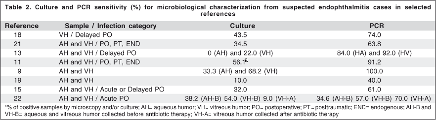
In clinical cases suspected of endophthalmitis, cultures show a positivity of about 25% to 56%(11-15). The low sensitivity of microbiological culture is due to various factors such as small quantity of specimen, fixation of microorganisms in solid surfaces (intraocular lens, lens fragments, capsule) and consequent decrease of cells in the vitreous/aqueous humor, use of antibiotics before the collection of clinical material and the presence of fastidious microorganisms such as agents causing endophthalmitis(14). The application of molecular techniques based on PCR increases significantly the cases of endophthalmitis laboratorial confirmed by and has prompted their growing utilization in this setting. Therefore, in the present work, the results of studies that utilized molecular techniques for the laboratory microbiological characterization of bacterial endophthalmitis cases are compiled, serving as a reference for those who aim to implement or utilize this type of methodology in the routine practice of clinical ophthalmology services.
Molecular detection and identification in cases of bacterial endophthalmitis
Usually, the detection of bacteria of clinical interest by molecular biology laboratories is completed by the amplification of the 16S rRNA gene which codes for the small subunit of ribosomal RNA. Post-PCR analysis, utilizing hybridization with DNA probes, restriction analysis of PCR products by RFLP (restriction fragment length polymorphism), or direct sequencing of these products allow the species identification of the pathogen(9,11,14). Recently, a platform that utilizes mass spectrometry of ionized PCR products (including products of the 16S rRNA gene) was developed which is capable of integrating in the same system, identification of the microorganism, genotyping, and detection of virulence and antibiotic resistance genes(16).
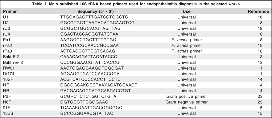
The 16S rRNA gene shows regions that are highly conserved among all bacterial species, and highly variable regions that permit the differentiation between species(17). Nine hypervariable regions are present while the 16S rRNA, which demonstrates the phylogenetic diversity among different bacterial species, allows their identification in most cases. By the utilization of universal primers that are complementary to the conserved regions flanking the variable regions of the gene, it is possible to carry out PCR directly on the vitreous and/or aqueous humors followed by sequencing of the amplified product. This allows the identification of the bacterial species causing the infection by alignment with sequences obtained from available databanks, usually BLAST/Genbank (http://www.ncbi.nlm.nih.gov/blast/). Table 1 shows the sequences of primers utilized in the core studies that evaluated the utilization of PCR for the diagnosis of bacterial endophthalmitis.
The use of PCR for bacterial detection in aqueous and vitreous humor from patients with suspected endophthalmitis has been described by several authors. The first study applied the PCR technique for the diagnosis of delayed postoperative endophthalmitis. Utilizing the nested PCR technique with universal primers and a combination of species-specific primers for Propionibacterium acnes, the method was capable of detecting bacterial DNA in 17 (74%) of 23 specimens of vitreous humor, while the culture was positive in 10 (43.5%) of these specimens. Bacterial DNA was detected by PCR in 8 specimens with a negative culture and in all cases where there was bacterial growth in culture(18).
Soon after, various authors showed how the utilization of PCR for the direct detection of pathogens from aqueous and vitreous humor samples could impact effectively the diagnosis of bacterial endophthalmitis, mainly by increasing significantly the number of cases that were characterized microbiologically with the use of PCR but that were shown to be negative by microscopy or culture(11,13-15,19-22) (Table 2). The nested PCR method was later used in specimens of vitreous (n=30) and aqueous (n=28) humor collected from 55 patients with clinical diagnosis of endophthalmitis(21). This study utilized universal primers for the bacterial 16S rRNA gene and a set of species-specific primers for P. acnes. Among the 58 samples included, 27 (46.5%) were positive by culture, 20 (34.5%) were positive for bacteria and 7 (12%) for fungi and 31 (53.5%) were negative after culture. On the other hand, PCR was capable of detecting bacterial DNA in 37 (63.8%) of the specimens tested, demonstrated 100% concordance with cases characterized microbiologically by culture, and was capable of detecting bacterial DNA in 44.7% of the specimens found negative for bacterial culture. Later studies were able to observe a sensitivity of detection of pathogens by PCR varying from 57% to 100% while sensitivity of the culture varied between 24% and 56%(11,13-15,22).
A multicenter study published recently where PCR was applied for the diagnosis of bacterial endophthalmitis, included 100 patients with acute endophthalmitis following cataract surgery who were treated in 4 academic hospitals in France(22). The specimens were divided into two groups, those collected at the time of admission and before the initiation of antibiotic therapy (n=76 for AH and n=38 for VH) and those collected at a second time (n=53 for AH and n=57 for VH) only in patients who required another intravitreal injection of antibiotic or were submitted to vitrectomy via pars plana. The sensitivity of the culture and PCR for the specimens collected before the initiation of antibiotic therapy was 38.2% and 34.6% for aqueous humor and 54.0% and 56.7% for vitreous humor, respectively. In this group, PCR was positive in specimens of aqueous humor in 9.7% and vitreous humor in 40.0% of cases showing negative cultures. For the specimens of aqueous humor collected after the administration of antibiotics, the sensitivity of PCR decreased to 23.4% and culture to 13.6%. However, for the specimens of vitreous humor included in this second group, PCR was positive in 70.1% and culture in 8.8% of the cases, where PCR was positive in 73.0% of the cases with negative cultures. When the PCR and culture results were evaluated in a subgroup of patients who had specimens of vitreous humor collected at the time of admission as well after the use of antibiotic, positivity of PCR was 73.7% and of culture 58.8%, before treatment, while after the initiation of treatment, PCR was positive in 81.2% of the specimens tested and culture positive in only one case. Culture methods require viable microorganisms to be able to infer their development in a particular infection after their isolation. On the other hand, the detection of pathogens by PCR depends only on the presence of bacterial DNA in the specimen, whether or not there are viable microorganisms, characterizing one of the advantages of this technique in relation to culture, mostly in fluids where normal flora is not present.
The detection of bacterial DNA utilizing universal primers is useful as a rapid test in determining the etiology of an intraocular infection, but does not provide information in relation to Gram staining and identification of the species involved in each case. Anand et al.(11) submitted the PCR products of the 16S rRNA gene to hybridization with DNA probes capable of differentiating PCR products obtained from gram-positive versus gram-negative microorganisms. These authors evaluated 57 specimens (n=17 for AH and n=40 for VH) of patients with a clinical diagnosis of endophthalmitis. The positivity of the microbiological methods (culture and microscopy) was 56.1%, and that of PCR followed by hybridization was 91.2%, resulting in an increase of 35.1% positivity. The differentiation between gram-positive and gram-negative microorganisms, utilizing hybridization with gram-specific probes, showed 100% correlation when compared to the microscopy and culture results. Carroll et al.(23) with the aim of also differentiating gram-positive from gram-negative microorganisms, standardized a nested PCR reaction, utilizing gram-specific primers. The reaction consisted in the use of a pair of universal primers in the first reaction (uniplex), and a combination of universal and gram-specific primers (nested primers) for the second round of the reaction. After the first round, two reactions were prepared, a uniplex reaction for the detection of gram-negative pathogens, whose amplification product is about 985 bp, and a multiplex PCR reaction for the detection of any bacteria present in the specimen (nested universal primer) and detection of the main gram-positive pathogens, whose products are respectively 1025 bp and 355 bp. The method was standardized using laboratory strains of the principal microorganisms involved in cases of endophthalmitis, showing excellent sensitivity of detection, where it is 10 fg of DNA for gram-negative microorganisms and 100 fg to 1 pg of DNA for gram-positive microorganisms. The technique was applied in 4 cases suspected of endophthalmitis and showed 100% concordance when compared to the results of specimens that were positive on microscopy and culture. In addition, the time estimated to obtain the result after receiving the specimen was 3 h 50 min. Multiplex PCR was also utilized for the etiological diagnosis of bacterial and fungal endophthalmitis, using universal primers for bacteria (16S rRNA) and fungi (28S rRNA) and a mixture of species-specific primers for P. acnes(19). In this work, 60% of cases suspected of endophthalmitis were positive. Bacterial DNA was detected in 40% of cases, and fungal DNA in 6.6%, and the detection of specific product for P. acnes occurred in 13.3%. The etiology of the infection can be established 5 to 6 h after receiving the specimens.
With the aim of identifying the species involved in cases of bacterial endophthalmitis, the RFLP technique was standardized for the identification of 14 bacterial species involved in cases of endophthalmitis, utilizing the restriction pattern of 16S rRNA amplicons for identification, after digestion with nine restriction endonucleases(9,14). In the first study, laboratory strains isolated from cases of endophthalmitis and keratitis and ATCC (American Type Culture Collection) standard strains were utilized for the creation of restriction pattern controls, to be used later in species identification. The amplification of the products was done by the nested PCR method, utilizing two mixtures of distinct universal primers, and the products submitted to RFLP. PCR-RFLP was capable of differentiating the products of 13 bacterial species tested, where E. coli and S. marcescens showed identical restriction profiles. After standardization, the method was applied in two cases suspected of endophthalmitis (one of them with a positive culture for E. coli) where PCR was positive in both, and the restriction profile allowed the identification of the two specimens, one as E. coli/S. marcescens (the same that was culture positive) and the other as coagulase-negative Staphylococcus. Later, this method was applied in 37 specimens of aqueous (n=15) and vitreous (n=22) humor collected from patients with clinical signs of endophthalmitis. PCR was capable of detecting bacterial DNA in all the specimens, while the culture was positive in 15 specimens of vitreous humor and 5 of aqueous humor. A subgroup of 18 paired specimens (nine patients) was evaluated utilizing the microbiological data of identification by RFLP and sequencing. The culture was positive for 5 (55.0%) patients (5 VH and 1 AH specimens), and PCR positive for all specimens. For the vitreous humor specimens that were positive in culture, the results of sequencing and PCR-RFLP were 100% concordant.
The sequencing of PCR products of the 16S rRNA gene for bacterial identification has been utilized by various authors(9,13,15,22,24), showing that it may be the most applicable method, by supplying excellent results in identification with less complexity and time in relation to others post-PCR analysis techniques. Not only the sequencing of the 16S rRNA gene has been used for the identification of bacteria involved in cases of endophthalmitis. The sodA (superoxide dismutase) gene was utilized as an alternative for the identification of species of Streptoccoccus, in cases where sequencing of the 16S rRNA gene was not sufficient for identification(15,22).
By representing a technique with greater sensitivity and by allowing the detection of pathogens that are difficult or impossible to recover in culture, PCR was also already applied for the elucidation of some cases of bacterial postoperative endophthalmitis including: one case of delayed endophthalmitis (5 years after phacoemulsification) caused by Staphylococcus spp. with negative bacterial culture(25), one case of endophthalmitis caused by Corynebacterium macginleyi also with negative culture growth(26) and two cases of delayed endophthalmitis due to Propionibacterium acnes, both with negative cultures(27,28). It was also applied in cases of endogenous endophthalmitis including: one case of endophthalmitis due to Neisseria meningitidis but without associated meningitis(29), one other case of bilateral endogenous endophthalmitis caused by N. meningitidis in a 13-year-old patient, previously healthy, who showed negative cultures of vitreous and aqueous humor and blood, and positive culture for N. meningitidis in an atypical skin lesion(30), one case of endophthalmitis due to Bartonella henselae with negatives culture and Warthin-Starry silver impregnation in an patient with a history of cat scratch disease, diagnosed and treated three years prior to the presentation of signs of intraocular infection(31), and one case of endophthalmitis caused by Serratia marcescens, also with negative cultures in a patient submitted previously to resection of a prostatic adenocarcinoma(32).
Clinical correlation
Contamination of the anterior chamber by microorganisms of the conjunctival or palpebral flora can occur during intraocular surgery, which can vary from 2.0% to 46.25%(33-34). Thus, bacterial DNA can be detected in a small number of specimens of aqueous or vitreous humor collected from patients without evidence of intraocular infection included in control groups(9). The degree of contamination of the anterior chamber has not yet been evaluated utilizing PCR, and theoretically, it is expected that it is greater in relation to studies that utilized culture for this evaluation. Therefore, it is important that for each standardized method, the number of false-positive results is evaluated (examining a significant control group) in order to determine the specificity and negative predictive value of the method for the clinical scenario where it is applied. Studies involving the utilization of quantitative real-time PCR, which can determine a cut-off value for the differentiation between a true infection and a simple post-surgical contamination, would lead to a great impact on its application in the diagnosis of bacterial endophthalmitis in patients who develop signs of intraocular infection few days after surgery.
The absence of bacterial growth during endophthalmitis or in cases diagnosed based on clinical evidence is known to be a predictive factor for a good prognosis(35). In relation to this aspect, how should the PCR results be interpreted by the clinician? Taking into consideration the high sensitivity of detection of the technique, can we infer a prognosis of the infection in specimens collected after the first intravitreal injection of antibiotic? Could bacterial DNA continue to be detected, and for how long, after the initiation of treatment? Should the PCR results be utilized for defining the resolution or not of the infection? Some of these questions remain to be answered. Recently reported findings(22), discussed above, show that bacterial DNA can be detected in specimens of vitreous humor after intravitreal injection of antibiotic. However, it is not possible to infer the time interval between the initiation of treatment and the detection of bacterial DNA by PCR, and also what is the clinical importance for specimens with positivity after treatment. In some cases, the use of only one intravitreal injection was shown to be insufficient for eliminating bacteria completely. Cultures of aqueous and vitreous humor carried out after the initiation of treatment were positive in 13.6% and 8.8% of the cases evaluated. Taking into consideration that PCR technique does not need the microorganisms to be viable for detection, in some of these cases, mainly those with positive PCR and negative culture, DNA detected could be that of microorganisms already killed by the antibiotic. The analysis of these results for the prognosis disease evaluation and for determining adequate treatment time may not be ideal. In these cases, critical clinical evaluation should guide the medical therapy.
Future perspectives
The utilization of molecular methods has been explored in ophthalmology field, especially for the diagnosis of endophthalmitis, because they represent a diagnostic approach with a marked increase in positivity in relation to conventional methods. Due to the small quantity of specimen collected, and consequently less quantity of microorganisms detected in the aqueous and vitreous humors, the nested PCR technique is indicated for the diagnosis of endophthalmitis, by increasing substantially the sensitivity of bacterial DNA detection(23). The amount of bacterial DNA that can be detected by nested PCR can be as low as 1 fg(14). Therefore, the real-time PCR technology could be a potential technique for use in ophthalmology. Real-time PCR combines amplification and detection of a DNA sequence target by detection using specific fluorochrome-labeled probes, or based on the determination of denaturation temperature of a double-stranded DNA sequence ("melting temperature" - Tm) labeled with an intercalating fluorescent substance(36). In this manner, it has high sensitivity and eliminates post-PCR steps, yielding results in less time. It has been utilized for the diagnosis of various ocular infections such as trachoma, keratitis by herpes virus and Acanthamoeba spp., adenoviral conjunctivitis and hemorrhagic conjunctivitis by coxsackievirus A24, adenoviral keratoconjunctivitis, and uveitis caused by herpes simplex virus, varicella zoster virus, cytomegalovirus, Toxoplasma gondii, and Treponema pallidum(37-45). Although it has not yet been applied in the diagnosis of endophthalmitis, the application of real-time PCR could be advantageous. It would be possible to obtain a more rapid result in relation to the etiology of the intraocular inflammatory process, permitting the introduction of an early more specific antibiotic therapy, improving the prognosis, reducing the toxicity of treatments, and preventing the inappropriate use of antibiotics, which would thereby minimize the potential emergence of resistant bacterial strains. In addition, due to the possibility of quantifying bacterial DNA present in the specimen, its application can contribute to the differentiation between true infection and a possible contamination of the anterior chamber by microorganisms present in the conjunctival flora in patients recently submitted to intraocular surgery.
CONCLUSION
An early and accurate diagnosis of endophthalmitis is an essential factor for therapeutic success. The detection of a microorganism in material from cases of intraocular inflammation and the confirmation that such inflammation is of infectious nature helps in determining the most appropriate and effective course of action for each patient. Molecular biology techniques applied in the laboratory elucidation of bacterial endophthalmitis have been shown to be effective, and the confirmation or elimination of the involvement of microorganisms as the causative agents, with greater sensitivity, makes it possible to determine a better clinical management for these cases. The use of such techniques increases substantially the laboratory confirmation of suspected endophthalmitis cases, with the special advantage to detect microorganisms that are difficult or impossible to culture.
Study method used
A search for secondary data were carried out in the PubMed databank utilizing the terms PCR and bacterial endophthalmitis, PCR and ocular infection, and PCR and intraocular infection. Only full articles in English were included. For articles in other languages, only the abstracts were consulted.
REFERENCES
1. Callegan MC, Engelbert M, Parke DW 2nd, Jett BD, Gilmore MS. Bacterial endophthalmitis: epidemiology, therapeutics, and bacterium-host interactions. Clin Microbiol Rev. 2002;15(1):111-24.
2. Kinnear FB, Kirkness CM. Advances in rapid laboratory diagnosis of infectious endophthalmitis. J Hosp Infect. 1995;(30 Suppl):253-61.
3. Taban M, Behrens A, Newcomb RL, Nobe MY, McDonnell PJ. Incidence of acute endophthalmitis following penetrating keratoplasty: a systematic review. Arch Ophthalmol. 2005;123(5):605-9.
4. Han DP, Wisniewski SR, Wilson LA, Barza M, Vine AK, Doft BH, Kelsey SF. Spectrum and susceptibilities of microbiologic isolates in the Endophthalmitis Vitrectomy Study. Am J Ophthalmol. 1996;122(1):1-17. Erratum in: Am J Ophthalmol 1996;122(6):920.
5. Thompson ST, Parver LM, Enger CL, Meiler WF, Liggett PE. Infectious endophthalmitis after penetrating injuries with retained intraocular foreign bodies. National Eye Trauma System. Ophthalmology. 1993;100(10):1468-74.
6. Das T, Kunimoto DY, Sharma S, Jalali S, Majji AB, Nagaraja Rao T, Gopinathan U, Athmanathan S; Endophthalmitis Research Group. Relationship between clinical presentation and visual outcome in postoperative and posttraumatic endophthalmitis in south central India. Indian J Ophthalmol. 2005; 53(1):5-16. Comment in: Indian J Ophthalmol. 2007;55(2):159; author reply 159.
7. Jackson TL, Eykyn SJ, Graham EM, Stanford MR. Endogenous bacterial endophthalmitis: a 17-year prospective series and review of 267 reported cases. Surv Ophthalmol. 2003;48(4):403-23.
8. Speers DJ. Clinical applications of molecular biology for infectious diseases. Clin Biochem Rev. 2006;27(1):39-51.
9. Okhravi N, Adamson P, Carroll N, Dunlop A, Matheson MM, Towler HM, Lightman S. PCR-based evidence of bacterial involvement in eyes with suspected intraocular infection. Invest Ophthalmol Vis Sci. 2000; 41(11):3474-9.
10. Van Gelder RN. Applications of the polymerase chain reaction to diagnosis of ophthalmic disease. Surv Ophthalmol. 2001;46(3):248-58.
11. Anand AR, Madhavan HN, Therese KL. Use of polymerase chain reaction (PCR) and DNA probe hybridization to determine the Gram reaction of the infecting bacterium in the intraocular fluids of patients with endophthalmitis. J Infect. 2000;41(3):221-6.
12. Bispo PJM, Melo GB, d'Azevedo PA, Höfling-Lima AL, Yu MCZ, Pignatari AC. Endoftalmites bacterianas com culturas positivas: uma revisão de 6 anos. Arq Bras Oftalmolol. 2008;71(5):617-22.
13. Lohmann CP, Linde HJ, Reischl U. Improved detection of microorganisms by polymerase chain reaction in delayed endophthalmitis after cataract surgery. Ophthalmology. 2000;107(6):1047-51; discussion 1051-2.
14. Okhravi N, Adamson P, Matheson MM, Towler HM, Lightman S. PCR-RFLP-mediated detection and speciation of bacterial species causing endophthalmitis. Invest Ophthalmol Vis Sci. 2000;41(6):1438-47.
1 5. Chiquet C, Lina G, Benito Y, Cornut PL, Etienne J, Romanet JP, et al. Polymerase chain reaction identification in aqueous humor of patients with postoperative endophthalmitis. J Cataract Refract Surg. 2007;33(4):635-41.
16. Ecker DJ, Drader JJ, Gutierrez J, Gutierrez A, Hannis JC, Schink A, et al. The Ibis T5000 Universal Biosensor: An automated platform for pathogen identification and strain typing. J Assoc Lab Automat. 2006;11:341-51.
17. Van de Peer Y, Chapelle S, De Wachter R. A quantitative map of nucleotide substitution rates in bacterial rRNA. Nucleic Acids Res. 1996;24(17):3381-91.
18. Hykin PG, Tobal K, McIntyre G, Matheson MM, Towler HM, Lightman SL. The diagnosis of delayed post-operative endophthalmitis by polymerase chain reaction of bacterial DNA in vitreous samples. J Med Microbiol. 1994;40(6): 408-15.
19. Bagyalakshmi R, Madhavan HN, Therese KL. Development and application of multiplex polymerase chain reaction for the etiological diagnosis of infectious endophthalmitis. J Postgrad Med. 2006;52(3):179-82.
20. Lohmann CP, Heeb M, Linde HJ, Gabel VP, Reischl U. Diagnosis of infectious endophthalmitis after cataract surgery by polymerase chain reaction. J Cataract Refract Surg. 1998;24(6):821-6.
21. Therese KL, Anand AR, Madhavan HN. Polymerase chain reaction in the diagnosis of bacterial endophthalmitis. Br J Ophthalmol. 1998;82(9):1078-82.
22. Chiquet C, Cornut PL, Benito Y, Thuret G, Maurin M, Lafontaine PO, Pechinot A, Palombi K, Lina G, Bron A, Denis P, Carricajo A, Creuzot C, Romanet JP, Vandenesch F; French Institutional Endophthalmitis Study Group. Eubacterial PCR for bacterial detection and identification in 100 acute postcataract surgery endophthalmitis. Invest Ophthalmol Vis Sci. 2008;49(5):1971-8.
23. Carroll NM, Jaeger EE, Choudhury S, Dunlop AA, Matheson MM, Adamson P, et al. Detection of and discrimination between gram-positive and gram-negative bacteria in intraocular samples by using nested PCR. J Clin Microbiol. 2000; 38(5):1753-7.
24. Knox CM, Cevallos V, Margolis TP, Dean D. Identification of bacterial pathogens in patients with endophthalmitis by 16S ribosomal DNA typing. Am J Ophthalmol. 1999;128(4):511-2.
25. Mochizuki K, Murase H, Sawada A, Suzuki T. Detection of staphylococcus species by polymerase chain reaction in late-onset endophthalmitis after cataract surgery and posterior capsulotomy. Clin Experiment Ophthalmol. 2007; 35(9): 873-5.
26. Ferrer C, Ruiz-Moreno JM, Rodríguez A, Montero J, Alió JL. Postoperative Corynebacterium macginleyi endophthalmitis. J Cataract Refract Surg. 2004; 30(11):2441-4.
27. Buggage RR, Callanan DG, Shen DF, Chan CC. Propionibacterium acnes endophthalmitis diagnosed by microdissection and PCR. Br J Ophthalmol. 2003;87(9):1190-1. Erratum in: Br J Ophthalmol. 2003;87(11):1432.
28. Lai JY, Chen KH, Lin YC, Hsu WM, Lee SM. Propionibacterium acnes DNA from an explanted intraocular lens detected by polymerase chain reaction in a case of chronic pseudophakic endophthalmitis. J Cataract Refract Surg. 2006;32(3):522-5.
29. Kerkhoff FT, van der Zee A, Bergmans AM, Rothova A. Polymerase chain reaction detection of Neisseria meningitidis in the intraocular fluid of a patient with endogenous endophthalmitis but without associated meningitis. Ophthalmology. 2003;110(11):2134-6. Comment in: Ophthalmology. 2004;111(7):1432-3.
30. Frelich VS, Murray DL, Goei S, Nussbaum J, Butler H. Neisseria meningitidis endophthalmitis: use of polymerase chain reaction to support an etiologic diagnosis. Pediatr Infect Dis J. 2003;22(3):288-90.
31. Goldstein DA, Mouritsen L, Friedlander S, Tessler HH, Edward DP. Acute endogenous endophthalmitis due to Bartonella henselae. Clin Infect Dis. 2001; 33(5):718-21.
32. Asensio Sánchez VM, Pérez Flández FJ, Gil Fernández E. [Endogenous serratia marcescens endophthalmitis diagnosis with PCR]. Arch Soc Esp Oftalmol. 2003; 78(5):281-3. Spanish.
33. Bausz M, Fodor E, Resch MD, Kristof K. Bacterial contamination in the anterior chamber after povidone-iodine application and the effect of the lens implantation device. J Cataract Refract Surg. 2006;32(10):1691-5.
34. Srinivasan R, Tiroumal S, Kanungo R, Natarajan MK. Microbial contamination of the anterior chamber during phacoemulsification. J Cataract Refract Surg. 2002;28(12):2173-6.
35. Barza M, Pavan PR, Doft BH, Wisniewski SR, Wilson LA, Han DP, Kelsey SF. Evaluation of microbiological diagnostic techniques in postoperative endophthalmitis in the Endophthalmitis Vitrectomy Study. Arch Ophthalmol. 1997; 115(9):1142-50.
36. Klein D. Quantification using real-time PCR technology: applications and limitations. Trends Mol Med. 2002;8(6):257-260.
37. Thompson PP, Kowalski RP, Shanks RM, Gordon YJ. Validation of real-time PCR for laboratory diagnosis of Acanthamoeba keratitis. J Clin Microbiol. 2008; 46(10):3232-6.
38. Sugita S, Shimizu N, Watanabe K, Mizukami M, Morio T, Sugamoto Y, Mochizuki M. Use of multiplex PCR and real-time PCR to detect human herpes virus genome in ocular fluids of patients with uveitis. Br J Ophthalmol. 2008; 92(7):928-32.
39. Rothova A, de Boer JH, Ten Dam-van Loon NH, Postma G, de Visser L, Zuurveen SJ, et al. Usefulness of aqueous humor analysis for the diagnosis of posterior uveitis. Ophthalmology. 2008;115(2):306-11.
40. Lévêque N, Lahlou Amine I, Tcheng R, Falcon D, Rivat N, Dussart P, et al. Rapid diagnosis of acute hemorrhagic conjunctivitis due to coxsackievirus A24 variant by real-time one-step RT-PCR. J Virol Methods. 2007;142(1-2):89-94.
41. Müller M, Ewert I, Hansmann F, Tiemann C, Hagedorn HJ, Solbach W, et al. Detection of Treponema pallidum in the vitreous by PCR. Br J Ophthalmol. 2007;91(5):592-5.
42. Rajan MS, Pantelidis P, Tong CY, French GL, Graham EM, Stanford MR. Diagnosis of Treponema pallidum in vitreous samples using real time polymerase chain reaction.Br J Ophthalmol. 2006;90(5):647-8.
43. Koidl C, Bozic M, Mossböck G, Mühlbauer G, Berg J, Stöcher M, et al. Rapid diagnosis of adenoviral keratoconjunctivitis by a fully automated molecular assay. Ophthalmology. 2005;112(9):1521-8.
44. Simon A, Labalette P, Ordinaire I, Fréalle E, Dei-Cas E, Camus D, Delhaes L. Use of fluorescence resonance energy transfer hybridization probes to evaluate quantitative real-time PCR for diagnosis of ocular toxoplasmosis. J Clin Microbiol. 2004;42(8):3681-5.
45. Dworkin LL, Gibler TM, Van Gelder RN. Real-time quantitative polymerase chain reaction diagnosis of infectious posterior uveitis. Arch Ophthalmol. 2002; 120(11):1534-9.
 Correspondence address:
Paulo José Martins Bispo
Rua Leandro Dupret, 188 - Vila Clementino
São Paulo (SP) CEP 04025-010
E-mail: [email protected]
Recebido para publicação em 11.09.2008
Última versão recebida em 31.03.2009
Aprovação em 04.04.2009
Trabalho realizado na Universidade Federal de São Paulo - UNIFESP - São Paulo (SP) - Brazil

How to cite this article:
 ABO is licensed under a Creative Commons Attribution-NonComercial 4.0 Internacional.