Arq. Bras. Oftalmol. 2007; 70 (2): 10.1590/S0004-27492007000200017
Total: 2662
Otacílio de Oliveira Maia Júnior1; Walter Yukihiko Takahashi2; Yoshitaka Nakashima3; Hélio Paulo Primiano Júnior4; Beatriz Sayuri Takahashi4; Aloísio Fumio Nakashima5
DOI: 10.1590/S0004-27492007000200017
ABSTRACT
PURPOSE: To demonstrate optical coherence tomography efficacy to evaluate macular anatomical outcomes, in eyes with silicone oil-filled vitreous cavity after vitrectomy. METHODS: A descriptive observational study of 28 (twenty-eight) patients submitted to pars plana vitrectomy having silicone oil as vitreous substitute. The macular findings were observed by means of indirect binocular ophthalmoscope, biomicroscopy and optical coherence tomography examinations. RESULTS: During the follow-up period the retina remained attached in all patients. In some cases, intraretinal cysts, epiretinal membrane and lamellar macular hole were observed only by optical coherence tomography examination. CONCLUSION: Optical coherence tomography provided improved imaging of finer retinal structures in eyes with silicone oil-filled vitreous cavity. Therefore, optical coherence tomography examination should be systematically performed in eyes filled with silicone oil to recognize changes in retinal morphology.
Keywords: Silicone oil; Vitrectomy; Tomography, optical coherence; Retina; Retinal detachement
RESUMO
OBJETIVO: Demonstrar a eficácia da tomografia de coerência óptica na avaliação da estrutura anatômica macular em olhos com a cavidade vítrea preenchida por óleo de silicone. MÉTODOS: Estudo observacional descritivo de 28 (vinte e oito) pacientes submetidos a vitrectomia com utilização de óleo de silicone como substituto vítreo. Estes pacientes foram avaliados pela biomicroscopia, oftalmoscopia indireta e pela tomografia de coerência óptica. RESULTADO: Todos os pacientes apresentaram retina aplicada no pós-operatório. A realização da tomografia de coerência óptica não apresentou dificuldade técnica na sua execução. O "cisto" de retina, membrana epi-retiniana e buraco lamelar foram apenas detectados na tomografia de coerência óptica. CONCLUSÃO: A tomografia de coerência óptica demonstrou boa eficácia para detectar alterações maculares em olhos com óleo de silicone. Assim, evidenciamos que é factível a execução deste exame e que este pode nos ajudar a diagnosticar alterações subclínicas no pós-operatório nestes pacientes.
Descritores: Óleos de silicone; Vitrectomia; Tomografia de coerência óptica; Retina; Descolamento retiniano
INTRODUÇÃO
A tomografia de coerência óptica, também conhecida pela sigla em inglês OCT, é um método diagnóstico que permite fornecer imagens de alta resolução das estruturas retinianas seccionadas transversalmente, sendo uma tecnologia não invasiva e sem necessidade de contato para sua realização(1). O Stratus OCT, modelo disponível comercialmente, possui uma resolução de aproximadamente 8 a 10 µm(2-3). O princípio do OCT é baseado na interferometria de baixa coerência(4) e as alterações são observadas como diferença relativa de refletividade na interface óptica das estruturas examinadas(1,3).
Existe uma boa correlação entre imagens obtidas pelo OCT e a estrutura anatômica correspondente. Isto se deve pela diferença de refletividade das estruturas retinianas, que são representadas por uma escala de cores fornecidas pelo computador, na qual as cores brilhantes (vermelha e branca) representam alta refletividade e as escuras (azul e preta), baixa refletividade(5-6).
O OCT pode sofrer limitações por opacidades dos meios ópticos como edema corneano, catarata ou anormalidades no humor vítreo(7-8). O óleo de silicone é utilizado como substituto vítreo na cirurgia vítreo-retiniana para algumas situações de descolamento de retina, para manter-se aplicada até a formação de uma adesão forte de retinopexia (laserterapia)(9-10). A presença de óleo de silicone na cavidade vítrea pode aparentemente afetar a refletividade mensurada pelo OCT e alterar a qualidade dos "scans", diminuindo sua precisão(11).
O objetivo deste trabalho é demonstrar a eficácia do Stratus OCT em avaliar a estrutura macular de pacientes submetidos à cirurgia vítreo-retiniana e tendo óleo de silicone como agente temporário da retinopexia.
MÉTODOS
Realizado um estudo observacional descritivo, no qual foram incluídos pacientes submetidos à cirurgia vítreo-retiniana e uso de óleo de silicone de 5000 cps do Departamento de Oftalmologia do Hospital das Clínicas da Faculdade de Medicina da Universidade de São Paulo (HC-FMUSP).
Os exames realizados no pós-operatório foram a biomicroscopia da mácula, oftalmoscopia indireta e OCT "Stratus version 4.0 software" (Carl Zeiss Meditec). As imagens obtidas pelo OCT foram realizadas no programa de medida da espessura da retina ("retinal thickness") e o cursor foi colocado manualmente no centro da fóvea, quando a depressão foveal era visível ou na parte mais espessa no centro da mácula.
As variáveis quantitativas foram expressas em médias e desvios-padrão; as qualitativas calcularam-se freqüências absolutas e relativas. O estudo foi aprovado pela Comissão de Ética para Análise de Projetos e Pesquisa da Diretoria Clínica do HC-FMUSP.
RESULTADOS
Totalizaram 28 (vinte e oito) olhos de 28 pacientes, 75% dos pacientes foram do sexo masculino e 25% feminino. A média de idade foi de 41,3 ± 10,7 anos. Todos os pacientes tiveram a retina reaplicada após o tratamento cirúrgico. Em 82,1% dos casos, o OCT foi realizado no primeiro mês após a cirurgia, sendo que 71,4% foram na segunda semana pós-operatória. Foram selecionados 7 (sete) pacientes para ilustração, pois 14 casos possuem história clínica e resultado similar ao do caso 1, 3 pacientes ao do caso 3, 2 ao descrito no caso clínico 6 e 2 ao do 7.
Caso 1
JVS, sexo masculino, 40 anos, alto míope, no 30º dia de pós-operatório (DPO) de vitrectomia e implante de óleo de silicone por descolamento de retina (DR) total, com rotura posterior e proliferação vítreo-retiniana (PVR). Apresentou, no pós-operatório, mácula aplicada com rarefação difusa do epitélio pigmentado da retina (EPR). O OCT evidenciou as camadas retinianas compatíveis com a normalidade e uma espessura macular de 162 µm na região foveal (Figura 1).

Caso 2
SAE, sexo masculino, 33 anos, portador de coroideremia e retinosquise, submetido a vitrectomia e implante de óleo de silicone por DR secundário a rotura do folheto externo da retinosquise. Observou-se no pré-operatório, ao OCT, a separação das camadas intra-retinianas e o descolamento da retina neurosensorial do EPR (Figura 2A). No 20º DPO, o OCT evidenciava uma retina aplicada, já apresentando a depressão foveal (Figura 2B).
Caso 3
OAS, sexo masculino, 25 anos, portador de uveíte posterior por toxoplasmose tratada há aproximadamente um ano, evoluindo com PVR e DR total. Foi submetido à vitrectomia, sendo utilizado perfluorcarbono para aplicação retiniana, endolaser para realização de retinopexia e óleo de silicone. No 18º DPO apresentava retina aplicada e as roturas bloqueadas pelas marcas do endolaser. O OCT demonstrou uma estrutura retiniana compatível com a normalidade e, anterior à depressão foveal, uma estrutura com refletividade aumentada, sendo atribuída a uma gotícula de perfluorcarbono residual (Figura 3).

Caso 4
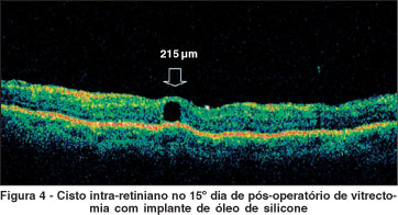
MLA, sexo masculino, 66 anos, portador de olho único, apresentou DR regmatogênico com mácula descolada. Foi submetido a tratamento cirúrgico com vitrectomia e implante de óleo de silicone. No 15º DPO, a retina mostrava-se aplicada, sem alterações maculares. O OCT evidenciou uma retina aplicada e um "cisto" perifoveal de 215 µm (Figura 4).
Caso 5
IIB, sexo masculino, 34 anos, vítima de trauma perfurante e corpo estranho intra-ocular (CEIO) no olho esquerdo há 8 meses, submetido à facectomia com implante de lente intra-ocular, vitrectomia e óleo de silicone, apresentando acuidade visual de 20/200. O exame biomicroscópico, prejudicado pela opacidade de cápsula posterior, evidenciou um edema de mácula. O OCT, além de demonstrar o espessamento macular (601 µm na região foveal), constatou uma estrutura de hiper-refletividade, anterior à membrana limitante interna, tendo como diagnóstico uma membrana epi-retiniana (MER) (Figura 5). Assim, foi programada uma intervenção cirúrgica para retirar o óleo de silicone, remover da MER e realizar uma injeção de triancinolona intravítrea para regressão do edema de macular.

Caso 6
ETA, sexo feminino, 46 anos, com baixa de acuidade visual progressiva há 10 meses e piora súbita há 5 dias. Apresentando DR total, foi submetida a vitrectomia com implante de óleo de silicone. No intra-operatório, foi visualizado um buraco macular. No 5º DPO, a suspeita durante a cirurgia foi confirmada na biomicroscopia. Entretanto, o OCT ainda evidenciou tecido retiniano na lesão, firmando diagnóstico de buraco lamelar (Figura 6).

Caso 7
RFM, sexo feminino, 56 anos, referindo baixa de AV de longa data no olho esquerdo que "sumiu" há 1 mês. Diagnosticado DR total, foi realizada vitrectomia com implante de óleo de silicone. No 20º DPO foi visualizado um buraco macular. O OCT confirmou o exame clínico, demonstrando ausência completa de tecido retiniano na lesão (Figura 7).

DISCUSSÃO
O mecanismo de funcionamento do OCT é similar ao da ultra-sonografia, diferindo que o OCT utiliza os reflexos de ondas luminosas infravermelhas ao invés de ondas acústicas(12). A existência de óleo de silicone representa um obstáculo na interpretação dos achados e na obtenção de imagens adequadas na ecografia B(13-14). O silicone intra-ocular causa um aumento aparente do diâmetro axial do olho, devido à menor velocidade do som neste meio (normal = 1584 m/s, silicone = 980 m/s)(13,15). Kumar et al., estudaram a influência da mudança de decúbito no resultado da ecografia em olhos com óleo de silicone, podendo facilitar os resultados(15). Apesar disto, estas imagens podem ser enganadoras e requerem experiência na interpretação(16).
Neste estudo, observou-se que as imagens obtidas pelo OCT, no pós-operatório de pacientes submetidos à cirurgia vítreo-retiniana e óleo de silicone como tamponante temporário, foram de boa qualidade, não apresentando dificuldades técnicas para execução e/ou interpretação dos resultados. As alterações refrativas induzidas pelo óleo de silicone intra-ocular são compensadas pelo OCT no momento de focar a estrutura retiniana, através de uma regulagem no monitor do aparelho. Esta hipermetropização é devida, fundamentalmente, ao maior índice de refração do silicone; variando, em média, de 4 a 7 dioptrias positivas(17-18).
Alguns autores mostraram um estudo com 14 pacientes submetidos a vitrectomia e óleo de silicone (como alternativa substituta para o gás) na cirurgia de buraco macular, sendo que o seguimento desde o 1º DPO foi realizado com OCT, tendo este um importante papel para detectar o fechamento do buraco, confirmando precocemente o sucesso cirúrgico(19). Neste estudo, observou-se na oftalmoscopia indireta a aplicação retiniana em todos os casos no pós-operatório. Na biomicroscopia de fundo foram constatados edema de mácula, gotícula residual de perfluorcarbono e buraco macular não vistos na oftalmoscopia indireta. O OCT, além de demonstrar o que a biomicroscopia de fundo e a oftalmoscopia indireta evidenciaram, também constatou a presença de um "cisto" intra-retiniano, membrana epi-retiniana e buraco lamelar, não detectados pelos outros métodos propedêuticos.
CONCLUSÃO
O Stratus OCT demonstrou uma boa eficácia para detectar alterações maculares em olhos com óleo de silicone, podendo ser útil em avaliações pós-operatórias. É um exame não invasivo, de fácil execução e interpretação, podendo ser incorporado ao arsenal propedêutico no seguimento das cirurgias vítreo-retinianas, mesmo com este tamponante temporário de retinopexia.
REFERÊNCIAS
1. Huang D, Swanson EA, Lin CP, Schuman JS, Stinson WG, Chang W, et al. Optical coherence tomography. Science. 1991 22;254(5035):1178-81.
2. Jaffe GJ, Caprioli J. Optical coherence tomography to detect and manage retinal disease and glaucoma. Am J Ophthalmol. 2004;137(1):156-69.
3. Hee MR, Puliafito CA, Wong C, Reichel E, Duker JS, Schuman JS, et al. Optical coeherence tomography of the human retina. Arch Ophthalmol. 1995; 113(3):325-32.
4. Fercher AF, Mengedoht K, Werner W. Eye-length measurement by interferometry with partially coherence light. Opt Lett. 1988;13:186-8.
5. Puliafito CA, Hee MR, Lin CP, Reichel E, Schuman JS, Duker JS, et al. Imaging of macular diseases with optical coherence tomography. Ophthalmology. 1995;102(2):217-29.
6. Toth CA, Narayan DG, Boppart SA, Hee MR, Fujimoto JG, Birngruber R, et al. A comparison of retinal morphology viewed by optical coherence tomography and by light microscopy. Arch Ophthalmol. 1997;115(11):1425-8. Erratum in: Arch Ophthalmol 1998;116(1):77.
7. Schuman JS, Pedut-Kloizman T, Hertzmark E, Hee MR, Wilkins JR, Coker JG, et al. Reproducibility of nerve fiber layer thickness measurements using optical coherence tomography. Ophthalmology. 1996;103(11):1889-98. Comment in: Ophthalmology. 1997;104(10):1530-1.
8. Puliafito CA, Hee MR, Schuman JS, Fujimoto JP. Interpretation of the OCT image. In: Puliafito CA, Hee MR, Schuman JS, Fujimoto JP, editors. Optical coherence tomography of ocular diseases. 2nd ed. Thorofare: Slack; 1996. p. 18-30.
9. Nehemy MB. Óleo de silicone em casos especiais de descolamento de retina. Rev Bras Oftalmol. 1988;47(6):331-7.
10. Castellarin A, Grigorian R, Bhagat N, Del Priore L, Zarbin MA. Vitrectomy with silicone oil infusion in severe diabetic retinopathy. Br J Ophthalmol. 2003;87(3):318-21. Comment in: Br J Ophthalmol. 2003;87(10):1303-4; author reply 1304-6.
11. Mastropasqua L, Carpineto P, Ciancaglini M, Falconio G, Harris A. Reproducibility of nerve fiber layer thickness measurements using optical coherence tomography in silicone oil-filled eyes. Ophthalmologica. 2001;215(2):91-6.
12. Medeiros FA. Comparação de métodos de imagem do disco óptico e da camada de fibras nervosas da retina para o diagnóstico do glaucoma [tese]. São Paulo. Universidade de São Paulo; 2005.
13. Fisher YL. Diagnostic ophthalmic ultrasonography. In: Tasman W, Jaeger EA, editors. Duane's Foundations of clinical ophthalmology. ed rev. Philadelphia: Lippincott-Raven; 1997. p. 1-10.
14. DiBernardo C, Schachat A, Fekrat S. Ophthalmic ultrasound: a diagnostic atlas. New York: Thieme; 1998.
15. Kumar A, Sharma N, Singh R. Prone position ultrasonography in silicone filled eyes. Acta Ophthalmol Scand. 1998;76(4):496-8.
16. Corrêa ZMS, Marcon IM. A ultra-sonografia diagnóstica (modo B) na avaliação pós-operatória das cirurgias vítreo-retinianas. Arq Bras Oftalmol. 2003;66 (1):33-8.
17. Nehemy MB, Nehemy ALG, Hilgert AHG, Garcia GFG. Alterações refrativas induzidas pelo óleo de silicone intraocular. Arq Bras Ofalmol. 1990;53(4):175-8.
18. Hotta K, Sugitani A. Refractive changes in silicone oil-filled pseudophakic eyes. Retina. 2005;25(2):167-70.
19. Jumper JM, Gallemore RP, McCuen BW 2nd, Toth CA. Features of macular hole closure in the early postoperative period using optical coherence tomography. Retina. 2000;20(3):232-7.
Endereço para correspondência:
Otacílio de Oliveira Maia Júnior.
Rua Machado Bittencourt, 29 - Apto. 53
São Paulo (SP) CEP 04044-000
E-mail: [email protected]
Recebido para publicação em 25.04.2006
Última versão recebida em 27.07.2006
Aprovação em 02.08.2006
Trabalho desenvolvido no Departamento de Oftalmologia do Hospital das Clínicas da Faculdade de Medicina da Universidade de São Paulo - USP - São Paulo (SP) - Brasil.

How to cite this article:
 ABO is licensed under a Creative Commons Attribution-NonComercial 4.0 Internacional.

